
Barrett oesophagus
I'll read the medical library skill first, then search for Barrett oesophagus content in parallel.
Reading File
Now let me search the library and PubMed in parallel:
Finding Sources
Searching PubMed
"Barrett esophagus"[MeSH Terms] AND management
Excellent sources found. Now let me read the most comprehensive ones in parallel - Yamada's Gastroenterology, Robbins Pathology, Goldman-Cecil Medicine, and Clinical Gastrointestinal Endoscopy:
Reading File
Reading File
Reading File
Reading File
Excellent content. Now let me fetch the Robbins pathology images and read more of the Goldman-Cecil management sections and Endoscopy surveillance content in parallel:
Loading Image
Reading File
Reading File
Now let me fetch the Barrett's endoscopy and histology images from Robbins, and read the final part of the Goldman-Cecil treatment section:
Loading Image
Loading Image
Reading File
Let me fetch the management flowchart and the dysplasia image from Robbins:
Loading Image
I now have comprehensive, well-illustrated content from multiple authoritative textbooks. Here is the full answer:
Barrett Oesophagus
Definition
Barrett oesophagus (BE) is an acquired condition in which the normal stratified squamous epithelium of the distal oesophagus is replaced by metaplastic columnar epithelium containing intestinal-type goblet cells. It is the consequence of chronic gastro-oesophageal reflux disease (GORD/GERD) causing repeated mucosal injury. Its clinical importance lies entirely in its well-recognised association with oesophageal adenocarcinoma.
- Robbins, Cotran & Kumar Pathologic Basis of Disease, p. 708
- Goldman-Cecil Medicine, p. 1432
Epidemiology
- Found in approximately 5-15% of patients undergoing endoscopy for GORD symptoms; population prevalence ~1.3-1.6%.
- Approximately 45% of patients have no reflux symptoms at the time of diagnosis.
- Predominantly affects middle-aged White men (40-60 years), though ~25% of patients are women or under 50.
- Prevalence plateaus between the 7th and 9th decades.
- Risk factors: chronic/frequent GERD, male sex, older age, central obesity (waist circumference is an independent risk factor even after adjusting for BMI and GERD), smoking, hiatal hernia, European descent.
Pathobiology
Patients who develop BE typically have:
- Greater oesophageal acid and bile exposure (confirmed on 24-hour pH monitoring)
- Hiatal hernias that are longer and associated with larger defects than in patients without BE
The cell of origin of the columnar metaplasia remains debated. Candidates include:
- Dedifferentiation of squamous epithelium
- Stem cells from the basal layer of the oesophageal epithelium
- Oesophageal submucosal glands
- Gastric cardia
- Residual embryonal stem cells at the gastro-oesophageal junction
The transcription factor CDX2, inducible by both acid and bile salts, plays a key role in promoting columnar epithelium development at the gastro-oesophageal junction. A small subset of patients may have an inherited predisposition.
Adipokines also modulate risk: higher ghrelin levels are associated with increased BE risk in the general population, while leptin is positively linked to GORD symptoms but inversely to BE among GORD controls.
Morphology
Gross / Endoscopic
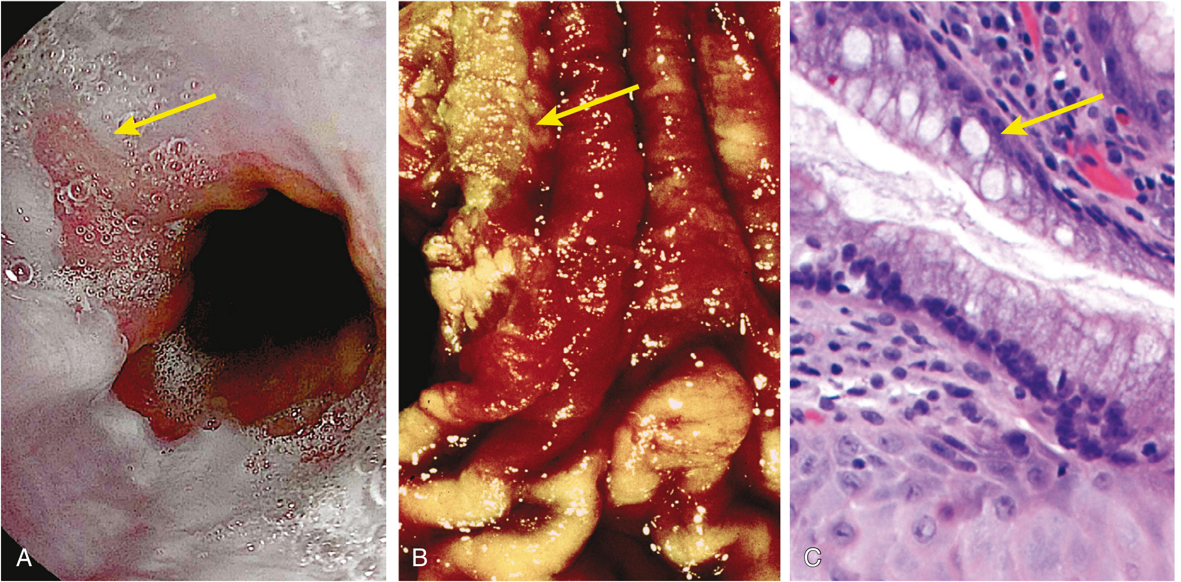
Barrett oesophagus appears endoscopically as tongues of red, velvety (salmon-pink) mucosa extending upward from the gastro-oesophageal junction (GEJ), alternating with the pale residual squamous mucosa proximally and tan gastric mucosa distally.
Fig. 17.9 (Robbins) - Barrett oesophagus: endoscopic view, gross specimen, and histology showing goblet cells (arrow)
Segment classification:
| Type | Length | Risk |
|---|---|---|
| Long-segment BE | ≥ 3 cm | Higher risk of dysplasia/carcinoma |
| Short-segment BE | < 3 cm | Lower risk; may lack GERD symptoms |
Microscopic
- Intestinal-type metaplasia: squamous epithelium replaced by columnar cells.
- Goblet cells are diagnostic - they contain mucus vacuoles that stain pale blue and give a wine-goblet shape to the remaining cytoplasm (Fig. 17.9C above).
- Non-goblet columnar cells (gastric-type foveolar cells) are also present; whether they alone suffice for diagnosis is debated internationally (the UK and Japan do not require goblet cells).
Endoscopic Landmarks (Diagnosis)
The ACG and AGA define BE as endoscopically recognisable columnar metaplasia confirmed pathologically by intestinal metaplasia (goblet cells).
Key anatomical markers:
- GEJ (gastro-oesophageal junction): defined in North America as the most proximal extent of gastric folds.
- SCJ / Z-line (squamocolumnar junction): junction of squamous and columnar mucosa; normally coincides with GEJ but is displaced proximally in BE.
- Diagnosis requires displacement of the SCJ ≥ 1 cm proximal to the GEJ plus intestinal metaplasia on biopsy.
- Prague C & M criteria are used to standardise grading of the Barrett segment extent.
Dysplasia
Dysplasia in BE is classified as low-grade (LGD) or high-grade (HGD).
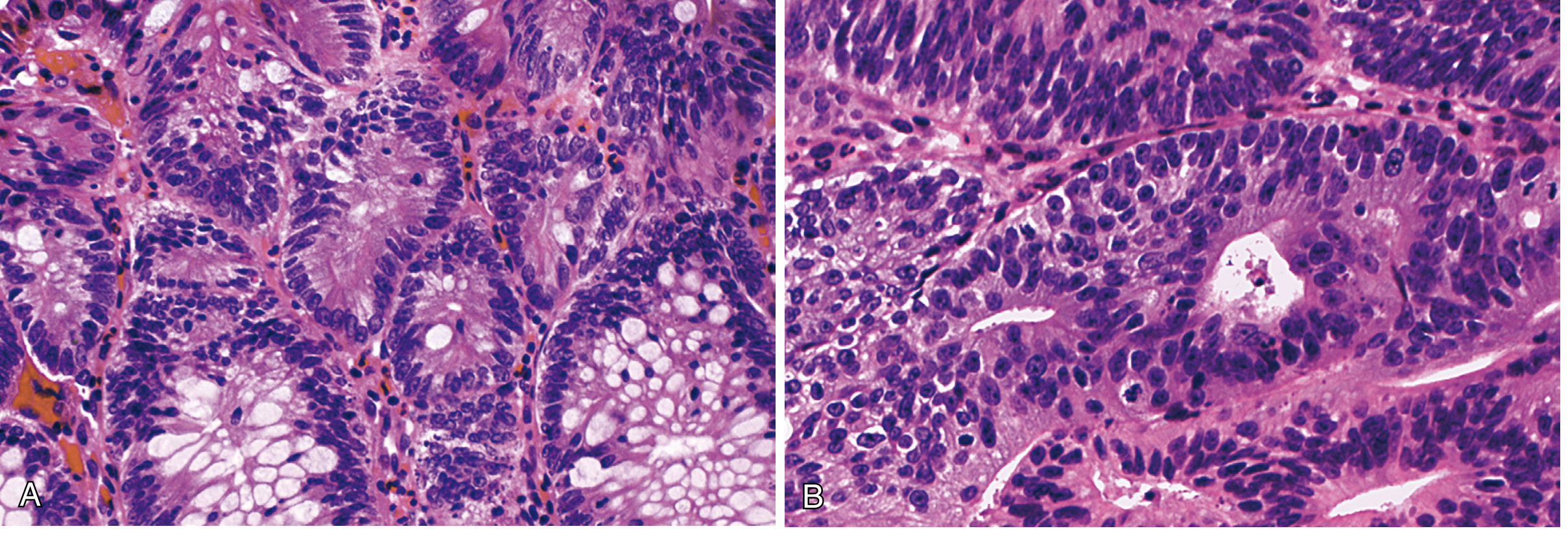
Features of dysplasia (both grades):
- Atypical mitoses
- Nuclear hyperchromasia, irregularly clumped chromatin
- Increased nuclear-to-cytoplasmic ratio
- Failure of epithelial cells to mature as they migrate to the surface
- Gland budding, irregular shapes, cellular crowding
Fig. 17.10 (Robbins) - LGD (A, left: abrupt transition from metaplasia to dysplasia with nuclear elongation) and HGD (B, right: severe nuclear pleomorphism, loss of polarity)
Genomic sequencing has identified driver mutations in cancer genes in BE biopsies; oncogenic mutations are more numerous in specimens showing dysplasia. Risk of progression is highest with goblet cells present, long-segment disease, prolonged symptoms, older age, and European ethnicity.
Cancer Risk
- Annual risk of oesophageal adenocarcinoma in BE: ~0.1 to 0.3% per year.
- Most esophageal adenocarcinomas arise in association with BE, but most individuals with BE will never develop cancer.
- Obesity is directly associated with progression to adenocarcinoma in BE patients; lower adiponectin and higher leptin are proposed markers of progression.
Clinical Features
BE is clinically difficult to distinguish from uncomplicated GERD. Paradoxically, some patients with BE have impaired sensitivity to oesophageal acid and may lack GERD symptoms entirely. Features that raise suspicion include:
- Onset of reflux symptoms at an earlier age
- Longer duration and greater severity (especially nocturnal) of reflux symptoms
- Prior GERD complications (esophagitis, ulceration, stricture, bleeding)
Non-Endoscopic Screening
The Cytosponge-trefoil factor 3 (TFF3) device is an emerging non-endoscopic tool: a mesh sponge in a soluble gelatin capsule is swallowed, then retrieved ~5 minutes later to collect cells for TFF3 immunohistochemistry. A randomised trial showed it significantly increased early diagnosis of BE.
Treatment
Medical
- Proton pump inhibitors (PPIs): cornerstone of therapy; reliably relieve symptoms and heal oesophagitis, but do not clearly induce regression of Barrett histology. Abnormal acid exposure persists in ~25% of patients despite twice-daily PPI.
- PPI + aspirin (300-325 mg/day): combination reduces progression to high-grade dysplasia, adenocarcinoma, or death.
- Anti-reflux surgery: same indications as for GERD without BE; should not be regarded as a superior cancer-prevention strategy.
Surveillance
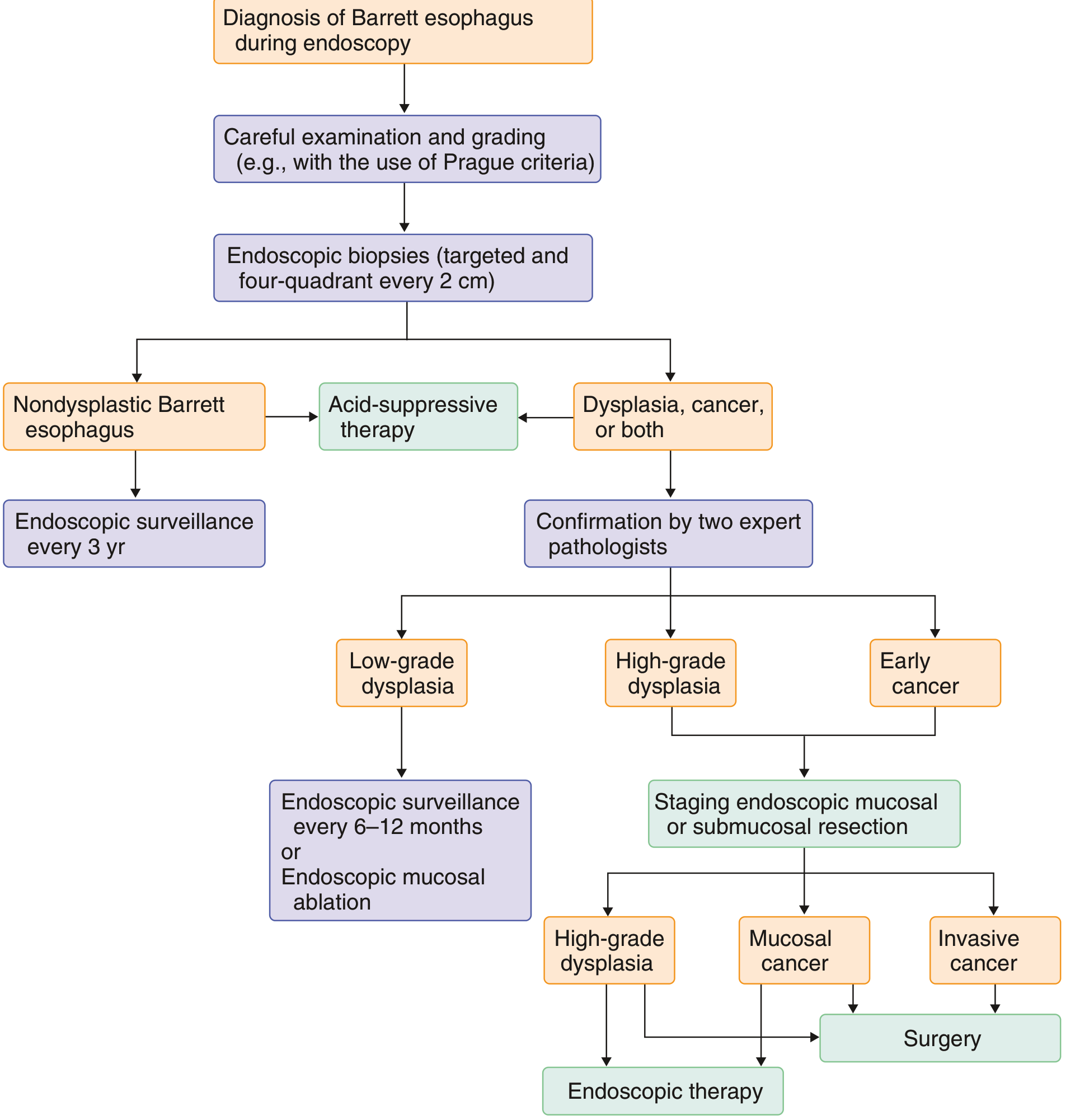
Systematic four-quadrant biopsies every 2 cm along the entire Barrett segment, after controlling reflux-related inflammation with antisecretory therapy.
| Dysplasia Grade | Recommended Action |
|---|---|
| No dysplasia | Surveillance endoscopy every 3-5 years |
| Low-grade dysplasia (LGD) | Confirm with expert GI pathologist; repeat endoscopy in 6 months; then surveillance every 6-12 months OR radiofrequency ablation (RFA) |
| High-grade dysplasia (HGD) | Confirm with expert GI pathologist; endoscopic ablation (RFA ± EMR) preferred over surgery |
| Visible mucosal abnormality | Endoscopic mucosal resection (EMR) for staging |
| Intramucosal carcinoma (T1a) | Endoscopic therapy (EMR/ablation) |
| Invasive cancer (>T1a) / failed endoscopic therapy | Surgical oesophagectomy |
Advanced imaging techniques such as chromoendoscopy and narrow-band imaging (NBI) increase the diagnostic yield of surveillance biopsies and are becoming standard practice.
Prognosis
With appropriate surveillance, most patients with BE will never develop oesophageal adenocarcinoma and will die of other causes. The annual cancer incidence is low (~0.1-0.3%), though individual risk varies by segment length, presence of dysplasia, and patient demographics.
Recent Evidence (2024)
A 2024 network meta-analysis comparing interventions for BE (PMID 38709808) and a systematic review and meta-analysis on argon plasma coagulation (APC) for BE ablation (PMID 37800316) provide updated comparative data on endoscopic ablative strategies, with RFA remaining the dominant modality and APC showing efficacy in selected settings.
Sources: Robbins, Cotran & Kumar Pathologic Basis of Disease | Goldman-Cecil Medicine | Robbins & Kumar Basic Pathology | Clinical Gastrointestinal Endoscopy (3e) | Yamada's Textbook of Gastroenterology (7e)
This is a shared conversation. Sign in to Orris to start your own chat.