
A 45 year old man has pain in his knee around 3 month
Where exactly is the knee pain located? · What is the nature of the pain / associated symptoms? · Any relevant history?
A 45 year old man has pain in his knee since 3 month , he has history of fall on tiles strech in his leg on palpatation slightly muscle type should around knee what is diagnosis and management
knee ligament anatomy diagram medial lateral collateral
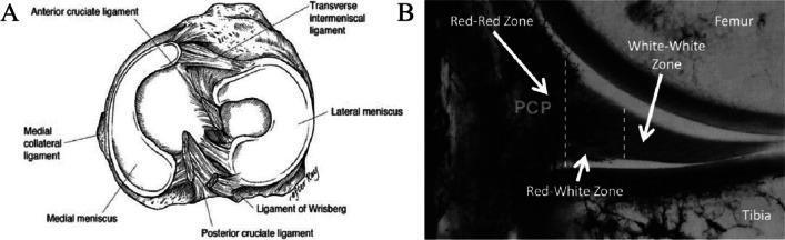
This dual-panel educational figure illustrates the gross and microvascular anatomy of the human knee meniscus. Panel A is a superior-view anatomical diagram of the tibial plateau, highlighting the C-shaped medial meniscus and circular lateral meniscus. Key supporting structures are labeled, including the anterior and posterior cruciate ligaments, the transverse intermeniscal ligament, the medial collateral ligament, and the ligament of Wrisberg. Panel B is a frontal-section diagnostic image of the knee's medial compartment following India ink perfusion to demonstrate vascularity zones. It identifies three distinct regions critical for surgical repair assessment: the peripheral 'Red-Red Zone' (highly vascularized, appearing dark from ink perfusion), the central 'White-White Zone' (avascular, appearing bright), and the transitional 'Red-White Zone' located between them. The image serves to educate on the spatial relationship between meniscal fibrocartilage, ligamentous attachments, and the graded blood supply that dictates the intrinsic healing potential of meniscal tears.
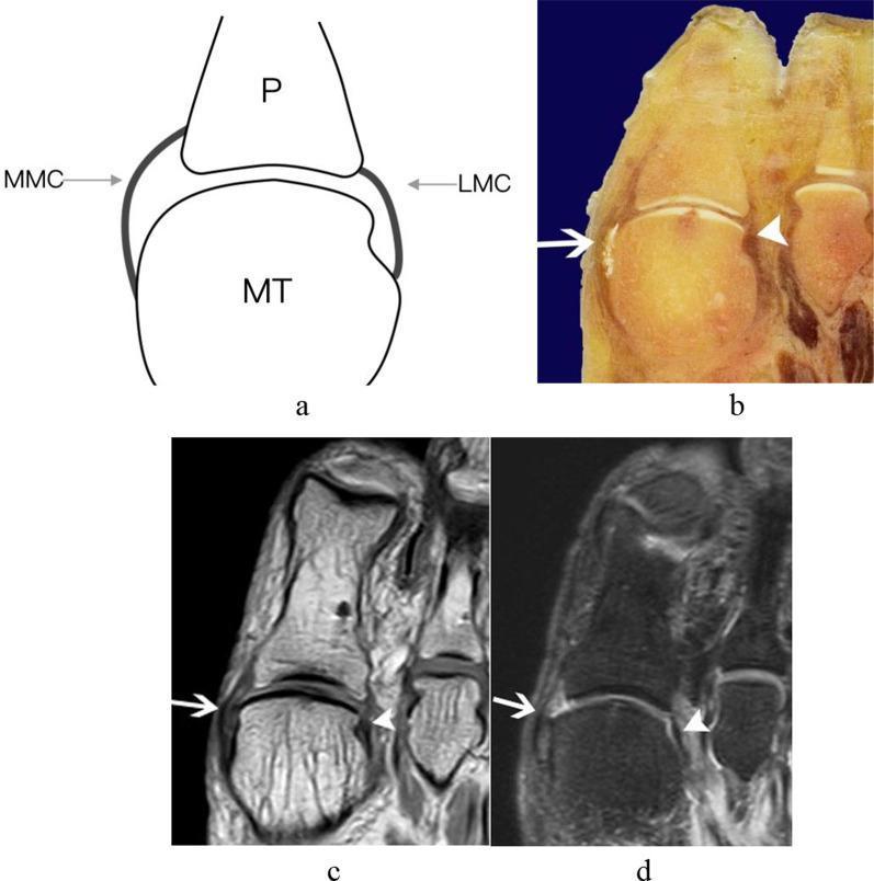
This composite educational resource depicts the anatomy of the medial (MMC) and lateral main collateral ligaments (LMC) of the first metatarsophalangeal joint (MTPJ) using four distinct modalities. (a) A transverse schematic diagram illustrates the origin of the ligaments on the metatarsal head (MT) and their insertion on the proximal phalangeal base (P). (b) A transverse anatomic slice of a cadaveric specimen shows the physical structure of these ligaments as supportive bands flanking the joint. (c) A transverse T1-weighted image (T1WI) and (d) a transverse T2-SPAIR MRI sequence demonstrate the clinical presentation of these structures. In both MRI modalities, the ligaments are visualized as distinct, curved, hypointense (dark) signal bands. White arrows indicate the medial collateral ligament, while white arrowheads point to the lateral collateral ligament. The collection serves to correlate gross anatomy with radiological findings, highlighting the ligaments' role in stabilizing the first MTPJ and their typical low-signal appearance on standard MRI sequences used for musculoskeletal assessment.
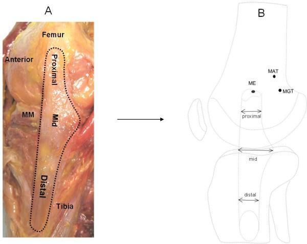
This composite image illustrates the anatomy and morphometry of the superficial medial collateral ligament (sMCL) of the knee. Image A is a clinical photograph of a right cadaveric knee dissection in the medial view. The sMCL is outlined and partitioned into proximal, mid, and distal segments. It demonstrates the ligament's broad, fan-like structure originating from the femur and inserting onto the tibia, noting its close anatomical relationship with the medial meniscus (MM). Image B is a corresponding schematic diagram used for quantitative morphometric analysis. It identifies three key femoral landmarks: the medial epicondyle (ME), medial adductor tubercle (MAT), and medial gastrocnemius tubercle (MGT). Arrows indicate the anteroposterior width measurements at the proximal, mid, and distal levels of the sMCL. The diagram highlights the characteristic triangular or hourglass morphology where the mid-portion at the joint line is significantly wider than the proximal and distal segments. This educational material is designed for orthopedic and anatomical studies focusing on knee ligamentous stability and surgical landmarks for MCL reconstruction.
Clinical Assessment: 45-Year-Old Male with 3 Months of Knee Pain
Likely Diagnosis
Reasoning from the history:
| Feature | Significance |
|---|---|
| Fall on tiles | Traumatic mechanism - sudden forced leg extension/flexion |
| Stretch in the leg | Suggests eccentric muscle overload - classic for quadriceps or hamstring strain |
| Pain around the knee (periarticular) | Not isolated joint line - suggests muscle/tendon/ligament involvement |
| Slight muscular tightness/tenderness on palpation | Residual muscle strain / partial tear with ongoing spasm |
| 3-month duration | Chronic/subacute stage - inadequately managed acute injury |
Differential Diagnoses to consider:
- Quadriceps muscle strain (most likely - fall + stretch mechanism, tenderness around patella/above knee)
- Medial Collateral Ligament (MCL) sprain - if medial side tenderness
- Meniscus injury - if there was rotational component to the fall
- Patellofemoral pain syndrome - secondary to altered gait/loading after initial injury
- Pes anserine bursitis - in a 45-year-old man, common cause of medial knee pain
Investigations
First Line:
- X-ray knee (AP + lateral views) - to rule out fracture, avulsion injury, or degenerative changes
- Ultrasound of knee - cost-effective for assessing muscle/tendon partial tears, ligament integrity, bursitis
Second Line (if needed):
- MRI knee - gold standard for soft tissue injuries; best sensitivity/specificity for ligament tears, meniscal tears, muscle pathology. Not urgent but very useful at 3 months if no improvement.
- CBC, ESR, CRP, uric acid - to rule out inflammatory/infective arthritis
Management
Phase 1 - Acute / Subacute Pain Relief:
- NSAIDs (e.g., Ibuprofen 400-600 mg TDS with meals, or Naproxen 500 mg BD) - for pain and inflammation. This is important for recovery and patient satisfaction, per Rosen's Emergency Medicine.
- Muscle relaxants (e.g., Cyclobenzaprine or Methocarbamol) - if significant muscle spasm is present on examination
- Ice application for 15-20 minutes, 3-4 times/day (first 48 hours of acute flares)
- Avoid compressive wraps that may worsen distal venous stasis
Phase 2 - Rehabilitation (most important at 3 months):
- Range of motion (ROM) exercises - gentle passive then active knee flexion/extension
- Quadriceps strengthening - straight leg raises, wall slides, terminal knee extensions
- Hamstring stretching - seated towel stretch
- Proprioception training - single leg stance, balance board
- Gradual weight-bearing progression - full weight-bearing as tolerated
"Complete recovery from a muscle strain can take weeks to months. Any patient with significant injury should be referred for physical therapy." - Rosen's Emergency Medicine
Phase 3 - Referral:
- Orthopedic / Sports Medicine referral if:
- No improvement after 6 weeks of physiotherapy
- MRI shows complete ligament or tendon tear
- Suspicion of meniscal tear (locking, giving way, positive McMurray's test)
Red Flags to Watch For:
- Inability to fully extend the knee (suggests quadriceps tendon rupture - needs urgent surgical repair)
- Joint effusion / locking / giving way (meniscal or cruciate ligament pathology)
- Night pain or systemic symptoms (consider malignancy or infection)
Summary
| Most Likely Diagnosis | Chronic periarticular muscle strain (quadriceps/hamstrings) post-traumatic, possibly with ligament sprain |
| Key Investigation | X-ray first; MRI if no improvement |
| Key Treatment | NSAIDs + physiotherapy (strengthening & stretching) |
| Referral | Sports medicine / orthopedics if not improving |
X ray showing no fracture
knee soft tissue injury MRI ligament sprain
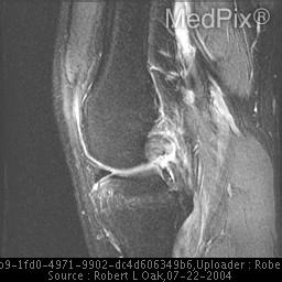
**Imaging Modality:** Sagittal T2-weighted fat-suppressed Magnetic Resonance Imaging (MRI). **Anatomical Region:** Lateral-to-midline view of the human knee joint, including the distal femur, proximal tibia, patella, and posterior soft tissue structures. **Observed Pathology:** The image demonstrates a posterior cruciate ligament (PCL) injury. The PCL appears thickened with heterogeneous, increased intrasubstance signal intensity, consistent with a partial-thickness tear or high-grade sprain. While the ligamentous fibers maintain some continuity, there is significant blurring of the normal low-signal (dark) morphology. **Characteristic Visual Features:** - **Signal Abnormality:** Elevated T2 signal within the PCL fibers indicating edema and hemorrhage. - **Morphology:** Loss of the characteristic "hockey stick" or crisp curvilinear black appearance of a healthy PCL. - **Associated Findings:** Mild joint effusion is visible in the suprapatellar bursa. The surrounding soft tissues and posterior capsule show reactive hyperintensity. **Key Diagnostic Features:** Intrasubstance signal changes in the PCL on fluid-sensitive sequences are the primary diagnostic indicators for distinguishing ligamentous strain or partial rupture from a complete discontinuity (full-thickness tear).
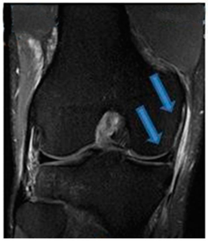
This diagnostic image is a coronal T2-weighted fluid-sensitive MRI of the human knee, specifically focusing on the medial compartment structures. The image shows the distal femur, proximal tibia, and the intervening joint space. Large blue arrows indicate pathology in the medial aspect of the joint. One arrow points to the medial collateral ligament (MCL), which exhibits thickening and increased signal intensity, suggesting a high-grade sprain or tear. The second arrow points to the medial meniscus, where focal high-signal intensity is visible within the posterior horn, characteristic of a meniscal tear. Periligamentous and soft tissue edema is observed as bright (hyperintense) areas surrounding these structures. This visual representation is clinically significant for diagnosing O'Donoghue's triad components, demonstrating the radiological features of acute ligamentous and fibrocartilaginous injury in a musculoskeletal context. The image serves as an educational tool for identifying abnormal signal variations and structural disruptions in orthopedic radiology.
Updated Assessment: X-Ray Normal - No Fracture
What This Tells Us
- No bony fracture or avulsion
- No significant osteoarthritis (if joint space preserved)
- The problem is purely soft tissue - muscle, tendon, ligament, bursa, or meniscus
Revised Diagnosis
Most Likely: Chronic Soft Tissue Injury of the Knee
- Traumatic fall with leg stretch mechanism
- 3 months duration (subacute-to-chronic)
- Periarticular muscle tightness and tenderness
- Normal X-ray
| Diagnosis | Key Feature |
|---|---|
| Quadriceps/hamstring muscle strain (Grade I-II) | Tenderness above/below knee, pain on resisted extension/flexion |
| Medial Collateral Ligament (MCL) sprain | Medial knee tenderness, valgus stress test positive |
| Meniscal injury | Joint line tenderness, positive McMurray's or Thessaly test |
| Pes anserine bursitis | Medial knee pain 2-3 cm below joint line, common in middle-aged men |
| Patellofemoral pain syndrome | Anterior knee pain, crepitus, J-sign |
Next Step: MRI Knee (NOW STRONGLY INDICATED)
- Best sensitivity and specificity for all soft tissue structures
- Will identify: muscle tears, ligament sprains/tears, meniscal tears, cartilage damage, bone marrow edema (occult fracture)
- Will guide whether conservative management or surgery is needed
Management Plan (Normal X-Ray Confirmed)
1. Pharmacological
| Drug | Dose | Duration |
|---|---|---|
| Ibuprofen 400 mg TDS or Naproxen 500 mg BD | With food | 2-4 weeks |
| Paracetamol 500-1000 mg TDS | Adjunct | As needed |
| Muscle relaxant (Cyclobenzaprine 5 mg TDS) | If spasm present | Short course only |
| Topical diclofenac gel | Over painful area | 2-4 weeks |
2. Physical Therapy (Core of Treatment at 3 Months)
- Quadriceps strengthening: straight leg raises, wall squats, terminal knee extensions
- Hamstring stretching: towel stretches, supine hamstring stretch
- Calf raises for posterior chain stability
- Proprioception: single leg balance, wobble board
- Ice massage after exercise sessions (10-15 min)
- Avoid: deep squats, running, jumping until pain-free
3. Supportive Measures
- Knee sleeve/brace - provides compression and proprioceptive feedback
- Activity modification - avoid aggravating activities, continue gentle walking
- Weight management - even small weight loss reduces knee load significantly
4. Investigations Still Needed
- MRI knee - to definitively characterize the soft tissue injury
- Consider ultrasound - cheaper, real-time assessment of tendons and ligaments
5. Referral Criteria
- MRI shows complete ligament/tendon tear or significant meniscal tear
- No improvement after 6-8 weeks of physiotherapy
- Joint effusion, locking, or giving way develops
Key Message
Normal X-ray + post-traumatic knee pain at 3 months = soft tissue injury that needs MRI + physiotherapy. This is not an emergency but is undertreated - the 3-month duration suggests the initial injury was not properly rehabilitated.
I precribe him tab aceclofenac,pcm and serratiopeptidase Bd, tab diclo Bd, omni gel and crep bandage, tab pantop and tab lycopene