
Classify odontogenic tumors as a long and describe all related tumors as a classification for long question in university exam of bds 3rd yr oral pathology
ameloblastoma follicular plexiform histology jaw
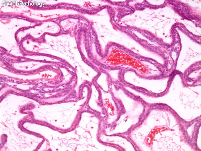
This histopathology slide, stained with Hematoxylin and Eosin and viewed under brightfield light microscopy at low to mid magnification, demonstrates classic plexiform ameloblastoma architecture within the jaw. Neoplastic odontogenic epithelium forms interconnected cords and anastomosing strands embedded in a loose, highly vascular stroma of fibrous connective tissue. The epithelial cords are composed of basaloid cells with peripheral cells often lacking the typical palisading and reverse nuclear polarity; within the cords, cells are more loosely arranged, resembling an inner stellate reticulum. Cystic change is uncommon in this plexiform subtype. The overall pattern shows benign cytology but a propensity for local invasion and bone destruction clinically. These features support the diagnosis of ameloblastoma, plexiform type, and help distinguish it from other odontogenic tumors and jaw cysts. Diagnostic significance includes confirming a locally aggressive, benign neoplasm that necessitates wide surgical excision with clear margins to minimize recurrence. Clinically, this pattern correlates with gradual cortical expansion and radiographic lytic lesions in the mandible or maxilla. In educational settings, the image is valuable for teaching odontogenic histology, subtyping ameloblastoma, and illustrating variations among follicular, plexiform, and desmoplastic patterns. Correlation with radiographs (multilocular radiolucencies) and clinical jaw swelling reinforces a multidisciplinary management approach.
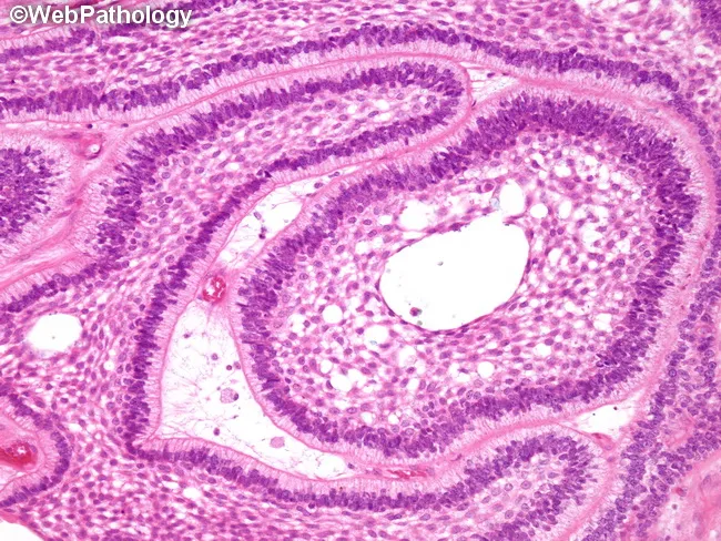
Imaging modality: Light microscopy of hematoxylin and eosin stained paraffin sections of an odontogenic jaw tumor. Anatomical localization: mandible or maxilla, commonly the posterior mandible; histologic plane is a transverse section through dental lamina–like islands within dense fibrous stroma. In this specimen, classic follicular ameloblastoma pattern dominates, with round to ovoid nests of odontogenic epithelium exhibiting peripheral palisading of columnar cells and reverse nuclear polarity away from the basement membrane. The peripheral layer rests on a basement membrane and displays elongated, polarized nuclei; centrally, loosely arranged polygonal to stellate reticulum–like cells form loosely cohesive islands. The islands are separated by mature fibrous connective tissue stroma, without marked keratinization. The overall architecture mimics the enamel organ of developing tooth, hence the term follicular. Additional patterns such as plexiform growth may be present in other areas, but not evident here. Clinically, ameloblastomas are benign but locally aggressive, with potential for local invasion and recurrence if incompletely excised. The image provides diagnostic evidence for odontogenic tumor identification, distinguishing from cystic lesions and other jaw tumors. Relevant differential diagnoses include plexiform ameloblastoma, acanthomatous type, and odontogenic keratocyst. This histology supports surgical planning, prognosis assessment, and educational discussion of jaw tumor pathology.
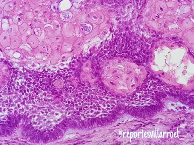
Imaging modality and technique: Light microscopy of hematoxylin and eosin (H&E) stained paraffin-embedded jaw lesion tissue, sectioned for routine histology. Anatomical localization: odontogenic tumor of the mandible, with epithelial islands embedded in a dense fibrous stroma consistent with ameloblastoma. The image demonstrates the acanthomatous variant, in which extensive squamous metaplasia occurs within the central portion of the epithelial islands, accompanied by keratin formation in the stellate reticulum. Peripheral ameloblast‑like cells show palisaded, polarized basilar nuclei; the central cells resemble the loosely arranged, eosinophilic stellate reticulum. The surrounding stroma is dense and vascular, with inflammatory cells in some fields. Notable architectural diversity is evident: nests and cords display follicular and plexiform arrangements, with focal desmoplastic areas and possible granular cell change in other regions. Such histology reflects intricate tumor biology while retaining characteristic features of ameloblastoma. Diagnostic relevance centers on distinguishing this entity from squamous odontogenic tumor and squamous cell carcinoma, as management and prognosis differ markedly. Key differentiators include peripheral palisading and reverse polarity, central stellate reticulum with metaplasia, and lack of overt malignant cytology. Clinically, recognizing acanthomatous ameloblastoma guides surgical planning and follow-up in jaw pathology and head‑neck oncology. This image serves as a reference for education and research.
odontogenic tumor classification odontoma compound complex
Classification of Odontogenic Tumors
(Long Question - BDS 3rd Year Oral Pathology)
INTRODUCTION
- Robbins & Cotran Pathologic Basis of Disease, p. 688
- Scott-Brown's Otorhinolaryngology Head & Neck Surgery, p. 453
WHO CLASSIFICATION OF ODONTOGENIC TUMORS
A. BENIGN ODONTOGENIC TUMORS
I. Tumors Arising from Odontogenic Epithelium Only
- Ameloblastoma (conventional, unicystic, extraosseous)
- Squamous Odontogenic Tumor
- Calcifying Epithelial Odontogenic Tumor (CEOT / Pindborg Tumor)
- Adenomatoid Odontogenic Tumor (AOT)
- Keratocystic Odontogenic Tumor (KCOT / Odontogenic Keratocyst)
II. Tumors of Odontogenic Epithelium AND Ectomesenchyme (Mixed)
- Ameloblastic Fibroma
- Ameloblastic Fibro-dentinoma / Fibro-odontoma
- Complex Odontoma
- Compound Odontoma
- Calcifying Cystic Odontogenic Tumor (Gorlin Cyst)
- Dentinogenic Ghost Cell Tumor
- Odontoameloblastoma
III. Tumors Arising from Odontogenic Ectomesenchyme Only
- Odontogenic Fibroma
- Odontogenic Myxoma
- Benign Cementoblastoma
B. MALIGNANT ODONTOGENIC TUMORS
Odontogenic Carcinomas:
- Metastasizing (Malignant) Ameloblastoma
- Ameloblastic Carcinoma (primary and secondary/dedifferentiated types)
- Primary Intraosseous Squamous Cell Carcinoma (solid type; arising from KCOT; arising from other cysts)
- Clear Cell Odontogenic Carcinoma
- Ghost Cell Odontogenic Carcinoma
- Sclerosing Odontogenic Carcinoma
Odontogenic Sarcomas:
-
Ameloblastic Fibrosarcoma
-
Ameloblastic Fibro-dentino-sarcoma and Fibro-odontosarcoma
-
Scott-Brown's Otorhinolaryngology, Table 25.5 (WHO modification)
-
Robbins & Cotran, Table 16.4, p. 688
DETAILED DESCRIPTION OF INDIVIDUAL TUMORS
1. AMELOBLASTOMA
| Type | Key Features |
|---|---|
| Solid/Multicystic (Conventional) | Most common; locally invasive; high recurrence |
| Unicystic Ameloblastoma | Luminal, intraluminal, intramural subtypes |
| Peripheral (Extraosseous) | Soft tissue only; least aggressive |
| Desmoplastic | Anterior jaws; fibrous stroma |
| Odontoameloblastoma | Contains dysplastic dentine and enamel |
- Origin: Rests of dental lamina
- Site: Mandibular molar-ramus region most common; mandible:maxilla = 5:1
- Age/Sex: Mean age 37 years; M:F = 1.2:1
- Clinical features: Painless slow-growing jaw swelling; may cause facial deformity, tooth loosening, cortical expansion, eggshell crackling, jaw fracture
- Radiology: Multilocular "soap-bubble" or "honeycomb" radiolucency; scalloped margins; root resorption
- Histology:
- Odontogenic epithelium arranged in islands or strands in a fibrous stroma
- Follicular pattern: Islands with peripheral palisading of columnar ameloblast-like cells showing reverse polarity (nuclei away from basement membrane); central stellate reticulum-like cells
- Plexiform pattern: Anastomosing cords/strands of odontogenic epithelium; loose vascular stroma; cystic degeneration less common
- Other variants: acanthomatous (squamous metaplasia), granular cell, basal cell, desmoplastic
- Treatment: Wide local resection with 1-1.5 cm margins; high recurrence with curettage alone
- Malignant forms: Metastasizing ameloblastoma (typical histology but distant metastases) and ameloblastic carcinoma (cellular atypia present)
- Scott-Brown's, p. 454
- Robbins & Cotran, p. 688
- Cummings Otolaryngology, p. 1616
2. ADENOMATOID ODONTOGENIC TUMOR (AOT)
- Nickname: "Two-thirds tumor" - 2/3 in anterior maxilla, 2/3 in females, 2/3 in second decade
- Origin: Reduced enamel epithelium or dental lamina
- Site: Anterior maxilla most common; often associated with an unerupted canine (in a follicular/dentigerous relationship)
- Clinical features: Slow-growing, painless swelling; well-circumscribed
- Radiology: Well-defined unilocular radiolucency encasing an unerupted tooth; may show fine calcifications ("snowflake" pattern)
- Histology: Whorled masses or rosettes of cuboidal/columnar epithelial cells forming duct-like structures; eosinophilic material in the lumina; amyloid-like material; scattered calcifications
- Behavior: Benign; very low recurrence
- Treatment: Conservative enucleation; excellent prognosis
3. CALCIFYING EPITHELIAL ODONTOGENIC TUMOR (CEOT / Pindborg Tumor)
- Origin: Stratum intermedium of the enamel organ
- Site: Posterior mandible most common; often associated with an unerupted tooth
- Age/Sex: Adults (30-50 years); no sex predilection
- Clinical features: Slow-growing jaw swelling; may erode cortical bone
- Radiology: Mixed radiolucent-radiopaque lesion; "driven snow" appearance (scattered calcifications around unerupted tooth)
- Histology: Sheets and strands of polyhedral epithelial cells with distinct intercellular bridges; Liesegang ring calcifications (concentric calcifications); homogeneous eosinophilic amyloid-like material (stains with Congo red); nuclear pleomorphism without mitoses
- Behavior: Locally invasive; recurrence possible
- Treatment: Conservative resection
4. SQUAMOUS ODONTOGENIC TUMOR (SOT)
- Origin: Rests of Malassez in the periodontal ligament
- Site: Anterior mandible and maxilla; associated with erupted teeth
- Clinical features: Painful or painless lesion; can cause tooth loosening
- Radiology: Triangular radiolucency along the lateral aspect of a tooth root
- Histology: Islands of well-differentiated squamous epithelium within a mature fibrous stroma; no peripheral palisading; no stellate reticulum (distinguishes from ameloblastoma); microcyst formation common
- Behavior: Benign; locally aggressive
- Treatment: Conservative surgical removal
5. KERATOCYSTIC ODONTOGENIC TUMOR (KCOT / Odontogenic Keratocyst)
- Origin: Remnants of the dental lamina; PTCH1 gene mutation implicated
- Site: Mandibular ramus and posterior body most common; mandible:maxilla = 3:1
- Age/Sex: Peak in 2nd-3rd decades; can occur at any age; slight male predominance
- Clinical features: Slow-growing; anterior-posterior growth within medullary bone without obvious cortical expansion; may be associated with unerupted tooth
- Radiology: Well-defined unilocular (or multilocular) radiolucency; scalloped margins; may cause bowing of inferior border of mandible; cyst lumen contains creamy keratin material
- Histology:
- Thin, uniform, parakeratinized epithelial lining (6-8 cells thick)
- Flat epithelial-connective tissue interface (no rete ridges)
- Palisaded basal cell layer with hyperchromatic nuclei (resembles basal cell carcinoma)
- Corrugated parakeratinized surface
- Satellite/daughter cysts and odontogenic epithelial rests in wall
- Behavior: High recurrence rate (25-60%) due to satellite cysts, thin fragile wall, and mitotic activity; can transform to SCC
- Association: Multiple KCOTs are a hallmark of Nevoid Basal Cell Carcinoma Syndrome (Gorlin-Goltz syndrome) - also includes multiple basal cell carcinomas, bifid ribs, calcification of falx cerebri, frontal bossing
- Treatment: Enucleation + curettage + peripheral ostectomy; chemical fixation with Carnoy's solution; decompression/marsupializaton for large lesions; resection for recurrent/aggressive cases
6. CALCIFYING CYSTIC ODONTOGENIC TUMOR (CCOT / Gorlin Cyst)
- Origin: Odontogenic epithelium
- Site: Anterior jaws; equal maxilla and mandible distribution
- Age: Wide age range; bimodal peak (2nd decade and over 60 years)
- Clinical features: Painless swelling; can be intraosseous or extraosseous
- Radiology: Well-defined radiolucency with variable calcifications; may be associated with unerupted tooth or odontoma
- Histology: Ameloblastoma-like epithelial lining; characteristic ghost cells - pale, eosinophilic, anucleate epithelial cells; dystrophic calcification of ghost cells; fibrous connective tissue wall
- Behavior: Benign; low recurrence; the solid neoplastic counterpart is called Dentinogenic Ghost Cell Tumor
- Treatment: Conservative enucleation
7. AMELOBLASTIC FIBROMA (AF)
- Origin: Both odontogenic epithelium and ectomesenchyme (true mixed tumor)
- Site: Posterior mandible
- Age/Sex: Peak age 15 years; no sex predilection; mainly in children/young adults
- Clinical features: Painless swelling; may be associated with impacted tooth
- Radiology: Well-demarcated uni- or multilocular radiolucency with sclerotic rim
- Histology: Strands and islands of odontogenic epithelium with peripheral palisading (resembling tooth germ) embedded in cell-rich ectomesenchyme resembling dental papilla (immature, myxoid stroma - key distinction from ameloblastoma which has mature fibrous stroma)
- Behavior: Benign; recurrence after incomplete removal; rare malignant transformation to ameloblastic fibrosarcoma
- Variants: When dentine is present: Ameloblastic Fibro-dentinoma (AFD); when enamel + dentine: Ameloblastic Fibro-odontoma (AFO)
- Treatment: Enucleation and curettage; generally curative
8. ODONTOMA
A. Compound Odontoma
- Site: Anterior maxilla most common
- Age: Children and adolescents; most common odontogenic hamartoma
- Clinical features: Usually diagnosed when it impedes eruption of an adjacent tooth or displaces teeth
- Radiology: Multiple small tooth-like structures (denticles/odontoids) within a fibrous sac (radiolucent peripheral zone)
- Histology: Multiple miniature well-differentiated teeth (denticles) in an organized arrangement; all dental tissues present in proper relationship
- Treatment: Surgical removal; growth ceases once mature
B. Complex Odontoma
- Site: Posterior mandible most common
- Clinical features: Painless; may impede tooth eruption
- Radiology: Radiopaque mass with surrounding radiolucent halo; no recognizable tooth structure
- Histology: Haphazardly arranged mass of enamel, dentine, cementum, and pulp tissue - no tooth-like organization; represents the more disorganized end of the odontoma spectrum
- Treatment: Surgical removal; excellent prognosis
9. ODONTOGENIC MYXOMA (OM) / FIBROMYXOMA
- Origin: Uncertain; resembles dental papilla/follicle; may arise from myofibroblasts
- Site: Posterior mandible most common (2/3 cases); mandible > maxilla
- Age/Sex: Peak 15-35 years; more common in females
- Clinical features: Slowly increasing, painless jaw swelling; can cause tooth mobility, root resorption; in maxillary lesions may cause nasal obstruction
- Radiology: Multilocular "soap-bubble" or "honeycomb" radiolucency (similar appearance to ameloblastoma and KCOT - important differential)
- Histology: Bland rounded, spindle-shaped, or angular cells evenly spaced in abundant mucinous (myxoid) matrix; finely fibrillar matrix; occasional scattered islands of odontogenic epithelium; no significant cytological atypia; more collagenous examples = fibromyxoma
- Behavior: Locally infiltrative; recurrence 10-33%; no metastases
- Treatment: Conservative curettage for small lesions; radical resection for large/diffuse lesions; frozen section assessment of margins
10. BENIGN CEMENTOBLASTOMA
- Origin: Cementoblasts (from the periodontal ligament)
- Site: Mandibular first molar region most commonly; fused to tooth root
- Age: Young adults; peak around 20 years of age; no sex predilection
- Clinical features: Pain and swelling in the affected jaw region; cortical expansion
- Radiology: Well-defined radiopaque mass attached to (fused with) the root of a tooth; surrounded by a thin radiolucent halo
- Histology: Sheets of cementum-like material with prominent reversal lines (resembling Paget's disease pattern); active cementoblasts at the periphery; fibrovascular marrow spaces
- Behavior: Benign; slow-growing; the tooth must be extracted with the tumor (as it is fused to the root)
- Treatment: Surgical removal including the associated tooth; recurrence is uncommon
11. ODONTOGENIC FIBROMA
- Origin: Odontogenic ectomesenchyme; derived from the dental follicle (simple/epithelium-poor type) or periodontal ligament (complex/epithelium-rich type)
- Site: Premolar region of mandible; intraosseous or peripheral (gingival)
- Age/Sex: Adults; M:F = 1:3
- Clinical features: Slow-growing, painless swelling with cortical expansion
- Radiology: Unilocular radiolucency with well-defined sclerotic border; may have spotted radiopacities
- Histology: Cellular fibroblastic connective tissue containing islands and strands of inactive-looking odontogenic epithelium; foci of calcified material (metaplastic cementum/osteoid/dentine)
- Treatment: Simple enucleation; curative
12. MALIGNANT ODONTOGENIC TUMORS (Brief Overview)
| Tumor | Features |
|---|---|
| Metastasizing Ameloblastoma | Histologically benign ameloblastoma but with proven distant metastases (usually lung); retrospective diagnosis |
| Ameloblastic Carcinoma | Ameloblastoma with cytological atypia (mitoses, nuclear pleomorphism); may be primary or secondary (arising in a pre-existing ameloblastoma) |
| Primary Intraosseous SCC | SCC arising within the jaw with no primary elsewhere; may arise from KCOT or other cysts |
| Clear Cell Odontogenic Carcinoma | Sheets of clear cells; infiltrative; can metastasize |
| Ghost Cell Odontogenic Carcinoma | Malignant counterpart of CCOT; atypical cells with ghost cell formation |
| Ameloblastic Fibrosarcoma | Malignant transformation of ameloblastic fibroma; sarcomatous mesenchymal component with benign epithelial component |
SUMMARY TABLE
| Category | Tumor | Origin | Key Feature |
|---|---|---|---|
| Epithelial | Ameloblastoma | Dental lamina/enamel organ | Most common true neoplasm; locally invasive |
| Epithelial | AOT | Reduced enamel epithelium | Anterior maxilla; "2/3 tumor"; duct-like structures |
| Epithelial | CEOT (Pindborg) | Stratum intermedium | Liesegang rings; amyloid; "driven snow" X-ray |
| Epithelial | SOT | Rests of Malassez | Lateral root radiolucency; no palisading |
| Epithelial | KCOT | Dental lamina; PTCH1 mutation | Parakeratinized lining; high recurrence; Gorlin syndrome |
| Epithelial | CCOT (Gorlin cyst) | Odontogenic epithelium | Ghost cells; variable calcification |
| Mixed | Ameloblastic fibroma | Epithelium + ectomesenchyme | Immature myxoid stroma; young patients |
| Mixed | Odontoma (compound) | All dental tissues | Multiple denticles; organized; hamartoma |
| Mixed | Odontoma (complex) | All dental tissues | Disorganized mass; hamartoma |
| Mesenchymal | Odontogenic myxoma | Ectomesenchyme | Myxoid matrix; multilocular X-ray; locally invasive |
| Mesenchymal | Cementoblastoma | Cementoblasts | Fused to root; radiopaque with halo; pain |
| Mesenchymal | Odontogenic fibroma | Dental follicle/PDL | Inactive epithelial islands; well-demarcated |
| Malignant | Ameloblastic carcinoma | Odontogenic epithelium | Atypia + ameloblastoma pattern |
| Malignant | Ameloblastic fibrosarcoma | Mixed | Sarcomatous stroma + benign epithelium |
IMPORTANT EXAM POINTS TO REMEMBER
- Most common odontogenic tumor overall = Odontoma (hamartoma)
- Most common true odontogenic neoplasm = Ameloblastoma
- Ameloblastoma histology: reverse polarity + peripheral palisading + central stellate reticulum
- KCOT association = Gorlin-Goltz syndrome (nevoid basal cell carcinoma syndrome)
- CEOT = Liesegang ring calcifications + amyloid-like material + Congo red positive
- AOT = "Two-thirds tumor" - anterior maxilla, females, second decade; duct-like structures
- Cementoblastoma = Only odontogenic tumor fused to the tooth root; must remove tooth with tumor
- Odontogenic myxoma = Most locally infiltrative mesenchymal tumor; multilocular like ameloblastoma
- Compound odontoma = organized (multiple denticles); Complex odontoma = disorganized mass
- Ghost cells = pathognomonic feature of CCOT (Gorlin Cyst) and dentinogenic ghost cell tumor
- Ameloblastic fibroma stroma = immature, cell-rich, myxoid (vs. ameloblastoma = mature fibrous stroma)
- Soap-bubble X-ray = ameloblastoma, KCOT, odontogenic myxoma (all three share this appearance)