
Make a long note on the topic vitreous hemorrhage for ophthalmology postgraduate exams
Vitreous hemorrhage: postgraduate exam topic map
- Definition and epidemiology
- Applied anatomy and pathogenesis
- Etiology
- Proliferative diabetic retinopathy
- Retinal tear and posterior vitreous detachment
- Retinal vein occlusion and other retinal neovascular disorders
- Trauma
- Retinal vasculitis, sickle cell disease, tumors, and systemic hematologic causes
- Clinical presentation and differential diagnosis
- History and ocular examination
- Investigations
- B-scan ultrasonography
- OCT, fluorescein angiography, and systemic work-up after media clears or as indicated
- Management
- Immediate assessment and precautions
- Observation and serial review
- Laser photocoagulation and anti-VEGF therapy for the underlying cause
- Indications, timing, and principles of pars plana vitrectomy
- Special situations
- Diabetic VH
- VH associated with retinal tear/detachment
- Traumatic VH
- Pediatric VH
- Complications, prognosis, and follow-up
- Exam-oriented algorithms, tables, and viva points
Brief overview
- Complete exam answer
- Etiology and pathogenesis
- Investigations and differential diagnosis
- Management and vitrectomy
Expand this into a complete long-answer exam note
"vitreous hemorrhage"
vitreous hemorrhage management retinal tear ultrasound vitrectomy AAO
PMID: 39172227
https://eyewiki.org/Vitreous_Hemorrhage
fundus photograph vitreous hemorrhage proliferative diabetic retinopathy
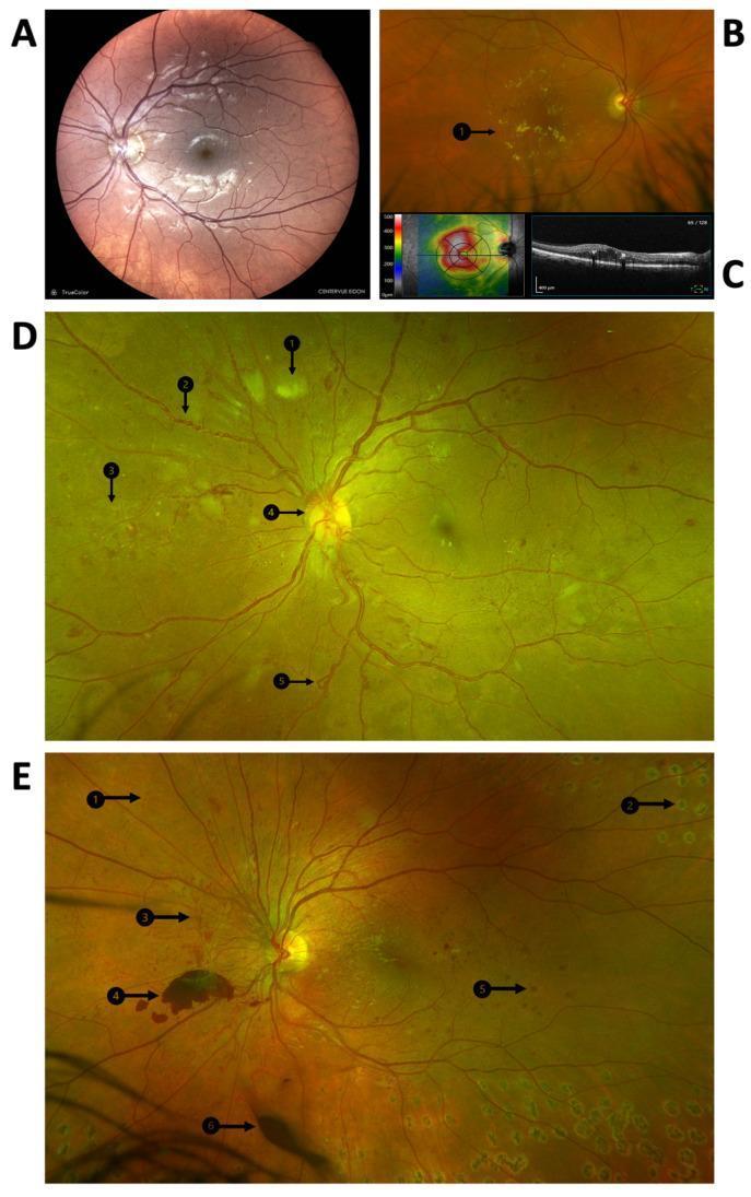
A multi-panel medical image illustrating the clinical manifestations and progression of diabetic retinopathy using fundus photography and Optical Coherence Tomography (OCT). Panel A displays a color fundus photograph of the posterior pole showing microaneurysms and dot hemorrhages indicative of diabetic maculopathy. Panel B highlights clinically significant macular edema (CSME) with visible hard exudates (B1). Panel C provides corresponding OCT imaging, including a retinal thickness heat map and a cross-sectional scan showing intraretinal fluid. Panels D and E are widefield fundus photographs demonstrating advanced proliferative diabetic retinopathy (PDR). Key pathological features identified with numbered arrows include cotton wool spots (D1), venous beading (D2), intraretinal microvascular abnormalities (IRMA) (D3/E1), neovascularization at the disc (D4), and venous loops (D5). Panel E further illustrates end-stage complications and treatment, showing laser photocoagulation scars (E2), retinal neovascularization (E3), pre-retinal hemorrhage (E4), blot hemorrhage (E5), and vitreous hemorrhage (E6). This compilation serves as an educational tool for identifying the transition from non-proliferative to proliferative diabetic disease.
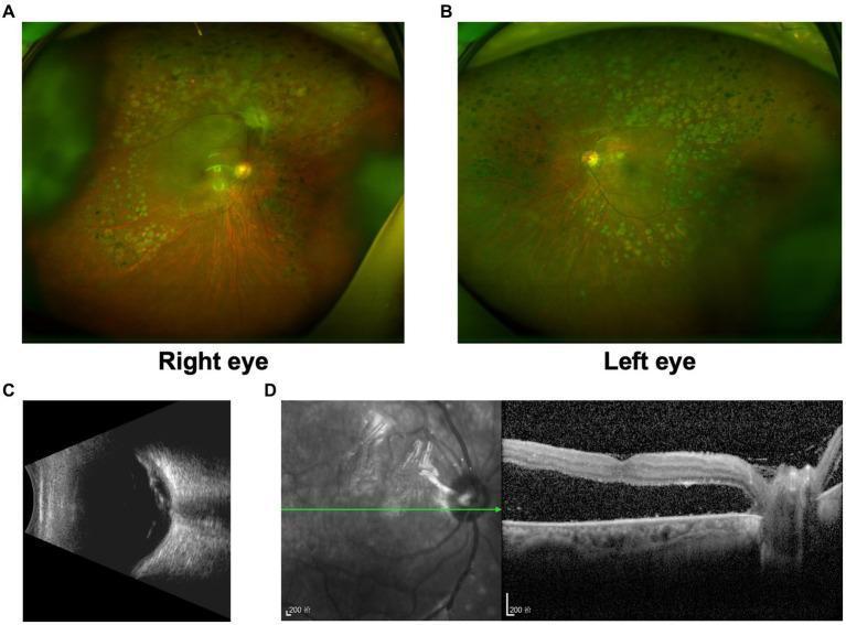
Multi-modal ophthalmic imaging of a patient with proliferative diabetic retinopathy (PDR). (A) Ultra-widefield fundus photograph of the right eye shows evidence of panretinal photocoagulation (PRP) scars in the periphery and tractional retinal detachment (TRD) involving the macula. (B) Ultra-widefield fundus photograph of the left eye demonstrates peripheral PRP laser scars with a flat, attached posterior pole. (C) B-scan ultrasound of the right eye illustrates a high-reflective undulating membrane in the vitreous cavity consistent with TRD, accompanied by low-reflective vitreous opacities suggesting hemorrhage. (D) Optical coherence tomography (OCT) of the right eye provides a cross-sectional view of the macula, revealing a thick, hyper-reflective epiretinal membrane causing significant traction and underlying subretinal fluid (retinal detachment). The images collectively demonstrate advanced diabetic eye disease and the structural complications necessitating surgical intervention.
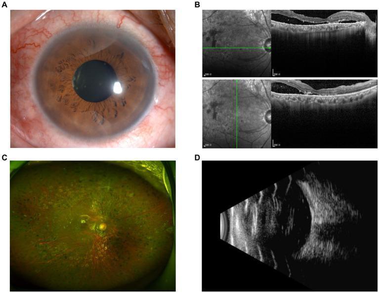
A multi-modal ophthalmological clinical panel displaying postoperative follow-up findings for a patient with a history of proliferative diabetic retinopathy. Panel A is an anterior segment photograph showing a clear cornea, intact iris with visible surgical peripheral iridectomy, and no acute inflammatory signs. Panel B displays Optical Coherence Tomography (OCT) scans alongside infrared reflectance imaging; the cross-sections demonstrate slight local subretinal fluid and retinal thickening in the macular region, indicating residual macular edema. Panel C is an ultra-widefield fundus photograph showing a mottled, greenish-yellow retinal appearance with laser photocoagulation scars in the periphery and visible retinal vasculature radiating from a defined optic disc, without evident gross retinal detachment. Panel D is a B-scan ocular ultrasound showing a well-attached posterior hyaloid/retinal interface and an acoustically empty vitreous cavity, confirming the absence of significant retinal detachment or dense vitreous hemorrhage. The composite serves as a comprehensive educational assessment of postoperative retinal stability and macular health following diabetic retinopathy surgery.
https://www.aao.org/education/image/b-scan-ultrasound
Vitreous Hemorrhage
Definition
Applied anatomy and sources of blood
- Retinal vessels, especially from a torn retinal vessel or retinal neovascularization.
- Optic-disc neovascularization.
- Ciliary body or pars plana, particularly after trauma, inflammation, or surgery.
- Choroidal circulation, usually in severe trauma or intraocular tumor.
- Breakthrough hemorrhage from preretinal or subretinal spaces.
Anatomical planes of hemorrhage
| Site | Appearance and clinical importance |
|---|---|
| Subhyaloid / retrohyaloid | Blood between posterior hyaloid and retina. Often sharply demarcated and boat-shaped when premacular. |
| Sub-internal limiting membrane hemorrhage | Dome-shaped or localized premacular collection. |
| Intravitreal hemorrhage | Diffuse red haze, dispersed red cells, or dense fundus-obscuring hemorrhage. |
| Preretinal hemorrhage | Lies anterior to retina and may coexist with VH. |
| Subretinal hemorrhage | Blood beneath retina, often associated with choroidal neovascularization, trauma, or retinal vascular disease. |
Epidemiology and major causes
- Proliferative diabetic retinopathy (PDR)
- Posterior vitreous detachment (PVD), with or without retinal tear
- Ocular trauma
Etiology
A. Retinal neovascularization
Causes
- Proliferative diabetic retinopathy
- Ischemic central or branch retinal vein occlusion
- Sickle-cell retinopathy
- Eales disease
- Retinopathy of prematurity
- Ocular ischemic syndrome
- Radiation retinopathy
- Retinal vasculitis, for example tubercular, sarcoid-related, or idiopathic vasculitis
- Retinal capillary hemangioma in von Hippel-Lindau disease
- Familial exudative vitreoretinopathy
PDR
- The Wills Eye Manual, p. 817-819
- Harrison's Principles of Internal Medicine, diabetic retinopathy section
B. Posterior vitreous detachment and retinal break
- Sudden onset floaters
- Photopsia
- Smoky vision or “shower of black dots”
- Acute painless visual reduction
- Pigment cells in the anterior vitreous, called Shafer sign or tobacco dust
- Peripheral retinal tear on indirect ophthalmoscopy with scleral depression
C. Retinal vascular disorders
- Branch retinal vein occlusion: VH usually results from peripheral retinal neovascularization in ischemic disease.
- Central retinal vein occlusion: May cause VH in proliferative or ischemic disease.
- Retinal arterial macroaneurysm: Rupture can produce multilayered hemorrhage, including preretinal and vitreous hemorrhage.
- Hypertensive retinopathy: Rarely causes VH directly, but vascular complications may coexist.
D. Trauma
Blunt trauma
- Avulsion of retinal vessels
- Retinal dialysis or retinal tear
- Choroidal rupture
- Traumatic retinal detachment
- Ciliary body injury
- Associated hyphema or angle recession
Penetrating trauma
- Open-globe injury
- Intraocular foreign body
- Retinal laceration or incarceration
- Endophthalmitis risk
E. Other ocular causes
- Wet age-related macular degeneration with breakthrough hemorrhage
- Retinal tears associated with lattice degeneration
- Choroidal melanoma or other intraocular tumor
- Toxocariasis
- Posterior uveitis and pars planitis
- Retinal cavernous hemangioma
- Coats disease
- Postoperative bleeding after vitreoretinal surgery, cataract surgery, glaucoma surgery, or intravitreal procedures
- Uveitis-glaucoma-hyphema syndrome
- Neovascular glaucoma
F. Systemic causes
- Diabetes mellitus
- Hypertension
- Sickle-cell disease
- Leukemia and other hematologic malignancies
- Thrombocytopenia
- Coagulation disorders
- Anticoagulant or antiplatelet therapy, usually contributory rather than the sole explanation
- Severe anemia or blood dyscrasia
- Subarachnoid hemorrhage, producing Terson syndrome
Terson syndrome
G. Pediatric causes
- Birth trauma
- Non-accidental injury, including shaken baby syndrome
- Retinopathy of prematurity
- Persistent fetal vasculature
- Familial exudative vitreoretinopathy
- X-linked retinoschisis
- Pars planitis
- Toxocariasis
- Coagulation disorders
- Leukemia
- Retinoblastoma or other masquerade syndromes
Pathogenesis
-
Bleeding from pathologic neovascularization
Fragile new vessels rupture spontaneously or after vitreous traction. -
Rupture of a normal retinal vessel during acute PVD
Vitreous traction may avulse a retinal vessel or create a retinal tear. -
Direct mechanical injury
Trauma may damage retina, choroid, ciliary body, or retinal vessels. -
Extension of adjacent hemorrhage
Blood may break through from preretinal, subretinal, or choroidal locations. -
Defective hemostasis
Blood dyscrasia or anticoagulation may increase the severity or persistence of bleeding.
Fate of blood in the vitreous
Clinical features
Symptoms
- Sudden, painless reduction of vision
- Floaters, black spots, cobwebs, haze, or “smoke”
- Red or dark visual haze
- Photopsia or floaters suggestive of acute PVD or retinal tear
- Visual-field defect or curtain may suggest retinal detachment
- Severe hemorrhage may result in hand-movements or perception-of-light vision only
Signs
Anterior segment
- Red blood cells in anterior vitreous
- Occasionally hyphema
- Iris or angle neovascularization
- Raised IOP due to ghost-cell or hemolytic glaucoma
- Hypotony in open-globe injury
- Associated signs of inflammation or trauma
Posterior segment
- Mild VH: partial fundal haze; retinal details may be visible.
- Dense VH: absent or markedly reduced red reflex; no fundus view.
- Preretinal hemorrhage: localized boat-shaped or D-shaped collection.
- Chronic VH: yellow-ochre discoloration due to hemoglobin degradation.
- Underlying retinal tear, detachment, neovascularization, fibrovascular proliferation, tumor, or vascular occlusion may be visible if the view permits.
Classification
1. According to severity
| Grade | Description |
|---|---|
| Mild | Red cells or small hemorrhage with fundus details visible |
| Moderate | Hazy view with partial obscuration of retina |
| Dense / severe | Fundus cannot be adequately visualized |
2. According to location
- Subhyaloid
- Sub-ILM
- Preretinal
- Intravitreal
- Combined intraocular hemorrhage
3. According to cause
- Proliferative / neovascular
- PVD-related
- Retinal tear or retinal detachment related
- Traumatic
- Inflammatory
- Tumor-related
- Hematologic or systemic
Differential diagnosis
| Condition | Distinguishing points |
|---|---|
| Vitritis | White inflammatory cells rather than red cells; may have uveitis, pain, photophobia, or chronic symptoms |
| Retinal detachment without VH | Flashes, floaters, curtain; retinal elevation visible if media clear |
| Posterior vitreous detachment | Floaters and photopsia but no blood unless retinal vessel or tear is involved |
| Asteroid hyalosis | Chronic, asymptomatic, sparkling white opacities |
| Vitreous degeneration / syneresis | No red cells, usually gradual floaters |
| Endophthalmitis | Pain, redness, hypopyon, marked inflammation, reduced red reflex |
| Choroidal detachment | Smooth peripheral elevations, often postoperative or hypotony related |
| Intraocular tumor | Mass lesion on B-scan, associated exudative detachment or sentinel vessels |
Evaluation
Objectives
- Confirm the presence and density of VH.
- Detect a retinal tear or retinal detachment.
- Identify the source of hemorrhage.
- Determine urgency of treatment.
- Assess the fellow eye.
- Evaluate systemic disease where relevant.
History
Ocular symptoms
- Exact onset and progression
- Floaters, flashes, curtain or visual-field defect
- Prior episodes of VH
- Pre-existing poor vision
- Pain, redness, photophobia
- Recent ocular surgery or intravitreal injection
- High myopia, previous retinal tear, retinal detachment, or laser treatment
Trauma
- Blunt or penetrating trauma
- Foreign body exposure, especially hammering, grinding, or metal work
- Recent assault or sports injury
Systemic history
- Diabetes and retinopathy status
- Hypertension
- Sickle-cell disease
- Coagulation disorder
- Leukemia or other hematological disease
- Anticoagulant and antiplatelet use
- Severe headache, loss of consciousness, or intracranial hemorrhage symptoms
Ocular examination
- Visual acuity in both eyes.
- Pupillary reactions, including RAPD.
- IOP.
- External examination for trauma, lid laceration, ecchymosis, or proptosis.
- Slit-lamp examination:
- Anterior chamber cells, flare, hyphema
- Iris neovascularization
- Red cells in anterior vitreous
- Lens status and pseudophakia
- Dilated indirect ophthalmoscopy in both eyes.
- Scleral depression, if safe and a retinal view is possible, to search for retinal breaks.
- Fellow-eye examination, which may reveal diabetic retinopathy, retinal neovascularization, sickle-cell changes, retinal tears, or other clues to cause.
Investigations
1. B-scan ultrasonography
It helps detect
- Dense VH
- Posterior vitreous detachment
- Retinal tear, though small tears can be missed
- Rhegmatogenous retinal detachment
- Tractional retinal detachment
- Choroidal detachment
- Intraocular foreign body
- Intraocular tumor
- Vitreoretinal membranes and traction
- Tintinalli's Emergency Medicine, vitreous hemorrhage ultrasonography section
- The Wills Eye Manual, p. 819-820
2. Optical coherence tomography
- Diabetic macular edema
- Macular traction
- Epiretinal membrane
- Subretinal fluid
- Premacular subhyaloid or sub-ILM hemorrhage
- Macular hole
3. Fundus fluorescein angiography
- Retinal or disc neovascularization
- Capillary non-perfusion
- Retinal vasculitis
- Proliferative diabetic retinopathy
- Ischemic retinal vein occlusion
4. Ocular CT
5. Systemic investigations
- Blood pressure measurement
- Blood glucose and HbA1c
- Complete blood count and platelet count
- PT/INR and aPTT when bleeding disorder or anticoagulant effect is suspected
- Sickle-cell testing where indicated
- Hematologic work-up if leukemia or blood dyscrasia is suspected
- Neuroimaging if Terson syndrome or intracranial hemorrhage is suspected
Management
Principles
- Treat VH as an urgent ophthalmic problem until retinal tear/detachment is excluded.
- Identify and treat the underlying pathology.
- Preserve vision by preventing or treating retinal detachment, neovascularization, traction, and secondary glaucoma.
- Use serial clinical and ultrasound evaluation if observation is selected.
- Perform pars plana vitrectomy when diagnostic, therapeutic, or rehabilitative benefit outweighs surgical risk.
Immediate management
- Explain retinal-detachment warning symptoms: new flashes, increase in floaters, curtain, worsening visual field, or sudden further visual decline.
- Avoid unnecessary exertion in the acute phase.
- Elevate the head during rest or sleep to allow blood to settle inferiorly and facilitate examination of the superior retina.
- Review urgently, often within days, based on severity and suspected cause.
- Manage systemic hypertension, diabetes, or coagulopathy in coordination with the appropriate physician.
- Do not discontinue anticoagulants or antiplatelet drugs without considering the indication and consulting the treating physician.
Observation
- Retinal examination is adequate and excludes tear/detachment.
- B-scan shows an attached retina without suspicious traction or mass.
- The cause is known and low risk.
- Hemorrhage is mild or clearing.
- The patient can return promptly for repeat examination.
Follow-up during observation
- Repeat dilated fundus examination with scleral depression as the blood clears.
- Repeat B-scan ultrasonography when the view remains inadequate.
- Monitor IOP.
- Assess for development of retinal detachment, traction, neovascularization, or non-clearing VH.
Cause-specific management
A. PVD with retinal tear
- Prompt laser photocoagulation around the retinal break if adequate visualization is possible.
- Cryotherapy may be used when laser uptake is not feasible, but is generally used selectively.
- If the break cannot be adequately treated because of dense VH, or there is a detachment, perform pars plana vitrectomy with endolaser and retinal-detachment repair as indicated.
B. Rhegmatogenous retinal detachment
- Pars plana vitrectomy
- Endolaser
- Gas tamponade
- Scleral buckling
- Silicone oil in selected complex cases
C. Proliferative diabetic retinopathy
- Panretinal photocoagulation (PRP) is the main treatment when the retina can be visualized.
- Intravitreal anti-VEGF therapy can cause rapid regression of neovascularization and may be used as an adjunct in selected situations.
- Dense VH that precludes PRP, recurrent VH, traction threatening or involving the macula, or tractional retinal detachment generally requires pars plana vitrectomy with endolaser PRP.
D. Retinal vein occlusion
- Treat retinal ischemia and neovascularization with PRP.
- Manage macular edema with anti-VEGF therapy where indicated.
- Vitrectomy is considered for dense non-clearing VH or associated traction/detachment.
E. Sickle-cell retinopathy and Eales disease
- Treat peripheral non-perfusion and neovascularization with sectoral scatter laser or PRP as appropriate.
- Control associated inflammation or systemic disease.
- Vitrectomy is indicated for non-clearing VH, retinal detachment, or significant traction.
F. Traumatic VH
- Exclude open globe, retinal tear, retinal dialysis, detachment, choroidal rupture, and intraocular foreign body.
- Urgent surgery is indicated for open globe or intraocular foreign body.
- Vitrectomy may be needed for non-clearing VH, retinal detachment, retinal incarceration, or proliferative vitreoretinopathy.
G. Terson syndrome
- Urgent neurologic management takes priority.
- Ophthalmic observation is possible if hemorrhage is clearing.
- Vitrectomy may be needed for dense persistent VH, particularly when visual rehabilitation is delayed.
Pars plana vitrectomy
Indications
Urgent or early vitrectomy
- Retinal detachment identified or strongly suspected
- Retinal break that cannot be treated adequately because of obscuring VH
- Dense unexplained VH with inadequate 360-degree retinal examination, especially if associated with PVD or suspicion of occult retinal tear
- Intraocular foreign body
- Open-globe trauma after primary repair, when indicated
- Endophthalmitis with dense media opacity
- VH associated with iris or angle neovascularization
- Ghost-cell glaucoma or hemolytic glaucoma not controlled medically
- Tractional retinal detachment threatening or involving the macula
Elective vitrectomy
- Dense non-clearing VH
- Recurrent VH that prevents treatment of the underlying proliferative retinopathy
- PDR with VH preventing PRP
- Persistent premacular hemorrhage causing significant visual disability
- Need for early visual rehabilitation in selected patients
Objectives of surgery
- Remove hemorrhagic vitreous
- Identify occult retinal breaks
- Relieve vitreoretinal traction
- Repair retinal tears/detachment
- Remove fibrovascular tissue where required
- Apply endolaser PRP or focal laser
- Use gas or silicone oil tamponade if indicated
Complications of vitrectomy
- Iatrogenic retinal break or retinal detachment
- Cataract progression, especially in phakic patients
- Endophthalmitis
- Recurrent VH
- Raised IOP or hypotony
- Cystoid macular edema
- Epiretinal membrane
- Need for repeat surgery
Complications of vitreous hemorrhage
- Missed retinal tear and rhegmatogenous retinal detachment
- Tractional retinal detachment, especially in PDR
- Persistent visual disability from non-clearing blood
- Secondary glaucoma
- Ghost-cell glaucoma
- Hemolytic glaucoma
- Neovascular glaucoma
- Proliferative vitreoretinopathy
- Epiretinal membrane and macular traction
- Hemosiderosis bulbi in chronic long-standing hemorrhage
- Amblyopia in children
- Complications due to underlying disease, such as diabetic macular edema or retinal ischemia
Prognosis
Favorable prognosis
- Mild hemorrhage
- Isolated PVD without retinal tear
- VH with attached retina and a treatable underlying lesion
- Rapid spontaneous clearing
- Timely PRP in proliferative retinal disease
Guarded or poor prognosis
- Dense long-standing VH
- Retinal detachment
- Advanced PDR with macular traction
- Severe ocular trauma
- Intraocular foreign body
- Neovascular glaucoma
- Optic neuropathy or extensive retinal ischemia
- Delayed detection of retinal tear or detachment
Practical clinical algorithm
Patient with acute painless visual haze or sudden floaters
- Visual acuity, pupils, IOP
- Slit lamp for cells, red blood cells, hyphema, iris neovascularization
- Dilated fundus examination and indirect ophthalmoscopy
If yes
- Identify retinal tear, detachment, PDR, vascular occlusion, trauma, or other source.
- Treat cause appropriately.
- Use laser for retinal breaks or PRP for neovascular disease when feasible.
If no
- Perform B-scan ultrasonography.
- If retinal detachment, retinal tear, mass, foreign body, or significant traction is found or suspected: urgent vitreoretinal referral and surgery as indicated.
- If retina appears attached but VH is dense and cause is unknown: close repeat review and serial ultrasound, with a low threshold for early vitrectomy.
- Diabetes, hypertension, blood dyscrasia, anticoagulants, sickle-cell disease, and neurologic symptoms where appropriate.
Short note: key exam points
- VH presents with sudden painless floaters, haze, cobwebs, or visual loss.
- The commonest causes are PDR, PVD with or without retinal tear, and trauma.
- A dense VH with no fundus view requires B-scan ultrasonography.
- A negative B-scan does not fully exclude a small retinal tear.
- The clinician must always consider retinal tear and retinal detachment.
- Examine the fellow eye, especially for PDR, retinal vascular disease, or sickle-cell retinopathy.
- PRP treats retinal neovascularization when media are sufficiently clear.
- Pars plana vitrectomy is indicated for retinal detachment or break, dense unexplained/non-clearing VH, VH preventing necessary retinal treatment, intraocular foreign body, selected traumatic cases, and VH with secondary glaucoma or tractional disease.
- In children, think of non-accidental injury, retinoblastoma, ROP, bleeding dyscrasia, and amblyopia.
Viva questions
Retinal tear with rhegmatogenous retinal detachment.
B-scan ultrasonography.
PDR, acute PVD with or without retinal tear, and ocular trauma.
Traction or spontaneous rupture of fragile retinal or disc neovascularization.
It treats retinal ischemia-driven neovascularization and reduces the risk of recurrent hemorrhage and tractional complications.
For retinal detachment/break, dense unexplained or non-clearing VH, inability to treat the causal retinal pathology, tractional disease, foreign body, and selected secondary glaucomas.
Intraocular hemorrhage, often preretinal or vitreous, associated with subarachnoid or intracranial hemorrhage.
Write a long note on traumatic glaucoma for ophthalmology pg examination
"traumatic glaucoma"
traumatic glaucoma angle recession hyphema management American Academy Ophthalmology
gonioscopy angle recession blunt trauma diagram glaucoma
https://www.aao.org/education/disease-review/secondary-glauc…
Traumatic Glaucoma
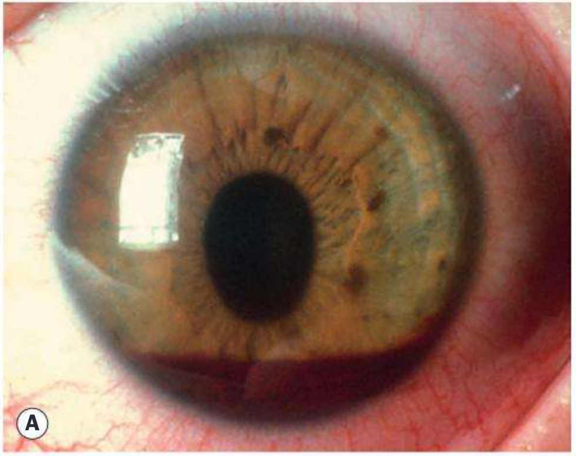
Definition
- Immediately after injury
- Days to weeks later
- Months to years, or even decades, after trauma
Importance
- The IOP rise may be acute and severe, threatening the optic nerve and cornea.
- It may occur long after an apparently trivial injury.
- Angle recession after blunt trauma can produce delayed chronic open-angle glaucoma.
- Associated injuries such as hyphema, lens dislocation, retinal tear, retinal detachment, and traumatic optic neuropathy may determine the final visual prognosis.
- Patients with traumatic hyphema require long-term surveillance for angle recession and glaucoma.
Etiology and classification
A. Closed-globe or blunt trauma
Early causes of raised IOP
- Hyphema with obstruction of trabecular meshwork by red blood cells
- Trabeculitis and traumatic iridocyclitis
- Direct trabecular meshwork injury
- Pigment dispersion and iris debris
- Pupillary block from lens subluxation, dislocation, vitreous prolapse, or swollen lens
- Lens particle glaucoma after traumatic capsular rupture
- Acute angle closure due to anterior lens displacement
- Retained intraocular material following trauma
Late causes
- Angle recession glaucoma
- Peripheral anterior synechiae causing secondary angle closure
- Ghost-cell glaucoma after vitreous hemorrhage
- Hemolytic glaucoma
- Lens-induced glaucoma
- Traumatic cataract with phacomorphic glaucoma
- Phacoantigenic glaucoma following rupture of the lens capsule
- Siderosis bulbi from a retained iron intraocular foreign body
- Epithelial or fibrous downgrowth after penetrating trauma
- Neovascular glaucoma secondary to retinal detachment, retinal ischemia, or severe ocular trauma
B. Open-globe trauma
- Lens capsule rupture with lens particle or phacoantigenic glaucoma
- Retained intraocular foreign body
- Epithelial downgrowth
- Peripheral anterior synechiae
- Vitreous incarceration in wound
- Post-traumatic inflammation
- Secondary angle closure
- Ghost-cell glaucoma after vitreous hemorrhage
- Secondary glaucoma after repair of globe injury
Pathophysiology
Mechanism of blunt trauma
- Tearing of iris sphincter
- Iridodialysis
- Hyphema from rupture of iris or ciliary-body vessels
- Separation of ciliary muscle fibers causing angle recession
- Cyclodialysis cleft
- Trabecular meshwork damage
- Zonular rupture and lens subluxation/dislocation
- Retinal and choroidal injury
Mechanisms of IOP elevation
1. Hyphema-associated glaucoma
Pathogenesis
- Mechanical blockage by erythrocytes
- Clot blocking the angle
- Inflammatory debris
- Trabecular meshwork injury
- Peripheral anterior synechiae
- Pupillary block in total hyphema
Rebleeding
Sickle-cell disease or trait
2. Traumatic iritis and trabeculitis
- Posterior synechiae
- Peripheral anterior synechiae
- Secondary angle closure
- Steroid-induced IOP elevation in susceptible patients
3. Angle-recession glaucoma
Definition
Gonioscopic features
- Broad, deepened ciliary-body band
- Irregular widening of angle recess
- Torn iris processes
- Increased trabecular pigmentation
- Associated peripheral anterior synechiae
- Comparison with the fellow eye is useful
Pathogenesis of delayed glaucoma
- Angle recession involving more than 180 degrees
- Traumatic hyphema
- High initial IOP
- Marked angle pigmentation
- Lens injury
- Absence of a cyclodialysis cleft
- Significant trabecular damage
4. Lens-related traumatic glaucoma
A. Lens subluxation or dislocation
- Pupillary block
- Anterior displacement of the lens with angle closure
- Direct lens-cornea contact with endothelial damage
- Vitreous prolapse causing block
B. Lens particle glaucoma
C. Phacoantigenic glaucoma
D. Phacomorphic glaucoma
5. Ghost-cell glaucoma
Pathogenesis
- History of trauma and vitreous hemorrhage
- Raised IOP
- Open angle
- Fine khaki or tan cells in anterior chamber
- Often a preceding delay of weeks after the hemorrhage
6. Hemolytic glaucoma
7. Glaucoma due to intraocular foreign body
8. Secondary angle-closure glaucoma after trauma
- Extensive peripheral anterior synechiae
- Pupillary block from lens dislocation or vitreous prolapse
- Total hyphema
- Iris bombe after severe inflammation
- Epithelial or fibrous downgrowth
- Neovascularization of the iris and angle
- Aqueous misdirection in complex postoperative traumatic eyes
Clinical features
Symptoms
- Blurred vision
- Ocular pain
- Headache
- Halos around lights
- Red eye
- Photophobia, if traumatic iritis is present
- Floaters or visual haze from hyphema or vitreous hemorrhage
- Nausea and vomiting in acute severe IOP elevation
Signs
External and anterior segment signs
- Periocular bruising, lid edema, or laceration
- Conjunctival chemosis or subconjunctival hemorrhage
- Corneal edema due to high IOP
- Corneal blood staining in prolonged total hyphema
- Hyphema or microhyphema
- Iris sphincter tear and traumatic mydriasis
- Iridodialysis
- Traumatic iritis
- Lens subluxation, phacodonesis, or lens dislocation
- Traumatic cataract
- Aphakia
- Vitreous in anterior chamber
- Wound leak or shallow anterior chamber in open-globe injury
Gonioscopy
- Angle recession
- Trabecular pigmentation
- Peripheral anterior synechiae
- Cyclodialysis cleft
- Iridodialysis
- Foreign material
- Blood or ghost cells in angle
Posterior segment examination
- Vitreous hemorrhage
- Commotio retinae
- Retinal tear or retinal detachment
- Choroidal rupture
- Traumatic optic neuropathy
- Retained intraocular foreign body
- Macular injury
Evaluation
Initial priorities
- Markedly reduced visual acuity
- Irregular or peaked pupil
- Shallow or flat anterior chamber
- Low IOP or hypotony
- Positive Seidel test
- Extrusion of uveal tissue or vitreous
- 360-degree subconjunctival hemorrhage
- Dense traumatic cataract or lens disruption
Ocular assessment after globe integrity is established
- Visual acuity in both eyes.
- Pupillary reactions and relative afferent pupillary defect.
- IOP measurement.
- Slit-lamp examination.
- Assessment and grading of hyphema.
- Corneal examination for blood staining.
- Lens position and integrity.
- Dilated fundus examination.
- Gonioscopy after the acute phase.
- Optic disc examination.
- Baseline automated visual field and OCT retinal nerve fiber layer assessment when feasible.
Investigations
- B-scan ultrasonography if the fundus is obscured by hyphema, vitreous hemorrhage, or media opacity.
- Orbital CT scan in penetrating trauma or suspected intraocular foreign body.
- Ultrasound biomicroscopy or anterior-segment OCT for angle recession, cyclodialysis cleft, zonular injury, lens position, and occult anterior segment pathology.
- Sickle-cell screening in appropriate patients with hyphema.
- Complete blood count and coagulation studies if bleeding disorder is suspected.
Management
General principles
- Treat acute ocular trauma as an emergency.
- Exclude open globe before any pressure-based examination.
- Identify the mechanism of raised IOP.
- Treat associated injuries.
- Reduce IOP promptly when the optic nerve or cornea is threatened.
- Arrange long-term monitoring for angle recession and glaucomatous optic neuropathy.
Management of traumatic hyphema
Conservative measures
- Protective rigid eye shield
- Restrict strenuous activity
- Head elevation, including during sleep
- Avoid eye rubbing and Valsalva maneuver
- Avoid aspirin and nonsteroidal anti-inflammatory drugs because of rebleeding risk
- Review anticoagulation only in coordination with the physician responsible for its indication
- Frequent examination during the period of risk of rebleeding
Medical treatment
- Topical corticosteroid to reduce inflammation
- Cycloplegic, commonly atropine in larger hyphema, to reduce discomfort, stabilize the blood-aqueous barrier, and reduce iris movement
- Topical beta-blocker
- Topical alpha-2 agonist
- Topical or systemic carbonic anhydrase inhibitor, where appropriate
- Hyperosmotic agent, such as mannitol, in severe uncontrolled IOP elevation
- Antifibrinolytic treatment may be considered in selected high-risk patients, according to local protocol
Important cautions
- Avoid miotics in traumatic hyphema because they can worsen inflammation, increase pupillary block risk, and disrupt the blood-aqueous barrier.
- Avoid carbonic anhydrase inhibitors, especially systemic acetazolamide, in patients with sickle-cell disease or trait unless specialist advice supports their use. Acidosis can promote sickling.
- In sickle-cell disease, lower thresholds for intervention are used because optic nerve and retinal ischemia may occur at comparatively modest IOP elevations.
Surgical treatment of hyphema
- Total hyphema
- Persistent uncontrolled IOP
- Corneal blood staining or high risk of staining
- Failure of hyphema to clear
- Recurrent or organized clot
- Threatened optic nerve damage
- Sickle-cell disease with uncontrolled IOP
Management of angle-recession glaucoma
Medical treatment
- Topical beta-blocker
- Alpha-2 agonist
- Topical carbonic anhydrase inhibitor
- Systemic carbonic anhydrase inhibitor if necessary and not contraindicated
- Prostaglandin analogue may be considered in chronic stable glaucoma, though inflammation should first be controlled
Laser treatment
Surgery
- Trabeculectomy, commonly with an antimetabolite
- Glaucoma drainage device
- Cyclodestructive procedure in eyes with poor visual potential or refractory glaucoma
Management of lens-related traumatic glaucoma
- Control acute IOP medically.
- Relieve pupillary block if present.
- Treat inflammation.
- Definitive management is usually lens extraction with appropriate intraocular lens planning or aphakic correction.
- Address associated vitreous prolapse, zonular weakness, capsular rupture, or retinal injury.
Management of ghost-cell and hemolytic glaucoma
- Topical and systemic IOP-lowering drugs as needed.
- Treat associated inflammation.
- Anterior chamber washout may be required in severe cases.
- Pars plana vitrectomy should be considered for persistent vitreous hemorrhage that continually supplies ghost cells.
- Identify and treat the source of vitreous hemorrhage, such as retinal tear, proliferative diabetic retinopathy, or trauma.
Management of foreign-body related glaucoma
- Urgent localization with CT when a foreign body is suspected.
- Surgical removal of the foreign body where indicated.
- Management of associated globe injury, cataract, retinal detachment, endophthalmitis risk, and siderosis.
- Standard IOP-lowering therapy plus definitive treatment of the underlying cause.
Follow-up
Follow-up should include
- IOP measurement
- Gonioscopy
- Optic disc examination
- Visual-field assessment
- OCT of retinal nerve fiber layer and ganglion cell complex where feasible
- Corneal examination
- Lens assessment
- Retinal examination
Complications
- Optic nerve damage and irreversible glaucomatous visual-field loss
- Corneal edema
- Corneal blood staining
- Peripheral anterior synechiae
- Chronic angle-recession glaucoma
- Traumatic cataract
- Lens dislocation
- Retinal tear or retinal detachment
- Vitreous hemorrhage and ghost-cell glaucoma
- Neovascular glaucoma
- Amblyopia in children
- Blind painful eye in severe neglected injury
Prognosis
- Type and severity of the original injury
- Degree and duration of IOP elevation
- Presence of hyphema and rebleeding
- Extent of angle recession
- Lens, retinal, and optic nerve injury
- Presence of open globe or intraocular foreign body
- Promptness of diagnosis and treatment
- Ability to maintain long-term follow-up
Exam-oriented table: timing and mechanism
| Timing after trauma | Important causes of glaucoma | Principal mechanism |
|---|---|---|
| Immediate to hours | Hyphema, traumatic iritis, direct trabecular injury, pigment/debris, lens dislocation | Trabecular obstruction, inflammation, pupillary block, angle closure |
| Days | Rebleed hyphema, persistent inflammation, pupillary block | RBC and clot obstruction, inflammatory debris, synechial changes |
| Weeks | Ghost-cell glaucoma, lens particle glaucoma, phacoantigenic glaucoma | Degenerated RBCs, lens material, immune inflammation |
| Months to years | Angle recession glaucoma, PAS, traumatic cataract, siderosis | Trabecular scarring, secondary angle closure, lens-related mechanisms, toxic trabeculopathy |
| Late after penetrating injury | Epithelial/fibrous downgrowth, retained foreign body | Progressive angle closure or trabecular dysfunction |
Key viva points
It is a tear between the longitudinal and circular fibers of the ciliary muscle after blunt trauma, producing abnormal widening of the ciliary-body band on gonioscopy.
Hyphema with obstruction of the trabecular meshwork by red blood cells, clot, inflammatory cells, and debris.
It may appear months, years, or decades after trauma.
No. Angle recession is common after blunt trauma, but only a minority develop chronic glaucoma.
Extensive recession, especially more than 180 degrees, hyphema, high initial IOP, angle pigmentation, and lens injury.
Secondary open-angle glaucoma caused by rigid degenerated erythrocytes entering the anterior chamber after vitreous hemorrhage and obstructing the trabecular meshwork.
They may worsen acidosis and promote sickling, increasing trabecular obstruction and ischemic complications.
After the acute phase, once hyphema and significant inflammation have resolved and open-globe injury has been excluded.
Annual lifelong IOP, optic nerve, and visual-field surveillance, particularly if angle recession is present.