
Embryology of the nervous system. What develops at what week of gestation?
Reading File
The nervous system develops from the neural plate starting around week 3, with neural tube closure by week 4. Major brain vesicles form in weeks 4-5, neuronal proliferation and migration occur during weeks 6-20, and myelination continues well into postnatal life.
Searching sources...
Finding Sources
Finding Sources
Finding Sources
Finding Sources
Finding Sources
Finding Sources
Finding Sources
Finding Sources
Excellent - I found the key embryology textbooks. Let me read the detailed content from Langman's and The Developing Human.
Reading File
Reading File
Let me find the main nervous system development chapter in Langman's and The Developing Human.
Finding Sources
Finding Sources
Now let me read the core sections from both textbooks.
Reading File
Reading File
Reading File
Reading File
Reading File
Reading File
Reading File
Reading File
Reading File
I now have comprehensive content from both Langman's and The Developing Human. Let me also fetch one of the key diagrams.
Loading Image
I now have all the information I need. Let me compile a comprehensive, well-organized answer.
Embryology of the Nervous System: Week-by-Week Development
Here is a detailed timeline drawn primarily from The Developing Human (Moore & Persaud) and Langman's Medical Embryology.
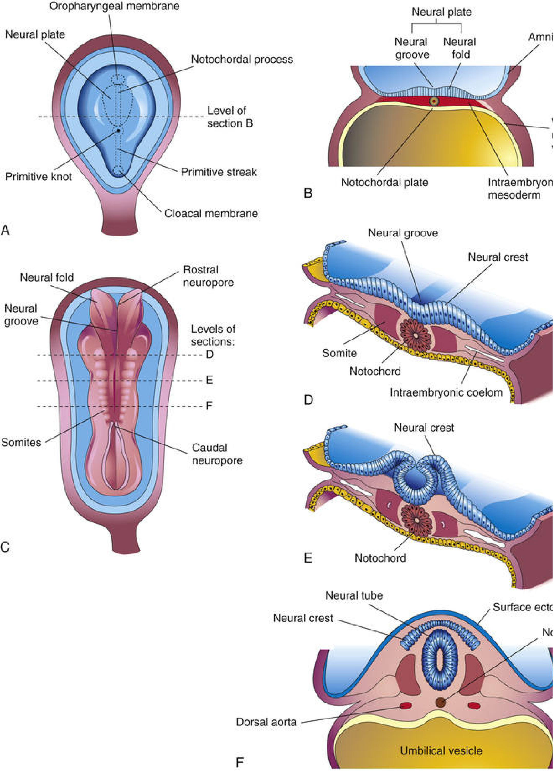
Week 3 - Neural Induction and the Neural Plate
- The neural plate appears around day 17-18 as a slipper-shaped thickening of dorsal ectoderm in the midline
- The notochord and paraxial mesenchyme induce the overlying ectoderm to form the neural plate (neural induction) via TGF-β family members, Wnts, SHH, and BMPs
- The lateral edges of the neural plate elevate to form neural folds flanking a central neural groove
- By day 20, the neural groove is deepening and somites are beginning to appear alongside it
Week 4 - Neural Tube Closure and Primary Brain Vesicles
This is the most critical week for CNS formation.
- Neural folds elevate and fuse to form the neural tube, beginning in the cervical region (~day 22) and proceeding both cranially and caudally
- Rostral (anterior) neuropore closes on day 25 (18-20 somite stage)
- Caudal (posterior) neuropore closes on day 27-28
- The cephalic end dilates into three primary brain vesicles:
- Prosencephalon (forebrain)
- Mesencephalon (midbrain)
- Rhombencephalon (hindbrain)
- Two brain flexures form: the cephalic flexure (in the midbrain region) and the cervical flexure (at the hindbrain-spinal cord junction)
- Neural crest cells delaminate from the edges of the closing neural folds and begin migrating. They will give rise to dorsal root ganglia, cranial sensory ganglia, autonomic ganglia, Schwann cells, chromaffin tissue, and many craniofacial structures
- The remainder of the neural tube caudal to the brain vesicles becomes the spinal cord; the lumen becomes the central canal
- The walls of the spinal cord differentiate into three zones: ventricular zone (neuroepithelium), mantle (intermediate) zone (future gray matter), and marginal zone (future white matter)
- The sulcus limitans divides the mantle layer into a dorsal alar plate (sensory) and ventral basal plate (motor)
Week 5 - Secondary Vesicles and Cerebral Hemisphere Primordia
- The three primary vesicles differentiate into five secondary vesicles:
| Primary Vesicle | Secondary Vesicles | Adult Derivatives |
|---|---|---|
| Prosencephalon | Telencephalon | Cerebral hemispheres, basal ganglia, olfactory bulbs, lateral ventricles |
| Prosencephalon | Diencephalon | Thalamus, hypothalamus, retina, 3rd ventricle |
| Mesencephalon | Mesencephalon (undivided) | Midbrain, superior/inferior colliculi, cerebral aqueduct |
| Rhombencephalon | Metencephalon | Pons, cerebellum, upper 4th ventricle |
| Rhombencephalon | Myelencephalon | Medulla oblongata, lower 4th ventricle |
- A pontine flexure develops, thinning the roof of the hindbrain (future 4th ventricle)
- The rhombencephalic isthmus separates the mesencephalon from the metencephalon
- Cerebral hemispheres begin as bilateral evaginations from the lateral wall of the telencephalon at the start of week 5
- Sympathetic ganglia begin forming as neural crest cells migrate lateral to the aorta during week 5
- The pituitary gland begins forming: Rathke's pouch (oral ectoderm upgrowth) meets the infundibulum (neuroectodermal downgrowth from the diencephalon floor) around week 5-6
Weeks 6-8 - Cortical Zones, Basal Ganglia, Cerebellum
- By the middle of the second month (week 6-7), the basal part of the cerebral hemisphere expands, forming the corpus striatum; fibers later divide it into caudate nucleus and lentiform nucleus (internal capsule formation)
- Choroid plexuses form in the lateral ventricles (week 6-7)
- The hippocampus thickens in the medial wall of the hemisphere
- The cerebellar plate (rhombic lips from the alar plates of the metencephalon) begins developing; midline fusion gives rise to the vermis
- Cortical zones of the cerebral wall are present: ventricular, subventricular, intermediate, subplate, and marginal zones
Weeks 8-20 - Neuronal Proliferation, Migration, and Cortical Layering
This period is defined by an explosive pace of neurogenesis and migration:
- Radial migration: Neurons born in the periventricular germinal matrix (ventricular and subventricular zones) migrate outward along radial glial fibers to form the cortical layers in an inside-out pattern (deep layers form first, superficial layers last)
- Cortical layers VI to I are laid down progressively from approximately weeks 8-20
- At peak activity, neurogenesis adds approximately 100,000 cells and 400,000 synapses per minute
- The cortex is initially smooth (lissencephalic); sulci and gyri begin appearing from around week 20 onwards, greatly increasing surface area
- The insula (buried cortex) becomes overgrown as the hemisphere expands, becoming hidden in the lateral sulcus
Weeks 8-12 - Spinal Cord, Meninges, and Peripheral Nervous System
- Spinal meninges differentiate from mesenchyme surrounding the neural tube (pia and arachnoid from neural crest-derived mesenchyme; dura from paraxial mesoderm)
- The spinal cord at this stage occupies the full length of the vertebral canal; it begins to lag behind vertebral column growth from about week 8 onward
- At birth, the conus medullaris is at the level of L3; in adulthood it rises to L1-L2
- This creates the cauda equina (roots of L2 and below) and the filum terminale
- Cranial nerves are organized and functional by the end of the embryonic period (week 8)
Weeks 16-24 - Gyri, Myelination Begins, Cerebellum Expands
- Primary sulci appear from about week 16-20 (central sulcus, lateral sulcus); secondary and tertiary sulci follow
- The corpus callosum begins forming by week 12 and is fully formed by week 20: the anterior commissure forms first, followed by the hippocampal commissure, then the corpus callosum proper (genu first, then body, then splenium)
- Myelination begins in the spinal cord around weeks 14-16 (motor pathways before sensory), then progressively ascends through the brainstem and into the cerebral hemispheres
- The cerebellum expands rapidly, and the external granule cell layer (from the rhombic lip) forms, then migrates inward to become the internal granule layer
Late Fetal and Postnatal - Myelination
- Myelination continues to be the dominant process in the late fetal and early postnatal period
- Peripheral nervous system myelin (by Schwann cells) begins around week 15
- CNS myelin (by oligodendrocytes) begins around week 18-20 and proceeds in a predictable sequence: spinal cord > brainstem > cerebellum > cerebrum
- The corticospinal tracts are not fully myelinated until the end of the second postnatal year, which explains the Babinski sign being normal in infants
- Cerebral cortex myelination is not complete until well into the second decade of life
Quick Reference Summary Table
| Gestational Age | Key Event |
|---|---|
| Day 17-18 (Wk 3) | Neural plate appears; neural induction by notochord |
| Day 20 (Wk 3) | Neural groove deepens; neural folds elevate |
| Day 22 (Wk 4) | Neural tube closure begins (cervical region); 3 primary vesicles form |
| Day 25 (Wk 4) | Rostral neuropore closes |
| Day 27-28 (Wk 4) | Caudal neuropore closes |
| Week 5 | 5 secondary vesicles; cerebral hemispheres begin; sympathetic ganglia form |
| Week 6 | Corpus striatum; choroid plexuses; hippocampus; Rathke's pouch meets infundibulum |
| Weeks 8-20 | Massive neuronal proliferation and radial migration; cortical layers laid down inside-out |
| Week 8 | End of embryonic period; cranial nerves organized |
| Week 12 | Corpus callosum begins; conus at L3 at birth |
| Weeks 16-20 | Primary sulci and gyri appear; myelination begins |
| Week 20+ | Secondary/tertiary sulcification; cerebellum expansion |
| Postnatal yrs 1-2 | Corticospinal tract myelination completes (Babinski disappears) |
| Adolescence | Cerebral cortex myelination completes |
Sources:
- The Developing Human: Clinically Oriented Embryology, Moore & Persaud (9th ed.), Ch. 17 - Nervous System, pp. 1017-1108
- Langman's Medical Embryology, Sadler (14th ed.), Ch. 18 - Central Nervous System, pp. 333-355
Embryology of the auditory system. What develops at what week of gestation?
The auditory system develops from multiple embryonic sources simultaneously: the otic placode (inner ear), the first pharyngeal arch and groove (middle and external ear), and neural crest cells. The otic vesicle forms by week 4-5, the cochlea coils by week 6-8, and the ossicles are distinct by week 8-9.
Searching sources...
Finding Sources
Finding Sources
Finding Sources
Finding Sources
Excellent - rich sources found. Let me read the key sections in parallel.
Reading File
Reading File
Reading File
Reading File
Reading File
Reading File
Reading File
Reading File
Reading File
Reading File
Reading File
Reading File
Loading Image
You've run out of free credits. Sign up for a free account to continue using Orris.
This is a shared conversation. Sign in to Orris to start your own chat.