

56de167b-cdd3-42d2-8d5b-6173fd146932.jpg
Differential diagnosis
analyse_media
Now let me consult the medical library for detailed information on this finding.
Reading File
Finding Sources
Finding Sources
Reading File
Reading File
Reading File
Reading File
Now let me fetch the textbook image of a urethral caruncle for visual comparison.
Loading Image
I now have comprehensive information to provide a detailed differential diagnosis.
Differential Diagnosis - Periurethral/Urethral Meatal Mass
Clinical findings from the image: A small, bright red, exophytic, polypoid/lobulated mass protruding from the urethral meatus, with surrounding pale, atrophic vulvovaginal mucosa and hypopigmentation consistent with hypoestrogenism.
1. Urethral Caruncle (Most Likely Diagnosis)
This is the top diagnosis. A urethral caruncle is an inflammatory lesion of the distal urethra, almost exclusively seen in postmenopausal women due to hypoestrogenism.
Key features supporting this diagnosis:
- Reddish, raspberry-like, friable exophytic mass at the posterior lip of the urethral meatus (the image shows a focal mass, not circumferential)
- Surrounding vulvovaginal atrophy (pale, dry mucosa visible)
- Sessile or pedunculated appearance
- Symptoms: dysuria, pain with intercourse, bloody spotting from mild trauma
Histology: Connective tissue rich in inflammatory cells and blood vessels, covered by epithelium
Textbook reference image for comparison:
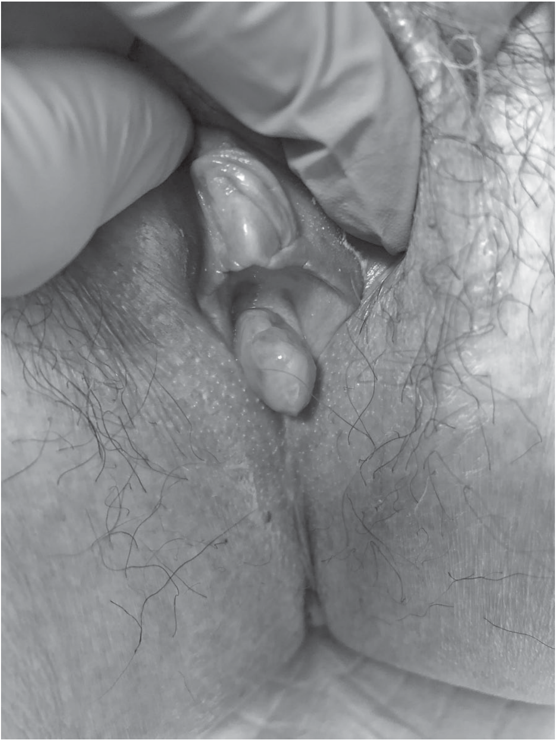
Figure 42-2, Smith and Tanagho's General Urology, 19th Ed. - The fleshy lobulated mass is emanating from the urethral meatus posteriorly. The meatal lumen is the sliver above the mass.
2. Urethral Prolapse
- Circumferential eversion of urethral mucosa at the meatus - appears as a beefy-red, doughnut-shaped lesion that completely surrounds the urethral meatus
- Two populations: postmenopausal women and prepubertal girls
- The lesion in the image appears focal/non-circumferential, making prolapse less likely
- Linked to estrogen deficiency and weakness of urethral smooth muscle attachments
Key distinguishing point: Prolapse is circumferential (360°); caruncle is focal, typically at the posterior lip. The image appearance is more consistent with a focal caruncle.
3. Atrophic Urethritis
- Very common in postmenopausal women
- Produces redness, eversion, and hypersensitivity at the urethral meatus - can mimic a caruncle
- Key difference: no discrete vascular mass - only mucosal eversion/redness without a distinct raised lesion
- Diagnosis confirmed by hypoestrogenic status and response to topical estrogen therapy
- Per [Smith and Tanagho's General Urology](Smith and Tanagho's General Urology, 19th Ed.): "Eversion of the posterior lip of the urinary meatus is evident in both conditions; however, a hypersensitive vascular mass is not present in atrophic urethritis"
4. Urethral Carcinoma
- Must be excluded when a lesion is atypical in appearance or behavior
- Presents as a periurethral mass with induration on palpation
- Associated with bleeding, urinary obstruction
- Much less common; biopsy required to exclude if any atypical features (firmness, irregular borders, rapid growth, failure to respond to treatment)
5. Skene Gland Cyst/Abscess
- Located at the urethral meatus (Skene glands open at the urethral orifice)
- Typically presents as a cystic/fluctuant mass rather than a solid vascular mass
- May become infected, causing pain and purulent discharge
6. Urethral Diverticulum
- Outpouching of urethral mucosa, typically on the posterior or lateral wall
- Usually presents as a suburethral mass, often with post-void dribbling and recurrent UTIs
- Less likely to appear as an isolated exophytic meatal lesion
7. Rare Mimics (to keep in mind for atypical cases)
| Entity | Clue |
|---|---|
| Primary urethral melanoma | Dark/pigmented lesion; confirmed by biopsy |
| Urethral lymphoma | Usually with systemic involvement |
| Intestinal metaplasia | Incidental histologic finding |
| Tuberculosis | History of TB exposure, other systemic features |
| Periurethral bulking agent granuloma | History of prior stress incontinence injections |
Summary and Recommended Approach
| Rank | Diagnosis | Key Differentiator |
|---|---|---|
| 1 | Urethral caruncle | Focal, posterior lip, postmenopausal, atrophic mucosa |
| 2 | Atrophic urethritis | No discrete mass, responds to topical estrogen |
| 3 | Urethral prolapse | Circumferential lesion, not focal |
| 4 | Urethral carcinoma | Induration, atypical features, no response to treatment |
| 5 | Skene gland cyst | Cystic/fluctuant rather than vascular |
Next steps: Initial conservative management with topical estrogen cream and sitz baths. If the lesion is large, painful, refractory, or atypical - excisional biopsy is warranted to exclude malignancy (carcinoma, melanoma, lymphoma can all mimic caruncle). - Campbell Walsh Wein Urology, 3-Volume Set and Smith and Tanagho's General Urology, 19th Ed.
This is a shared conversation. Sign in to Orris to start your own chat.