Cadaver as our first teacher

Reading File
Searching the Web

cadaver as first teacher medical education gross anatomy history significance

Finding Sources
Finding Sources
Searching PubMed

cadaver dissection anatomy medical education

Finding Sources
Reading Web Page

https://www.sciencedirect.com/science/article/pii/S128601152…

Searching Images

gross anatomy cadaver dissection medical students
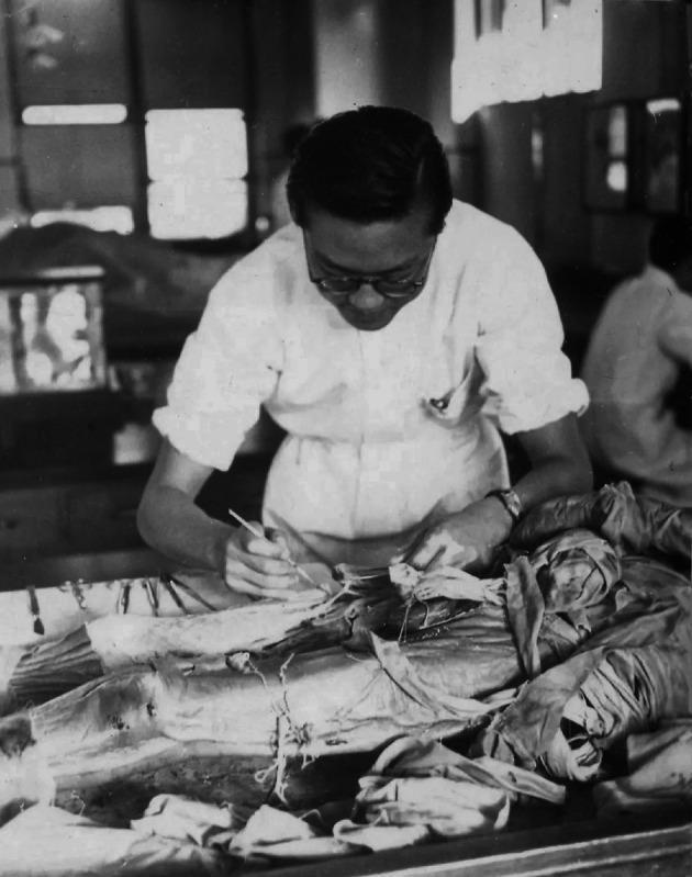
A historical black-and-white clinical photograph from 1941 depicting a medical student or physician, identified as Qi Fang, performing a gross anatomical dissection in a laboratory setting at Peking Union Medical College. The image serves as a documentation of medical education and anatomical study. The subject is wearing professional medical attire (white coat) and is focused on a partially dissected human cadaver positioned on a dissection table. The visible anatomical region involves the lower torso and proximal lower limb, with exposure of subcutaneous tissues and musculature. Various surgical instruments, including scalpels and forceps, are visible on the periphery of the table, used for precise tissue manipulation. This image illustrates the foundational medical education practice of cadaveric dissection for the study of human anatomy, reflecting the historical context of medical training in the mid-20th century.
A historical black-and-white clinical photograph from 1941 depicting a medical student or physician, identified as Qi Fang, performing a gross anatomical dissection in a laboratory setting at Peking Union Medical College. The image serves as a documentation of medical education and anatomical study. The subject is wearing professional medical attire (white coat) and is focused on a partially dissected human cadaver positioned on a dissection table. The visible anatomical region involves the lower torso and proximal lower limb, with exposure of subcutaneous tissues and musculature. Various surgical instruments, including scalpels and forceps, are visible on the periphery of the table, used for precise tissue manipulation. This image illustrates the foundational medical education practice of cadaveric dissection for the study of human anatomy, reflecting the historical context of medical training in the mid-20th century.

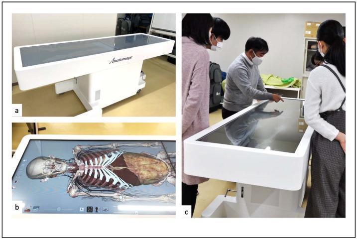
This composite educational graphic showcases the Anatomage Table™, a life-sized 3D virtual dissection and anatomy visualization tool. Image (a) displays the physical structure of the table in a horizontal orientation, featuring a large, touch-sensitive digital screen mounted on a white pedestal base with mobile casters. Image (b) provides a top-down view of the interface, demonstrating a high-resolution 3D anatomical rendering of a human cadaver in the supine position. The rendering highlights skeletal structures (cranium, rib cage), internal organs (liver, lungs), and the vascular system, emphasizing the tool's capacity for gross anatomy study. Image (c) illustrates the clinical application in an educational setting, showing a small group of medical students and an instructor interacting with the touch interface for collaborative learning. This technology serves as a digital alternative to traditional cadaveric dissection, allowing for the exploration of human anatomy through CT and MRI-based reconstructions. It is utilized in medical curricula for anatomy education, surgical planning, and physical examination training, offering a repeatable and cost-effective solution for medical residents and students.
This composite educational graphic showcases the Anatomage Table™, a life-sized 3D virtual dissection and anatomy visualization tool. Image (a) displays the physical structure of the table in a horizontal orientation, featuring a large, touch-sensitive digital screen mounted on a white pedestal base with mobile casters. Image (b) provides a top-down view of the interface, demonstrating a high-resolution 3D anatomical rendering of a human cadaver in the supine position. The rendering highlights skeletal structures (cranium, rib cage), internal organs (liver, lungs), and the vascular system, emphasizing the tool's capacity for gross anatomy study. Image (c) illustrates the clinical application in an educational setting, showing a small group of medical students and an instructor interacting with the touch interface for collaborative learning. This technology serves as a digital alternative to traditional cadaveric dissection, allowing for the exploration of human anatomy through CT and MRI-based reconstructions. It is utilized in medical curricula for anatomy education, surgical planning, and physical examination training, offering a repeatable and cost-effective solution for medical residents and students.

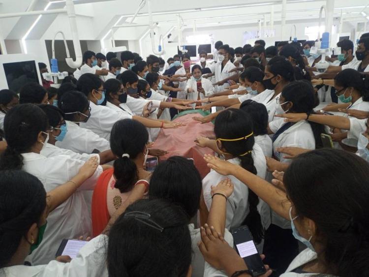
A clinical photograph depicting a large group of Phase I MBBS medical students participating in a formal 'Donor Oath' ceremony within an anatomy laboratory setting. The students, dressed in professional white coats and protective face masks, are gathered in a dense circle around a central dissection table covered with a reddish-pink cloth, likely containing a human cadaver. The individuals are performing a synchronized gesture, extending their right arms horizontally over the table, symbolizing their commitment to medical ethics, bioethics, and the respectful treatment of anatomical donors. The background shows specialized medical education infrastructure, including overhead surgical lighting and laboratory monitors. This image illustrates the integration of the Attitude, Ethics, and Communication (AETCOM) module in medical curricula, specifically focusing on humanizing anatomy education and instilling the principles of beneficence and non-maleficence in early undergraduate medical training.
A clinical photograph depicting a large group of Phase I MBBS medical students participating in a formal 'Donor Oath' ceremony within an anatomy laboratory setting. The students, dressed in professional white coats and protective face masks, are gathered in a dense circle around a central dissection table covered with a reddish-pink cloth, likely containing a human cadaver. The individuals are performing a synchronized gesture, extending their right arms horizontally over the table, symbolizing their commitment to medical ethics, bioethics, and the respectful treatment of anatomical donors. The background shows specialized medical education infrastructure, including overhead surgical lighting and laboratory monitors. This image illustrates the integration of the Attitude, Ethics, and Communication (AETCOM) module in medical curricula, specifically focusing on humanizing anatomy education and instilling the principles of beneficence and non-maleficence in early undergraduate medical training.

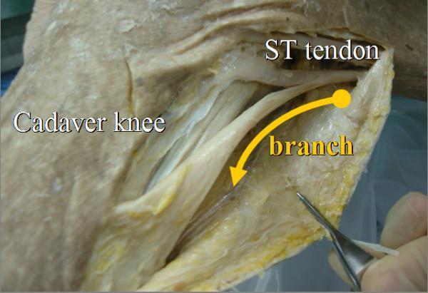
This clinical photograph shows a gross anatomical dissection of a right cadaver knee, specifically focusing on the medial aspect. The primary structure identified is the semitendinosus (ST) tendon, which appears as a prominent, white, fibrous band of connective tissue. An arrow and label highlight a secondary 'branch' of the ST tendon, which is shown diverging from the main tendon body and extending toward the gastrocnemius muscle. The dissection reveals the surrounding soft tissue, characterized by yellowish adipose tissue and fascia with a coarse texture. Surgical forceps are visible in the lower right, being used to isolate and retract the thin tendon branch. This visual demonstrates the accessory attachments of the hamstring tendons, specifically the ST branch that can be utilized as an 'inducer graft' during anterior cruciate ligament (ACL) reconstruction to facilitate tendon regeneration at the donor site. The image is an educational resource for orthopedic surgeons and medical students studying surgical anatomy and hamstring graft harvesting techniques.

This clinical photograph shows a gross anatomical dissection of a right cadaver knee, specifically focusing on the medial aspect. The primary structure identified is the semitendinosus (ST) tendon, which appears as a prominent, white, fibrous band of connective tissue. An arrow and label highlight a secondary 'branch' of the ST tendon, which is shown diverging from the main tendon body and extending toward the gastrocnemius muscle. The dissection reveals the surrounding soft tissue, characterized by yellowish adipose tissue and fascia with a coarse texture. Surgical forceps are visible in the lower right, being used to isolate and retract the thin tendon branch. This visual demonstrates the accessory attachments of the hamstring tendons, specifically the ST branch that can be utilized as an 'inducer graft' during anterior cruciate ligament (ACL) reconstruction to facilitate tendon regeneration at the donor site. The image is an educational resource for orthopedic surgeons and medical students studying surgical anatomy and hamstring graft harvesting techniques.

Cadaver as Our First Teacher

The phrase "cadaver as our first teacher" captures one of medicine's most profound traditions - the idea that the donated human body is the original, irreplaceable educator of every medical student.

Historical Roots

Formal anatomical dissection dates to Alexandria (~300 BCE) with Herophilus and Erasistratus, but the modern era began with Andreas Vesalius (1543), who revolutionized anatomy by personally dissecting cadavers and publishing De Humani Corporis Fabrica. By the 18th century, cadaver-based teaching had become a core pillar of medical education worldwide.
In India and many Asian medical programs, regional anatomy through cadaveric dissection has been considered a defining feature of the first-year MBBS curriculum, with students spending up to nine months dissecting and studying assigned cadavers.

What the Cadaver Teaches

The benefits of cadaveric dissection fall into three main domains (as described in Singapore Med J, Prakash et al., 2007):

1. Knowledge Acquisition and Integration

  • Three-dimensional spatial understanding of anatomy that no textbook, atlas, or digital model can fully replicate
  • Real exposure to anatomical variation - every human body differs, and students learn this firsthand as they move from table to table
  • Integration of structure with function - seeing how nerves, vessels, muscles, and organs relate to each other in situ
  • Understanding surgical planes, tissue layers, and fascial spaces that are the foundation of every clinical procedure

2. Clinical and Technical Skills

  • Development of manual dexterity through instrument handling - scalpels, forceps, dissecting probes
  • The principle: "better to learn on a cadaver how to use instruments than to experiment on patients"
  • Preparation for invasive procedures, surgical approaches, and procedural anatomy

3. Professional Attitudes and Ethics

Donor Oath Ceremony - Medical Students with Cadaver
Phase I MBBS students performing a Donor Oath ceremony around the dissection table - a ritual now integrated into AETCOM modules
  • The dissection room is often a student's first encounter with human death - a formative experience in developing compassion and the capacity to cope with mortality
  • It teaches the "compassionate detachment" essential for physicians dealing with death and bereavement
  • Gratitude to donors instills a sense of ethical responsibility that shapes the physician's entire career
  • Teamwork and collaborative learning emerge naturally in the dissection room

The "Nodal Point" Concept

The student-cadaver encounter has been described as the "nodal point" in medical education - the moment of transition from layperson to clinician. It marks the shift from absolute convergence (lay perspective) to comparative divergence (clinical perspective). This is where:
  • Students confront mortality for the first time in a controlled, educational setting
  • The abstract becomes tangible - anatomy moves from diagrams to reality
  • A deeply human bond forms between student and donor

The Gift of Body Donation

The body donor is called the "silent teacher" or "first teacher" because they give the ultimate, irreplaceable gift - their own body for science and the education of future physicians. This act:
  • Requires no direct interaction yet teaches more than any lecture
  • Demands the highest respect and gratitude from students and faculty
  • Is enshrined in legal frameworks (Anatomy Acts) that regulate ethical use of donated bodies

Modern Context and Debate

Historical cadaver dissection - Peking Union Medical College, 1941
A 1941 photograph of cadaveric dissection - the tradition spans generations of medical education
Recent evidence (Wilson et al., meta-analysis 2018) found no significant difference in test scores between traditional cadaver dissection and alternative methods (3D models, prosections, digital media). However, anatomy educators consistently argue that cadaveric dissection offers a multi-sensory experience - touch, smell, texture, weight, color - that no digital substitute provides.
The American College of Surgeons recommends a multimodal approach: cadaver labs supplemented with textbooks, multimedia modules, and a vertical integration model where anatomy is revisited throughout medical school and postgraduate training.
A 2025 study by Vadgaonkar et al. (Morphologie, 2025) specifically assessing "Knowledge, attitude, and perception of 'Cadaver as our First Teacher'" found that students widely acknowledged cadavers as their primary teacher in both teaching and research contexts.

Key Takeaway

The cadaver is not simply an anatomical specimen - it is a patient who lived, suffered, and chose to give. The dissection room is where medicine begins: where science meets humanity, where knowledge is earned through respect, and where every future physician first learns what it means to care for the human body. The cadaver truly is the first teacher - silent, generous, and irreplaceable.
This is a shared conversation. Sign in to Orris to start your own chat.