
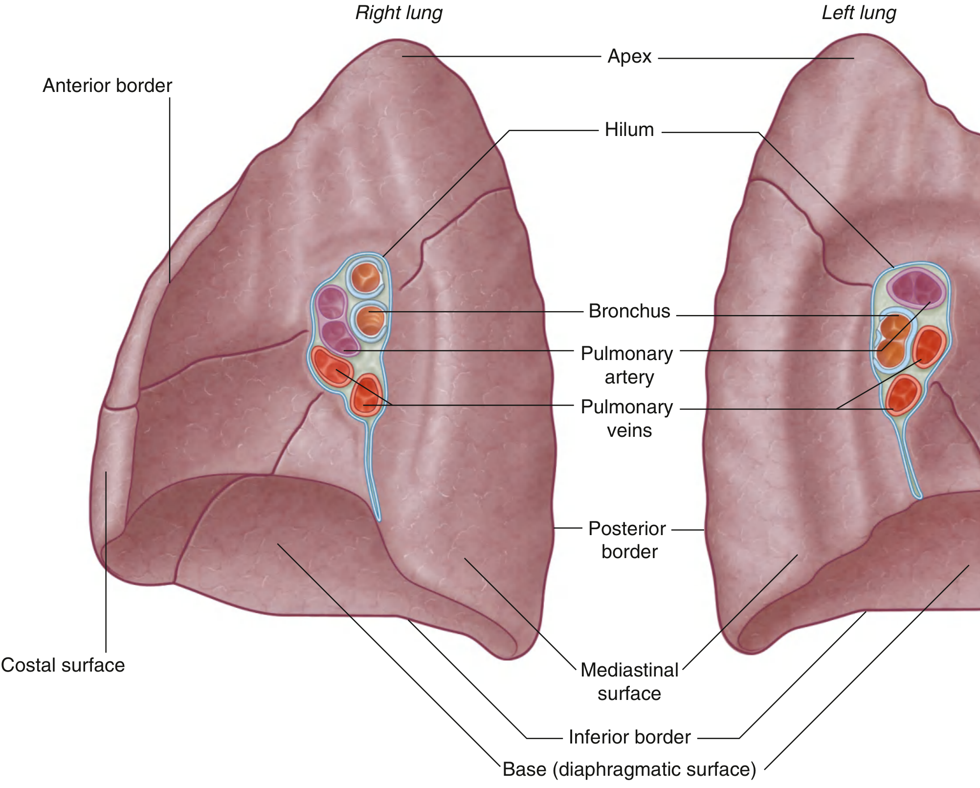
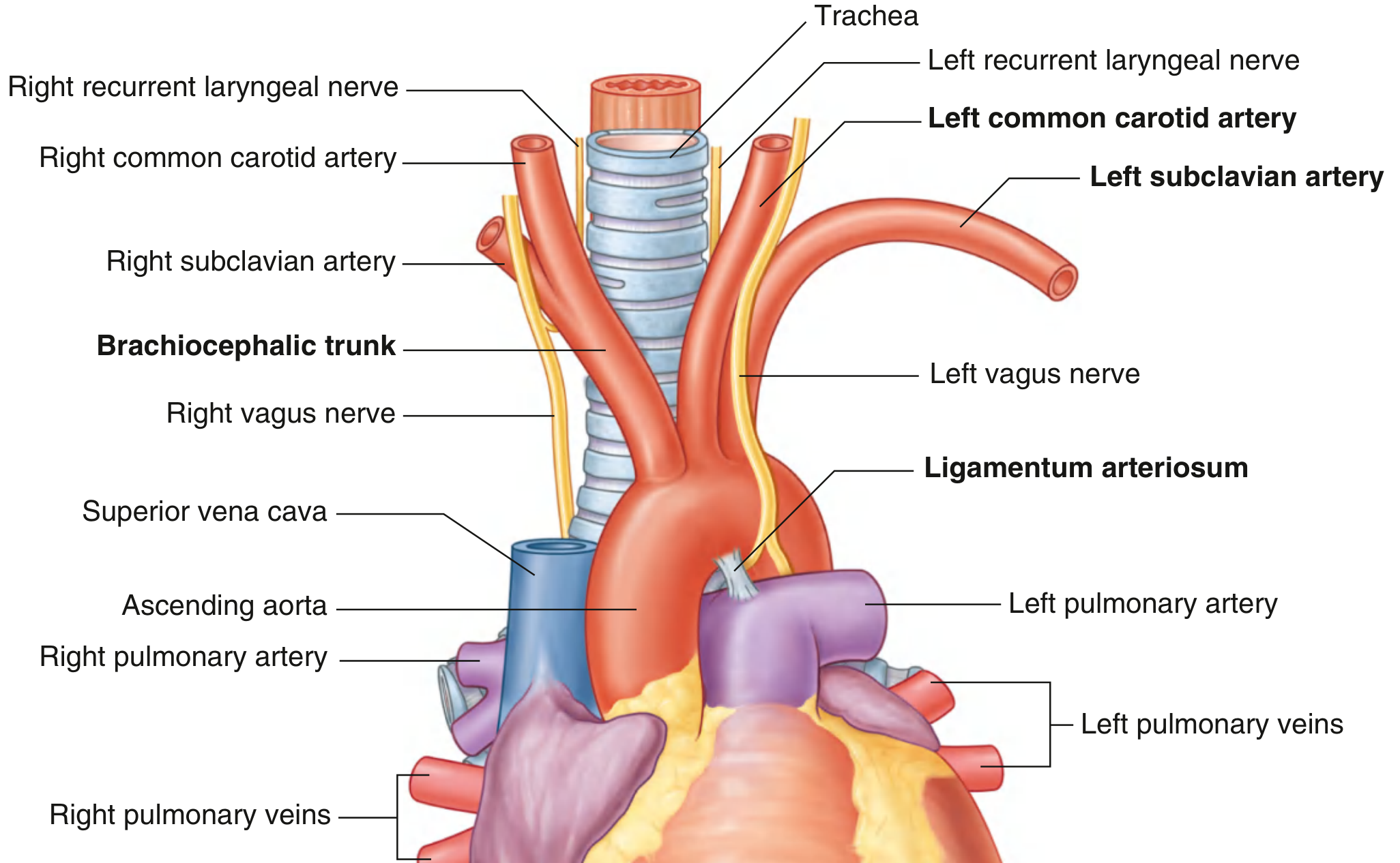
Hello everyone today I'll be demonstrating the specimens of lungs and heart for preparation of anatomy viva okay so here what we can see the specimens of the lungs one is right lung other is left lung we'll see how to identify which lung is right which lung is left and then we can see the specimens of the heart here okay okay so let's start with the lungs first so this is the lung of the right side okay so I'm holding it in the right hand like this vertically. You're looking from the top that's why you can see this narrow part that's the apex of the lung. In anatomical position you have to hold it in the right hand you can hold it like this with your palm towards the base or you all can hold it like this whichever way is comfortable okay so Why this lung is of the right side? You have to justify in three dimensions okay this upper part of the lung is referred to as the apex of the lung. Apex of the lung should be superior okay and then I've just tilted it now so that you can see the anterior and the posterior parts this border is referred to as the anterior border of the lung the anterior border is sharp and the posterior border is thick and rounded behind and this part is referred to as the hilum of the lung okay hilum of the lung is facing medially okay that's how I can say that this lung is of the right side okay if I try to hold it as if it's that of the left side see which point is going wrong the thick border is going on the anterior aspect okay the sharp border is coming behind this should not happen This sharp border should go anteriorly that's why this lung is of the right side okay and as everyone knows right lung has got three lobes but we don't say this point for identification of the side Why because as an abnormality (variation) the left lung may also have three lobes okay so that's why avoid saying this point okay if the examiner asks how many Lobes are there in then you can say the right lung has got three Lobes which are separated by Fissures okay this space here that's referred to as the horizontal Fissure this space here that's referred to as the oblique fissure okay so this lung is of the right side we saw the side determination points let's compare it with that of the left sided lung okay this lung is of the left side so hold it with the left hand and three side determination points we have to say one apex is on the superior aspect anterior border is sharp and hilum is on the medial aspect okay. Only these 3 points you supposed to say which covers the 3 dimensions okay when you say the Apex, superior / inferior dimension is covered when you say the sharp anterior border the anterior / posterior dimension is covered when you say hilum is medial the medial / lateral Dimension is covered okay so we can say this lung is of the left side okay and in left lung when we trace this anterior border down there is a tongue like projection that is seen in the lower part that's referred to as the lingula okay feature specific on the left lung okay and if we see the lobes there are 2 lobes separated by this fissure that's the oblique fissure okay so this lung is of the left side and this lung is of the right side okay so side determination is very important in lungs after that you may be asked about its various surfaces this surface is the costal surface this surface is the medial surface and below this is the region is referred to as the base okay base or the inferior surface okay and medial surface has got subdivisions anteriorly there is this mediastinal surface and behind this part is referred to as the vertebral surface in relation with the vertebral column okay and various relations of the mediastinal surface are important in the mediastinal surface of the right lung you can imagine the right lung here and the heart we can see suppose heart is here okay, so this is the right atrium of the heart right atrium of the heart comes in relation with the right lung okay there is one cardiac impression for the right atrium of the heart and in the right atrium we know there are openings of superior vena cava, inferior vena cava so those structures also come in the relations and then behind there is the trachea, the esophagus okay those structures are there and in the superior vena cava there is One vein which arches over this hilum and opens into the superior vena cava which vein is related here That's the arch of the azygous vein okay structure specific in the relation of right lung the arch of azygous vein is above the hilum and these structures we can see which are entering or exiting the Hilum How to identify these? Anterior most structure is always a vein okay so just remember these principles Anterior most structure is always a vein, inferior most structure is always a vein okay Wall of the bronchus will have the cartilage feeling okay there is a cartilage in the wall of the bronchus that we can feel okay so once we have identified the vein you can identify the bronchus with the cartilage and the remaining structure would be the artery. These both these are the branches of the right pulmonary artery that we can see okay so like this we can identify the structures of the hilum. Here the bronchus is dividing into two parts called as the eparterial bronchus and the hyparterial bronchus okay so these are the structures in the hilum. So for lung identification of the side, identification of the relations of the medial surface, structures at the hilum and there is one important structure here that's referred to as the pulmonary ligament okay lung is covered by pleura the parietal pleura and visceral pleura they unite at this point referred to as pulmonary ligament okay so there is a significance of this during increased Venuos return. When the inferior pulmonary vein expands this pulmonary ligament provides a space, it provides a dead space for expansion of this inferior pulmonary vein. Then let's see this left lung now side determination we had already done, surfaces are similar - costal surface, medial surface, inferior surface or the base then relations of the medial surface are slightly different this part is related with which part of the heart so if I keep the heart here in anatomical position okay this is the left lung this is the heart see which chamber of the heart is this? This is the left ventricle of the heart okay left ventricle of the heart comes in relation here okay so left ventricle of the heart is related here and which artery arises from the left ventricle? the aorta okay so the ascending aorta, the Arch of aorta the descending aorta all these structures are related in the left lung okay in the mediastinal surface of the left lung the structure arching over the hilum here is the arch of aorta. In the right lung the structure was the arch of azygous vein okay this one is the right lung above the hilum is arch of azygous vein okay and here is the arch of the aorta okay and Trachea and esophagus are common here as well okay and again multiple structures in the hilum are there so hilum structures may not be exactly same in all the lungs okay depending upon the section where the lung has been taken out. The root of the lung has to be cut to take the lungs out right so depending upon the different level of sections, structures size may be different. Here also apply the same principles now this part is anterior part this is a sharp anterior border the anterior most structure is a vein here is this is the inferior most structure is also vein okay so the left Superior pulmonary vein left inferior pulmonary vein structure having cartilage in the wall, that's the bronchus okay I can feel the cartilage here and the artery is the pulmonary artery okay and the shiny structure that we can see covering the lung is the pleura okay this is actually the visceral pleura which is Adherent to the lung. There are 2 layers of pleura called as parietal pleura and visceral pleura in between them there is a space called as pleural cavity there is an Applied Anatomy related to it called as pleural effusion that means the fluid collection in the pleural cavity okay this was about the lungs now we'll see the heart so here we can see the specimen of the heart. I'm holding it in the anatomical position so when I hold it in the anatomical position I'll just keep the lungs aside so that your view is much clear okay so now I'm I'm holding the heart in anatomical position you're looking from the top so the you can see the great vessels, the aorta, the pulmonary trunk okay the diaphragmatic surface of the heart I'm keeping it on the palm there's a narrow part called as the apex of the heart I'm keeping it between the thumb and the index finger okay so like this you have to hold the heart in anatomical position such that the apex is lying towards the left side okay so basic chambers of the heart you all know right this is the right atrium this one is the left atrium here okay and there are two ventricles right ventricle left ventricle okay. The apex of the heart is formed by this left ventricle and opposite the apex of the heart is the base of the heart which is formed by both the atrium okay this one is right atrium and left atrium both the atriums they form the base of the heart okay then in the external features there are grooves between the atrium and ventricle this is the atrio-ventricular groove also referred to as coronary sulcus because the coronary arteries lodges here. then we can see the arch of aorta with its branches. We can see three branches of arch of aorta, so if I'm just holding it, I've just turned around and now here this is the apex and we can see the arch of aorta with its three branches so name these three branches of arch of aorta is this one is the brachiocephalic trunk this one is left common carotid artery and this one is left subclavian artery okay. The three branches of arch of aorta are very important we need to identify that and this one is the descending aorta okay then interior of the chambers are important, interior of right atrium ventricles and all we'll see that before that, we can see the right atrium there is a projecting part called as the right auricle and in the left atrium there is a projecting part called as the left auricle okay this one is the left auricle and various borders and surfaces of the heart if we see, the right side extending from superior vena cava to the inferior vena is the right border of the heart okay this one is the superior vena cava from superior vena to the inferior vena cava is the right border of the heart okay and from the inferior vena cava to the apex of the heart that's the inferior border of the heart okay first understand the borders then I'll show you the surfaces which these borders are separating.
right lung and left lung lateral medial surface hilum fissure anatomy
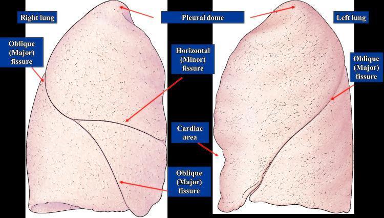
This anatomical diagram provides a lateral and medial perspective of the human right and left lungs, highlighting pulmonary lobar anatomy and surface landmarks. The right lung illustration demonstrates two fissures: the oblique (major) fissure, which runs diagonally, and the horizontal (minor) fissure, which branches anteriorly from the oblique fissure to separate the lung into three lobes (superior, middle, and inferior). The left lung illustration displays a single oblique (major) fissure, dividing it into two lobes (superior and inferior). Key anatomical features labeled include the 'Pleural dome' at the apex of both lungs and the 'Cardiac area'—a distinct indentation on the medial aspect of the left lung where the heart resides. The diagram effectively illustrates the morphological differences between the lungs, specifically the more complex fissural pattern of the right lung compared to the two-lobed structure of the left lung. This visual is designed for medical education regarding respiratory anatomy, thoracic landmarks, and surgical or diagnostic localization.
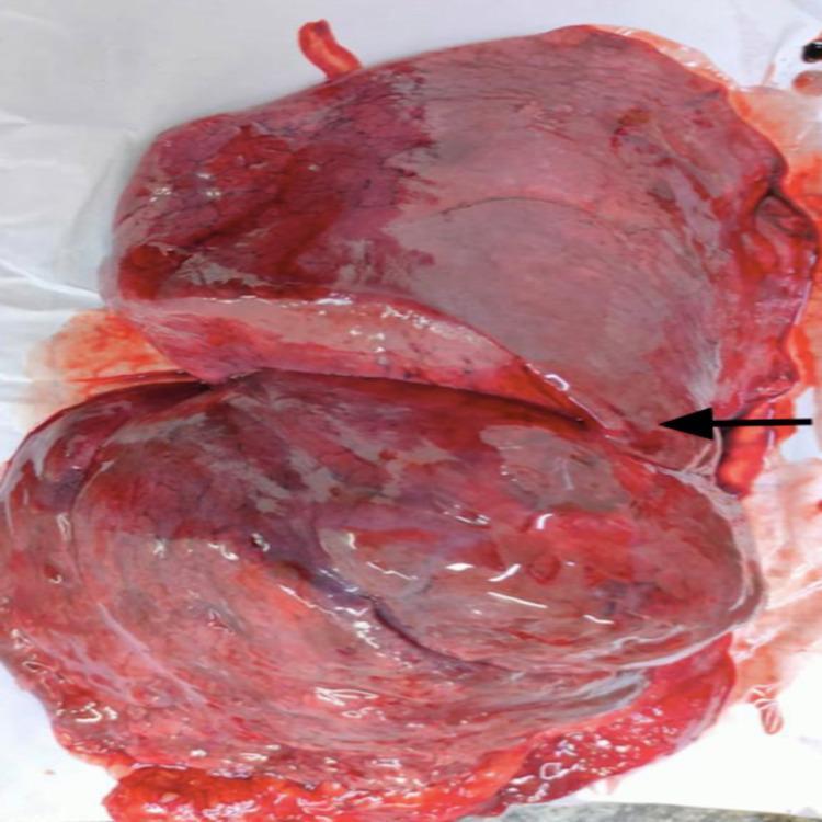
This clinical photograph shows a gross anatomical specimen of a right human lung, focusing on its lateral aspect. The lung exhibits a deep reddish-purple coloration and a moist, glistening pleural surface, characteristic of a fresh cadaveric or surgically resected specimen. A black arrow identifies an incomplete horizontal fissure. Rather than completely separating the superior lobe from the middle lobe, the fissure appears as a shallow, abbreviated groove that terminates before reaching the anterior border of the lung. This anatomical variation demonstrates partial fusion between the superior and middle lobes. The oblique fissure is also partially visible, separating the inferior lobe from the rest of the lung parenchyma. This image is used in medical education to illustrate normal anatomical variations in lung lobar anatomy and the clinical significance of fissure completeness for procedures such as lobectomies and pulmonary segmentectomies.
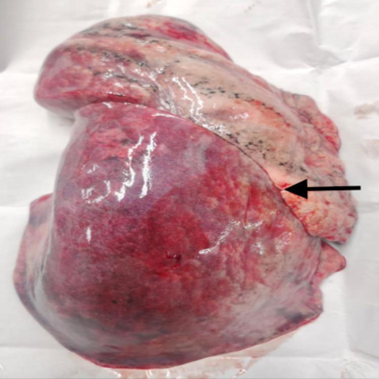
This clinical photograph displays a gross anatomical specimen of a human left lung, focusing on the lobar morphology and pleural surface. The primary feature is a well-defined, complete oblique fissure, marked by a black arrow, which extends across the lateral surface to anatomically separate the superior (upper) and inferior (lower) lobes. The lung parenchyma exhibits a mottled appearance with varied coloration: the upper lobe shows lighter pinkish tones interspersed with dark, anthracotic pigment spots typical of carbon particle deposition. In contrast, the lower lobe presents a deeper reddish-purple hue, likely indicating post-mortem congestion or hypostatic changes. The visceral pleura appears smooth and glistening. Small hexagonal outlines of pulmonary lobules are visible on the surface. This specimen serves as an educational reference for normal thoracic anatomy and common anatomical variations in pulmonary fissures, which are clinically significant for surgical resection, bronchoscopic interventions, and the localization of pulmonary pathology.
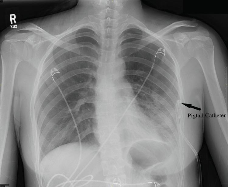
This anterior-posterior (AP) chest radiograph illustrates a large left-sided pneumothorax and its subsequent management. The left hemithorax exhibits a significant peripheral radiolucent area devoid of lung markings, indicating air within the pleural space. This results in medial retraction of the left lung towards the hilum, with a visible visceral pleural line. In contrast, the right lung remains fully expanded with normal bronchovascular markings reaching the chest wall. A radiopaque small-bore pigtail catheter is visible, inserted through the lateral left chest wall and terminating in a characteristic curled distal loop within the pleural space to facilitate air drainage and lung re-expansion. Radiopaque ECG leads are also present on the chest surface. This image is an educational example of diagnostic radiology in emergency medicine, demonstrating the visual characteristics of a simple pneumothorax and the correct positioning of a percutaneous pigtail catheter for thoracostomy.
heart anatomy chambers atria ventricles external gross specimen
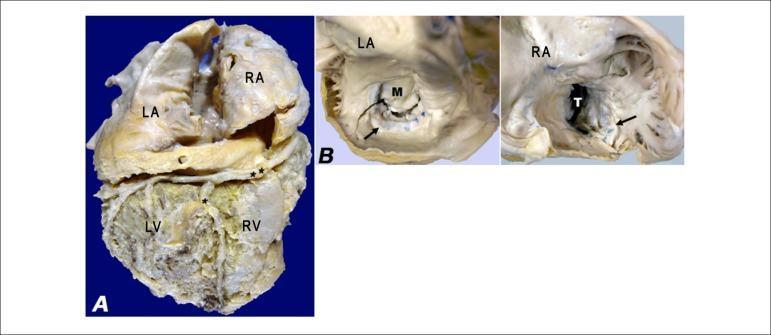
Two-panel clinical photograph of a gross pathology specimen illustrating cardiac hypertrophy and valvular changes. Panel A displays the diaphragmatic surface of the heart, showing massive dilatation of the left atrium (LA) and right atrium (RA) relative to the left ventricle (LV) and right ventricle (RV). The atrioventricular sulcus containing the right coronary artery (**) and the posterior interventricular sulcus containing the posterior interventricular branch (*) are visible. Panel B provides internal views of the left and right atria, highlighting bilateral endocardial thickening. Marked arrows indicate surgical sutures on the posterior cusps of the mitral (M) and tricuspid (T) valves from previous valvoplasties. The specimen demonstrates structural remodeling associated with chronic heart failure and restrictive cardiomyopathy, where the atrial chambers appear disproportionately enlarged compared to the ventricles. This image is educational for understanding cardiac gross anatomy, surgical landmarks in valvular repair, and pathological chamber remodeling.
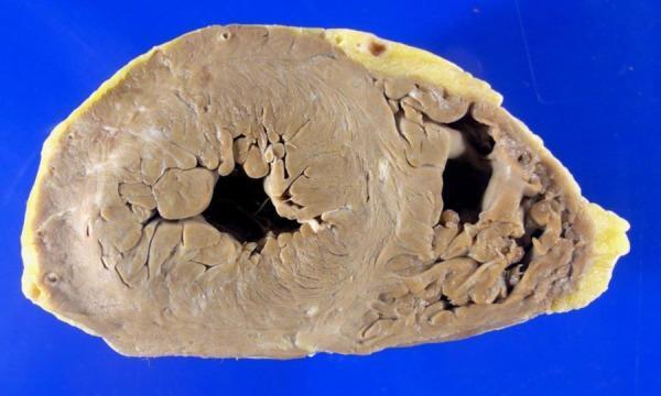
This clinical photograph displays a transverse cross-section (short-axis view) of a human heart specimen at the ventricular level. The specimen illustrates the gross anatomy of both the left and right ventricles. The left ventricular myocardium exhibits mild concentric hypertrophy, characterized by a thickened muscular wall without accompanying cavitary dilation. The myocardium possesses a uniform tan to light brown color and a firm, fibrous texture, showing no visual evidence of acute or chronic myocardial infarction (e.g., discoloration, softening, or white scarring). The endocardial surfaces and trabeculae carneae are clearly visible within the chambers. A thin layer of yellowish epicardial adipose tissue is present on the external surface of the heart. This specimen serves as an educational example of hypertensive or compensatory myocardial changes in the absence of ischemic heart disease.
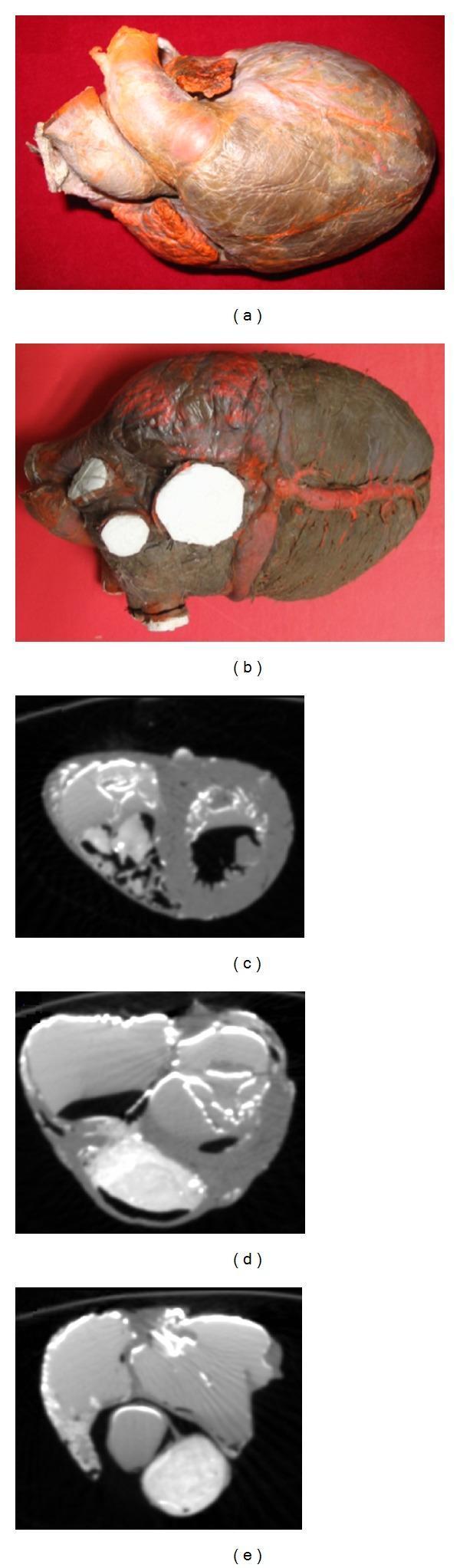
Educational panel illustrating human cardiac anatomy through gross specimen photography and Computed Tomography (CT). The upper section contains two clinical photographs of a human heart specimen: (a) a frontal view showing the anterior surface, large vessels (aorta and pulmonary artery), and surface musculature, and (b) a posterior view highlighting white circular patches where vessels were prepared and the darker diaphragmatic surface. The lower section features three axial CT slices demonstrating the heart's internal architecture across different planes: (c) at the ventricular level, showing the thicker left ventricular wall compared to the right; (d) a mid-level slice capturing the spatial transition between atria and ventricles; and (e) a superior slice focusing on the atrial chambers. The CT images utilize contrast-enhancing materials (lead oxide and gelatin) to delineate the endocardial borders and myocardial thickness, serving as a primary dataset for 3D anatomical reconstruction and fiber orientation modeling in cardiovascular research.
arch of aorta branches brachiocephalic left common carotid left subclavian
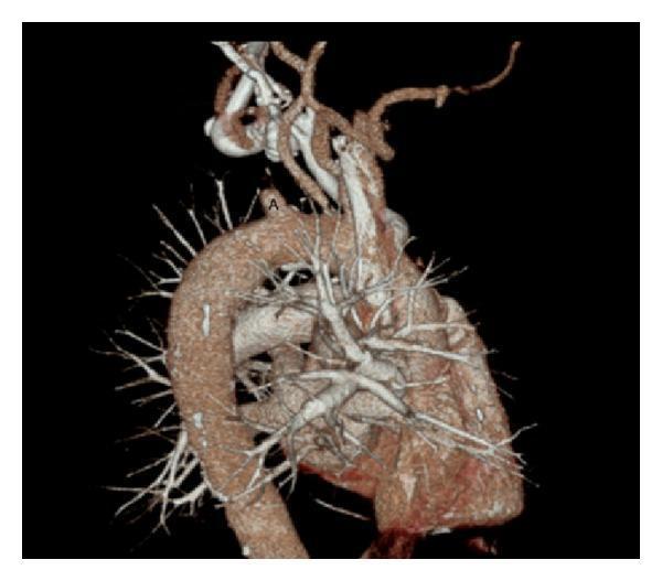
This diagnostic image is a 3D volume-rendered reconstruction of a computed tomography angiogram (CTA) of the chest, focusing on the thoracic aorta and its major branches. The aortic arch is clearly visualized, showing the origin of the great vessels. The brachiocephalic trunk and the left common carotid artery appear patent with normal contrast opacification. However, there is a significant vascular pathology at the origin of the left subclavian artery, labeled with 'A'. At this site, there is an abrupt termination of contrast enhancement, indicating a complete proximal occlusion of the left subclavian artery. The distal segment of the artery is not visualized via primary antegrade flow. The image also depicts the pulmonary arterial tree and smaller branching vessels. This finding is clinically significant for diagnosing subclavian steal syndrome or explaining significant inter-arm blood pressure discrepancies. The image serves as a clear educational example of arterial occlusion and the utility of 3D vascular reconstruction in identifying atherosclerotic or thrombotic vascular disease.
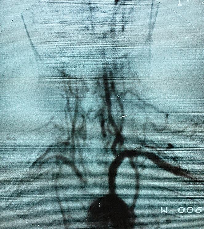
This diagnostic image is an arteriography of the aortic arch and supra-aortic trunks. The imaging captures the major arterial branches, including the brachiocephalic trunk, the left common carotid artery, and the left subclavian artery. Significant vascular pathology is demonstrated through several key visual features: there is a distinct proximal stenosis of the brachiocephalic trunk shortly after its origin from the aorta. Both the right and left common carotid arteries exhibit a markedly narrowed, 'filiform' (thread-like) appearance as they ascend the neck, indicating significant luminal reduction. Additionally, the right subclavian artery shows a decreased diameter in its distal segment compared to the more robust appearance of the left subclavian artery. These findings are characteristic of Takayasu arteritis, demonstrating the diffuse parietal thickening and resulting stenoses of large-vessel vasculitis. The image serves as a clinical example of large-vessel involvement in systemic inflammatory diseases, illustrating how arterial morphology changes under conditions of chronic wall inflammation and remodeling.
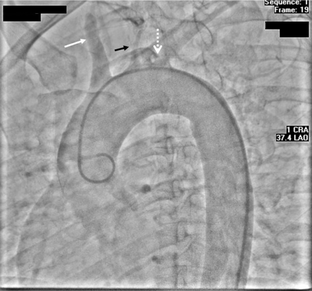
**Imaging Modality:** Digital Subtraction Angiogram (DSA), Aortic Arch Study. **Anatomical Region:** Aortic arch and supra-aortic branches, including the brachiocephalic trunk, left common carotid artery, and left subclavian artery. **Observed Pathology:** * **Brachiocephalic Artery:** Significant high-grade stenosis at the proximal segment (indicated by the white arrow). * **Left Common Carotid Artery:** Complete proximal occlusion with no distal contrast opacification (indicated by the solid black arrow). * **Left Subclavian Artery:** Complete proximal occlusion at the origin from the aortic arch (indicated by the dashed white arrow). **Characteristic Visual Features:** The image demonstrates a pigtail catheter positioned in the ascending aorta. There is a profound lack of normal contrast runoff into the primary arch vessels. The only remaining patent pathway for cerebral perfusion is via the severely stenosed right-sided brachiocephalic circulation. **Clinical Context:** These findings are characteristic of advanced aortoarteritis (such as Takayasu arteritis) or severe multi-vessel atherosclerotic disease, resulting in "pulseless" clinical presentation and critical cephalic ischemia. **Diagnostic Features:** Multi-vessel proximal occlusion/stenosis involving the great vessels of the aortic arch, visualized in a Left Anterior Oblique (LAO) cranially angulated projection.
coronary sulcus atrioventricular groove heart external anatomy
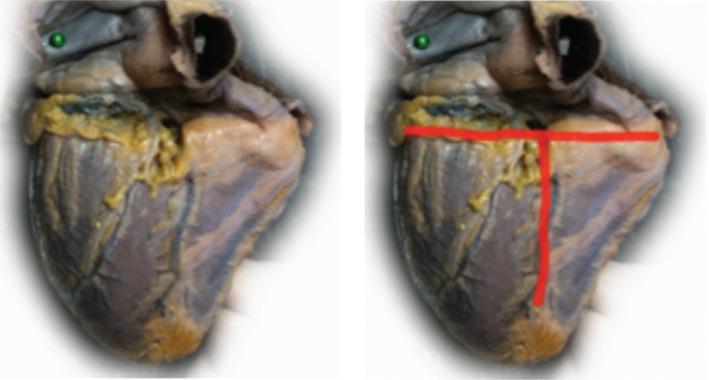
This clinical photograph shows a diaphragmatic (inferior) view of a human heart specimen. The image is presented in a side-by-side comparison format: the left panel displays the original gross specimen, while the right panel includes red annotations highlighting key anatomical landmarks. The cardiac surface exhibits a mottled reddish-brown myocardium with visible yellow epicardial adipose tissue concentrated along the sulci. The annotations demonstrate a 'T-shaped' intersection of major grooves: a horizontal red line identifies the coronary sulcus (atrioventricular groove), and a perpendicular vertical red line identifies the posterior interventricular sulcus. These landmarks delineate the boundaries between the atria and ventricles, and between the left and right ventricles, respectively. The educational focus is on the spatial orientation of cardiac surface anatomy and the identification of major coronary pathways as they appear from an inferior perspective.
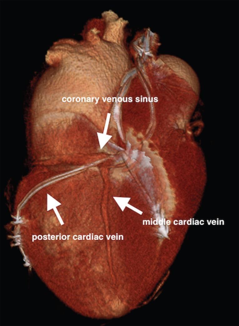
This diagnostic image is a 3D multi-slice computed tomography (MSCT) reconstruction of the heart, focusing on the cardiac venous anatomy and a cardiac resynchronization therapy (CRT) lead placement. The visualization highlights the coronary venous sinus, which extends across the posterior aspect of the atrioventricular groove. Labeled arrows identify the middle cardiac vein running along the posterior interventricular sulcus and the posterior cardiac vein positioned on the left ventricular wall. A notable clinical feature is the presence of a multipolar left ventricular lead successfully implanted within the posterior cardiac vein. The reconstruction demonstrates the complex anatomical relationships and distal anastomoses between these vessels. This clinical finding is significant for demonstrating a separate venous ostium and the use of the posterior vein as an alternative route for CRT lead placement when traditional cannulation of the coronary sinus is anatomically challenging.
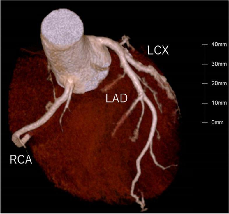
This 3D volume-rendered Computed Tomography Angiography (CTA) image provides a comprehensive visualization of the coronary arterial anatomy. The image displays the major coronary arteries originating from the ascending aorta and coursing over the epicardial surface of the heart. The Left Anterior Descending (LAD) artery is clearly visible descending along the anterior interventricular sulcus, exhibiting multiple diagonal branches. The Left Circumflex (LCX) artery is seen branching from the left main coronary artery and traveling along the left atrioventricular groove. The Right Coronary Artery (RCA) originates from the right coronary sinus and traverses the right atrioventricular groove. All visualized vessels appear patent with smooth luminal contours and no evidence of significant calcified or non-calcified atherosclerotic plaques or stenotic lesions. The myocardium is rendered in a dark, textured brown tone to provide anatomical contrast, and a 40mm scale bar is included for size reference. This diagnostic image is typical for assessing coronary artery disease (CAD) and evaluating coronary anatomy in a clinical setting.
medial surface right lung azygos vein hilum impression
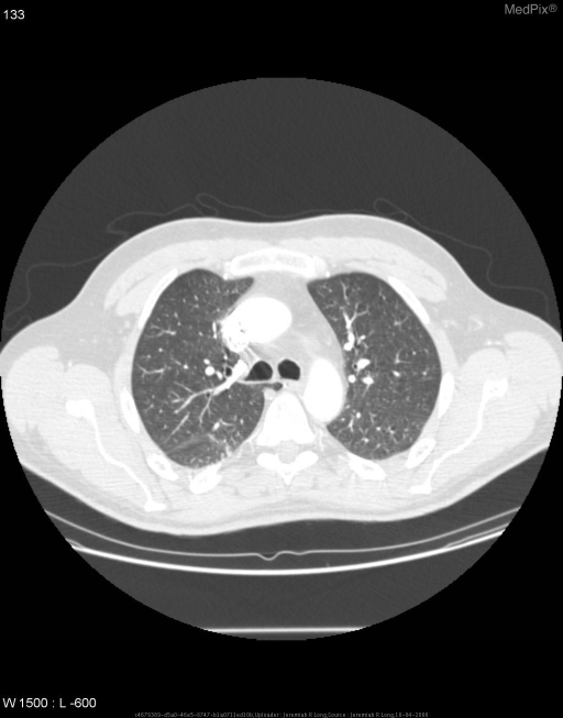
**Imaging Modality:** Computed Tomography (CT) of the chest, axial section, lung window. **Anatomical Region:** Upper thorax at the level of the great vessels and tracheal bifurcation. **Observed Pathology/Anatomic Variant:** Azygos fissure and azygos lobe of the right lung. **Characteristic Visual Features:** * **Azygos Fissure:** A fine, curvilinear high-attenuation line is visible traversing the right upper lung zone. This represents the invaginated mesoazygos, consisting of four layers of pleura (two visceral and two parietal). * **Azygos Vein:** At the medial/anterior termination of the fissure, a small, rounded soft tissue density is noted, representing the azygos vein in an anomalous cephalad position. * **Azygos Lobe:** The accessory fissure separates the superior-medial portion of the right upper lobe, creating an accessory "azygos lobe." **Key Diagnostic Features:** The pathognomonic "teardrop" appearance of the azygos vein at the end of the curvilinear fissure in the right apex. The underlying lung parenchyma within the accessory lobe demonstrates normal attenuation and vascularity. **Clinical Context:** This is a common incidental anatomical variant resulting from the failure of the azygos vein to migrate medially over the lung apex during embryogenesis.
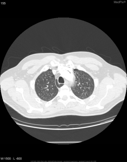
**Imaging Modality:** Computed Tomography (CT) of the chest, axial section, displayed in lung window settings. **Anatomical Region:** Upper thorax at the level of the aortic arch and tracheal bifurcation. **Observed Pathology/Anatomical Variant:** An **azygos lobe** is identified in the apex of the right lung. **Characteristic Visual Features:** - **Azygos Fissure:** A distinct, curvilinear thin line (pleural reflection) is visible traversing the right upper lung field. This fissure is composed of four layers of pleura (two visceral and two parietal). - **Azygos Vein:** At the inferior aspect of the fissure, a focal, teardrop-shaped density is noted, representing the abnormally positioned azygos vein. - **Lung Parenchyma:** The portion of the right lung located medial to the fissure constitutes the accessory azygos lobe. The surrounding lung parenchyma appears otherwise clear without evidence of consolidation or masses. **Key Diagnostic Features:** The pathognomonic finding of a high-riding azygos vein within a displaced pleural fissure, creating an accessory lobe in the right lung apex. This is a common incidental anatomical variant resulting from the failure of the azygos vein to migrate medially over the lung apex during development.
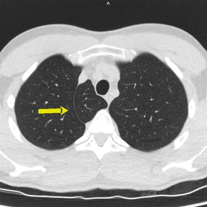
An axial CT scan of the chest at the level of the upper lobes, displaying a common anatomical variant known as an azygos lobe in the right lung. The image shows a characteristic azygos fissure (indicated by a yellow arrow) coursing through the right apical lung parenchyma. This fissure is formed by the anomalous lateral course of the azygos vein, which creates a 'mesoazygos' consisting of four layers of pleura. The resulting azygos lobe is a small, well-defined medial segment of the right upper lobe. The surrounding lung parenchyma appears normally aerated with standard bronchovascular markings. The central airway (trachea) and mediastinal structures are visible and midline. This diagnostic image serves as a classic educational example of a non-pathological thoracic variation that should be distinguished from pulmonary masses or apical scarring during radiological interpretation.
Anatomy Viva: Lungs and Heart - Lecture Summary & Study Notes
PART 1: THE LUNGS
Side Determination - The 3 Essential Points
| Dimension | Landmark | What to say |
|---|---|---|
| Superior/Inferior | Apex | Apex is superior |
| Anterior/Posterior | Anterior border | Anterior border is sharp; posterior border is thick and rounded |
| Medial/Lateral | Hilum | Hilum faces medially |
If you hold the wrong lung in the wrong hand, the sharp border comes posteriorly - that immediately tells you the identification is wrong.
Fissures
- Right lung (3 lobes): Oblique fissure + Horizontal fissure
- Oblique fissure separates the inferior lobe from the superior and middle lobes
- Horizontal fissure separates the superior lobe from the middle lobe
- Left lung (2 lobes): Oblique fissure only
- Separates superior and inferior lobes
Surfaces
- Costal surface - faces the ribs laterally
- Medial surface - faces the mediastinum; subdivided into:
- Mediastinal surface (anteriorly)
- Vertebral surface (posteriorly, in relation with vertebral column)
- Base (Diaphragmatic/Inferior surface) - rests on the diaphragm
Lingula (Left Lung - Specific Feature)
Hilum - Identifying Structures (Viva Key Points)
- 1 pulmonary artery
- 2 pulmonary veins (superior and inferior)
- 1 main bronchus
- Bronchial vessels, nerves, lymphatics
- Anteriormost structure = vein (pulmonary vein)
- Inferiormost structure = vein (inferior pulmonary vein)
- Structure with cartilage in the wall = bronchus (you can feel it)
- Remaining structure = pulmonary artery
- Pulmonary artery is superior
- Pulmonary veins are inferior
- Bronchi are somewhat posterior
Mediastinal Surface Relations
Right Lung:
- Right atrium - produces the cardiac impression
- Superior vena cava (SVC) and Inferior vena cava (IVC) - drain into right atrium
- Arch of azygous vein - arches over the hilum from behind (structure specific to right lung)
- Trachea and Esophagus - posterior relations
- Right phrenic nerve - anterior to root
Left Lung:
- Left ventricle - produces the cardiac impression (deeper and larger than right)
- Ascending aorta, Arch of aorta, Descending aorta - all related
- Arch of aorta - arches over the hilum from above (left lung equivalent of azygous arch)
- Trachea and Esophagus - posterior relations
- Left phrenic nerve - anterior to root
Exam mnemonics: Right lung = azygos arch; Left lung = aortic arch
Pulmonary Ligament
Pleura (Applied Anatomy)
- Two layers: Parietal pleura (lines thoracic wall) and Visceral pleura (adherent to lung surface - the shiny covering you see on specimens)
- Between them: Pleural cavity (a potential space)
- Pleural effusion = fluid collection in the pleural cavity (common exam point)
PART 2: THE HEART
Anatomical Position for Holding
- Diaphragmatic surface rests on the palm
- Apex points toward the left (between thumb and index finger)
- Great vessels (aorta, pulmonary trunk) are superior
Chambers
| Chamber | Position | Key Feature |
|---|---|---|
| Right atrium | Right side | Receives SVC + IVC; right auricle projects anteriorly |
| Left atrium | Posterior | Left auricle; receives 4 pulmonary veins; forms base |
| Right ventricle | Anterior surface | Gives rise to pulmonary trunk |
| Left ventricle | Left/posterior | Forms the apex; gives rise to aorta |
- Apex = formed by the left ventricle
- Base = formed by both atria (primarily left atrium)
Surfaces and Borders
- Right border = right atrium (from SVC to IVC)
- Inferior border = from IVC to apex
- Left border = left ventricle and left auricle
- Superior border = atria and great vessels
- Atrioventricular groove (Coronary sulcus) - separates atria from ventricles; lodges the coronary arteries
- Anterior interventricular sulcus - between right and left ventricles on the anterior surface
- Posterior interventricular sulcus - on the diaphragmatic surface
Arch of Aorta - Three Branches
- Brachiocephalic trunk - divides into right common carotid + right subclavian arteries
- Left common carotid artery
- Left subclavian artery
Lung Fissure Diagram (Both Lungs)
Quick Viva Checklist
- 3-point side determination (apex superior, sharp anterior border, hilum medial)
- Fissures: oblique (both), horizontal (right only)
- Lingula = left lung only
- Hilum structures: vein anterior, vein inferior, cartilage = bronchus, remaining = artery
- Eparterial bronchus on right only
- Right lung above hilum = arch of azygous vein; Left lung above hilum = arch of aorta
- Cardiac impression: right atrium on right; left ventricle on left
- Pulmonary ligament = inferior pleural fold, allows inferior pulmonary vein expansion
- Apex = left ventricle; Base = both atria
- Coronary sulcus = AV groove = lodges coronary arteries
- 3 arch of aorta branches: brachiocephalic trunk, left common carotid, left subclavian
- Ligamentum arteriosum = remnant of ductus arteriosus
- Right and left auricles are projections of the respective atria