
When patient seeing his visual field in a lower l- low- near the lower lid, see a white line which is irritated by what that means. It's a visual field problem, not the eyelid problem. Can you explain why, why, why, why it happens?
patient sees white line lower visual field near lower eyelid what causes it ophthalmology
inferior arcuate scotoma glaucoma visual field defect Bjerrum
nasal step visual field glaucoma retinal nerve fiber layer
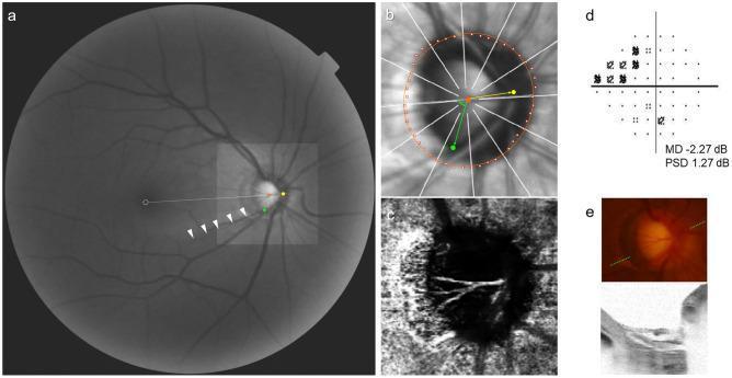
This composite diagnostic image illustrates an ophthalmic evaluation of an eye with a glaucoma-related initial nasal step (INS). Panel (a) is a red-free fundus photograph identifying key landmarks: the Bruch's membrane opening (BMO, red dots), central retinal vascular trunk (CRVT, yellow dot), and a retinal nerve fiber layer defect (RNFLD, white arrowheads) with its proximal margin marked (green dot). Panel (b) shows an infrared OCT image with a best-fitted ellipse for the BMO (orange) and geometric measurements relative to the fovea-BMO axis (white dotted line), including CRVT obliqueness (red angle) and RNFLD proximity (green angle). Panel (c) is an en face OCT angiography (OCTA) image of the choroidal layer, which demonstrates an intact microvascular network without focal dropout. Panel (d) displays Humphrey visual field results with a mean deviation of -2.27 dB, confirming a nasal step defect. Panel (e) includes a color disc photograph and a corresponding B-scan OCT image of the optic nerve head, showing no focal lamina cribrosa defects. This set demonstrates the spatial relationship between optic disc anatomy, vascular exit points, and functional visual field loss.
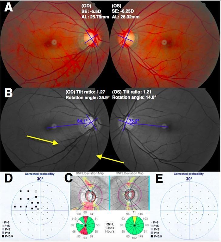
This composite ophthalmological image illustrates a comparative glaucoma case study focusing on the relationship between optic disc morphology and visual field (VF) loss in a myopic patient. (A) Color fundus photographs of the right (OD) and left (OS) eyes display bilateral optic disc tilting and peripapillary atrophy (PPA). (B) Red-free fundus images annotate disc tilt ratios and rotation angles, highlighting a more pronounced inferior rotation (25.9°) in the less myopic right eye. Yellow arrows indicate an inferotemporal retinal nerve fiber layer (RNFL) defect in the right eye. (C) SD-OCT RNFL deviation maps and clock-hour diagrams show significant inferotemporal thinning (indicated by red and yellow coding) in the right eye compared to the relatively normal thickness profile of the left eye. (D, E) Standard automated perimetry (Humphrey Visual Field) results show a corresponding superior nasal step defect in the right eye (D), while the left eye (E) remains largely within normal limits. The image demonstrates that in myopic glaucoma, greater optic disc rotation may correlate more strongly with glaucomatous damage than the absolute degree of axial myopia.
Why a Patient Sees a "White Line" at the Bottom of Their Visual Field - Not an Eyelid Problem
What the Patient is Actually Describing
The Anatomy Behind It: Retinal Nerve Fiber Layer (RNFL) Architecture
-
The retina is divided by a horizontal raphe - an imaginary horizontal line running through the fovea. Nerve fibers from the temporal retina above this line sweep up and around (superior arcuate bundle), and fibers below sweep down and around (inferior arcuate bundle), both joining the optic disc.
-
These two bundles never cross the horizontal raphe. This is the anatomical rule that governs all arcuate visual field defects.
-
When the inferior arcuate nerve fiber bundle is damaged (most commonly by glaucoma, but also ischemic optic neuropathy, or other optic nerve disease), a superior arcuate visual field defect results (remember: inferior retina = superior visual field, and vice versa).Conversely, when the superior arcuate bundle is damaged, an inferior arcuate visual field defect results - which is what the patient is experiencing when they describe "seeing a white line near the lower visual field."
Why It Looks Like a "White Line" - The Edge Phenomenon
- At the sharp edge of an arcuate scotoma, the brain perceives a bright, luminous border - this is called a positive scotoma edge or a form of phosphene at the defect margin.
- The patient looks down into their lower visual field and sees the bright "edge" of where their functional vision ends.
- This edge follows the horizontal raphe precisely - so it appears as a nearly horizontal white line, consistent with the architecture of the Bjerrum area (the arcuate zone of retinal nerve fibers, from 10-20° eccentricity from fixation).
The Classic Patterns (per Adams & Victor's Neurology)
"The classic finding in glaucoma is the Bjerrum field defect, consisting of an arcuate scotoma extending from the blind spot and sweeping around the macula to end in a horizontal line at the nasal equator. Other characteristic patterns are winged extensions from the blind spot (Seidel scotoma) and a narrowing of the superior nasal quadrant that may progress to a horizontal edge, corresponding to the horizontal raphe of the retina (nasal step)."
- Adams and Victor's Principles of Neurology, 12th Ed.
Why It Is "Irritated" (Made Worse / Triggered)
| Trigger | Reason |
|---|---|
| Bright light / glare | High luminance increases contrast at scotoma borders, making the edge more visible |
| Looking downward or at bright backgrounds | Moves the scotoma edge into the patient's attention zone |
| Fatigue or dim light | Reduces the normal visual signal in neighboring areas, making the defect edge more prominent by contrast |
| Eye movement (saccades) | Transient stimulation of the defect border can generate phosphene-like sensations |
Why NOT the Eyelid?
- Lid problems (ptosis, dermatochalasis) cause a mechanical shadow that moves with the lid and disappears when the lid is lifted. The "shadow" follows the lid margin physically.
- This white line does NOT move when the lid is lifted, does not change with lid position, and persists regardless of lid manipulation.
- The defect is fixed in visual space, not in physical space - it stays at the same location relative to where the eye is pointing, not relative to where the lid is.
Bottom Line
What is the treatment
Treatment of Glaucoma (Cause of That Inferior Visual Field White Line)
Step 1: Medications (First Line)
1. Prostaglandin Analogues - FIRST CHOICE
Latanoprost 0.005%, Bimatoprost 0.01/0.03%, Travoprost 0.004%, Tafluprost 0.0015% - all given once at night (q.h.s.)
- Mechanism: Increase drainage of aqueous humor through the uveoscleral pathway
- Most effective IOP-lowering agents; reduce IOP by ~25-35%
- Side effects: Eyelash growth (hypertrichosis), iris pigment darkening, periorbital skin darkening
- Contraindicated: Pregnancy, active uveitis, cystoid macular edema
2. Beta-Blockers
Timolol 0.25-0.5%, Levobunolol - once daily or twice daily (b.i.d.)
- Mechanism: Reduce aqueous humor production by ciliary body
- Avoid in: Asthma, COPD, heart block, bradyarrhythmia, heart failure, depression
3. Alpha-2 Agonists
Brimonidine 0.1%, 0.15%, 0.2% - b.i.d. to t.i.d.
- Mechanism: Reduce aqueous production AND increase uveoscleral outflow
- Contraindicated: Patients on MAO inhibitors (risk of hypertensive crisis); children under 5 (CNS/respiratory depression)
4. Topical Carbonic Anhydrase Inhibitors (CAIs)
Dorzolamide 2%, Brinzolamide 1% - b.i.d. to t.i.d.
- Mechanism: Reduce aqueous production by inhibiting carbonic anhydrase in the ciliary body
- Caution: Sulfa allergy; use carefully in corneal endothelial disease (Fuchs dystrophy)
5. Systemic CAIs (when drops are insufficient)
Acetazolamide 125-250 mg p.o. b.i.d.-q.i.d. or 500 mg sustained-release b.i.d.
- Reserved for acute situations or when topical therapy fails
- Monitor potassium; significant side effects (fatigue, nausea, paresthesias, rare aplastic anemia)
6. Miotics (older, less used)
Pilocarpine 1-4% q.i.d.
- Rarely used now due to poor tolerability (headache, accommodative spasm in young patients), risk of uveitis and retinal detachment
Step 2: Laser (Can Be Used as First Line or After Failed Medications)
Selective Laser Trabeculoplasty (SLT) - NOW RECOMMENDED AS FIRST LINE
- Applies laser to the trabecular meshwork to improve aqueous drainage
- The LiGHT trial (6-year results) showed SLT resulted in reduced progression rates, cost savings, and improved quality of life compared to topical medication - it is now recommended as first-line treatment for newly diagnosed open-angle glaucoma and ocular hypertension
- Especially suitable for patients at risk for poor medication compliance or with medication side effects
Argon Laser Trabeculoplasty (ALT)
- Older version of laser trabeculoplasty; less commonly used now
Laser Iridotomy
- Used specifically for angle-closure glaucoma (not open-angle)
- Creates a small hole at the base of the iris to widen the drainage angle
Step 3: Surgery (for Advanced, Progressive, or Uncontrolled Disease)
| Procedure | Notes |
|---|---|
| Trabeculectomy | Most common filtering surgery; creates a new drainage channel (bleb) under the conjunctiva |
| Tube shunt (drainage device) | Increasingly used; threshold for use has been lowered by many surgeons |
| MIGS (Minimally Invasive Glaucoma Surgery) | Newer options for mild-to-moderate disease; shorter healing, fewer complications. Includes trabecular micro-bypass devices (iStent), canaloplasty, subconjunctival microstents (XEN gel stent), trabectome ablation |
| Cyclo-photocoagulation | Laser ablation of the ciliary body to reduce aqueous production; for refractory cases |
| Phacoemulsification (cataract removal alone) | Associated with ~15% IOP reduction; offered when significant lens opacity is also present; can be combined with MIGS |
- Kanski's Clinical Ophthalmology notes that surgery gives more consistent IOP control because the result is lower and less fluctuating, and compliance is no longer a factor.
Key Points on Prognosis
- The scotoma (white line boundary) will NOT disappear - it is permanent damage. Treatment prevents it from spreading.
- Most patients with POAG will not go blind in their lifetime if treated adequately.
- Historically, the lifetime chance of bilateral blindness in a White population with POAG was 5-10% - this has improved with modern treatment.
- Causes of treatment failure include: wrong target pressure, poor compliance (~25% of patients), wide IOP fluctuations, and occasionally progression despite controlled IOP (possibly normal-tension glaucoma mechanism).
- Wills Eye Manual, pp. 555-558
- Kanski's Clinical Ophthalmology, 10th Ed., p. 385
- Histology: A Text and Atlas (Pawlina), p. 2391