
Acute and chronic smoker
Reading File
The query "acute and chronic smoker" is broad - it likely refers to the pathological/clinical effects of smoking (acute vs. chronic), commonly tested in pathology/respiratory medicine. Let me search the library in parallel.
Finding Sources
Finding Sources
The query "acute and chronic smoker" in medicine typically refers to the pathological changes seen in the airways/lungs. Let me search more specifically for the airway/lung changes in smokers.
Finding Sources
Finding Sources
Excellent - Robbins Pathology has the exact content. Let me read that section and also search for COPD/chronic bronchitis and the precursor lesions section.
Reading File
Finding Sources
Difference between a and c smoker in terms of duration packet etc
The user is asking about the difference between acute and chronic smokers in terms of duration and pack history - this is a pathology/clinical concept. Let me search the textbooks directly.
Searching sources...
Finding Sources
Finding Sources
Reading File
Finding Sources
Finding Sources
Finding Sources
Reading File
Reading File
Now I have solid textbook content. Let me also fetch the precursor lesion image from Robbins which is directly relevant.
Loading Image
Now I have everything needed for a thorough answer. Here is the complete comparison:
Acute vs. Chronic Smoker - Key Differences
Definition by Duration & Pack History
| Feature | Acute Smoker | Chronic Smoker |
|---|---|---|
| Duration of smoking | Recent/new onset (days to weeks to a few months) | Long-term, years to decades |
| Pack-year history | Low (< a few pack-years) | Heavy - typically ≥ 20 pack-years (often 40+ pack-years for COPD/bronchitis) |
| Pack-year formula | Packs per day × years smoked | Same formula, but accumulated over long periods |
A "pack-year" = smoking 1 pack (20 cigarettes) per day for 1 year. So 2 packs/day × 20 years = 40 pack-years.
Pathological Changes: Progression from Acute to Chronic
This is the core concept in pathology - smoking causes a stepwise progression of epithelial injury:
Acute Smoker - Early/Mild Changes
These are the earliest changes seen in smoking-damaged respiratory epithelium:
- Goblet cell hyperplasia - increase in mucus-secreting cells
- Basal (reserve) cell hyperplasia - proliferation of the regenerative layer
- Squamous metaplasia - normal ciliated pseudostratified epithelium replaced by squamous epithelium
These changes are potentially reversible if the person stops smoking early.
Chronic Smoker - Progressive/Severe Changes
With continued, long-term exposure:
4. Squamous dysplasia - disordered squamous epithelium, loss of nuclear polarity, nuclear hyperchromasia, pleomorphism, mitotic figures
5. Carcinoma in situ (CIS) - full thickness atypia, basement membrane still intact
6. Invasive squamous cell carcinoma - basement membrane disrupted, stromal invasion
These are illustrated in the Robbins histology image below:
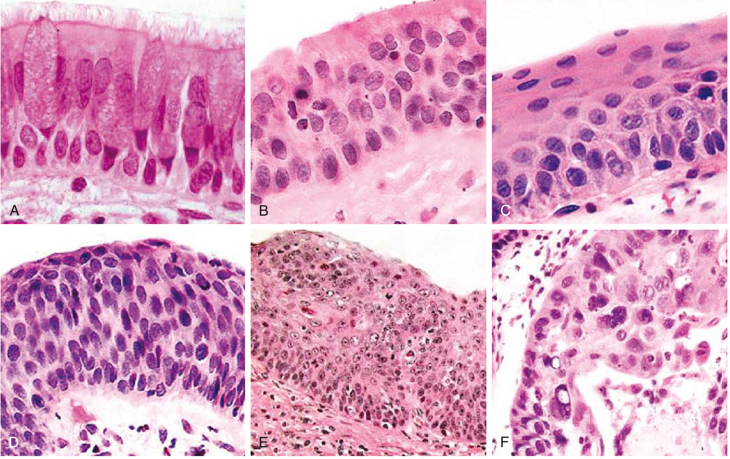
A-C = acute/early smoker changes; D-F = chronic/late smoker changes - Robbins & Kumar Basic Pathology
Specific Conditions Associated with Each
Acute Smoker
- Acute eosinophilic pneumonia - a subset is directly linked to recent onset of smoking (within weeks of starting)
- Ciliary dysfunction and impaired mucus clearance begin almost immediately
Chronic Smoker (≥ 20-40 pack-years)
- Chronic bronchitis - defined clinically as productive cough for ≥ 3 months in ≥ 2 consecutive years; histologically: enlarged mucous glands, Reid index > 0.4, goblet cell metaplasia, mucus plugging
- Emphysema - destruction of alveolar walls (centriacinar type, upper lobe dominant)
- COPD - FEV1/FVC < 0.7; most affected patients have ≥ 40 pack-year history
- Smoking-related ILDs - Respiratory bronchiolitis-ILD (RB-ILD), Desquamative Interstitial Pneumonia (DIP), Pulmonary Langerhans Cell Histiocytosis (PLCH)
- Lung cancer - squamous cell carcinoma and small cell carcinoma most tightly linked; screening recommended at ≥ 50 years old with ≥ 20 pack-year history
- Acquired CFTR dysfunction - leads to abnormal dehydrated mucus, worsening chronic bronchitis
Summary Table
| Parameter | Acute Smoker | Chronic Smoker |
|---|---|---|
| Duration | Days - months | Years - decades |
| Pack-years | < 5 | ≥ 20 (COPD/cancer risk threshold) |
| Epithelial change | Goblet hyperplasia, basal cell hyperplasia, squamous metaplasia | Dysplasia → CIS → carcinoma |
| Reversibility | Yes (if cessation is early) | Partially reversible at best; cancer risk remains |
| Associated disease | Acute eosinophilic pneumonia | COPD, chronic bronchitis, emphysema, lung cancer, RB-ILD, DIP |
| Reid index | Normal (≈ 0.4) | Elevated (> 0.4), proportional to severity |
| Cilia function | Early impairment | Severe, chronic impairment with recurrent infections |
- Robbins & Kumar Basic Pathology (eFig. 11.6; p. 487)
- Robbins, Cotran & Kumar Pathologic Basis of Disease (p. 637)
- Murray & Nadel's Textbook of Respiratory Medicine
This is a shared conversation. Sign in to Orris to start your own chat.