
Otalgia
I'll look up otalgia in the medical library right away.
Searching sources...
Reading File
Finding Sources
Finding Sources
Searching PubMed
otalgia ear pain diagnosis management
Good results. Now let me read the key sections from Scott-Brown's (the most detailed ENT reference) and Cummings/Tintinalli in parallel.
Reading File
Reading File
Reading File
Good content. Let me now read the detailed sections on primary otalgia, referred otalgia, and the nerve anatomy/causes from Scott-Brown's.
Reading File
Reading File
Reading File
Reading File
Now let me fetch the key anatomy diagram and get the section on specific referred otalgia causes (pharyngeal, cervical, neuralgias).
Loading Image
Reading File
I now have comprehensive content from the textbooks. Let me compile a thorough answer.
Otalgia (Ear Pain)
Otalgia is pain perceived in or around the ear. It is classified into two fundamental types:
- Primary (otogenic) otalgia - pain arising from disease within the ear itself
- Secondary (referred) otalgia - pain arising from a remote structure whose sensory nerve supply converges with that of the ear
In children, otalgia is predominantly otogenic. In adults, referred otalgia is the more common cause. - Scott-Brown's Otorhinolaryngology Head & Neck Surgery, Vol. 2
Sensory Nerve Anatomy - The Basis of Referred Otalgia
The ear has an unusually complex sensory supply from 4 cranial nerves and 2 cervical nerves. Any pathology within the sensory territory of these nerves can refer pain to the ear:
| Nerve | Area Supplied | Key Referred Structures |
|---|---|---|
| CN V3 (auriculotemporal br.) | Lateral TM, EAM, lateral pinna above meatus | Teeth, TMJ, sinuses, oral cavity |
| CN VII (facial) | Postero-inferior TM, meatal skin, concha | Parotid, middle ear |
| CN IX (glossopharyngeal) | Middle ear (Jacobson's nerve), tonsils | Pharynx, tonsils, tongue base |
| CN X (vagus - Arnold's nerve) | Postero-inferior TM, concha, meatal skin | Larynx, hypopharynx, oesophagus, even cardiac |
| C2, C3 (greater auricular, lesser occipital) | Cranial surface of pinna, lateral pinna below meatus | Cervical spine, cervical muscles |
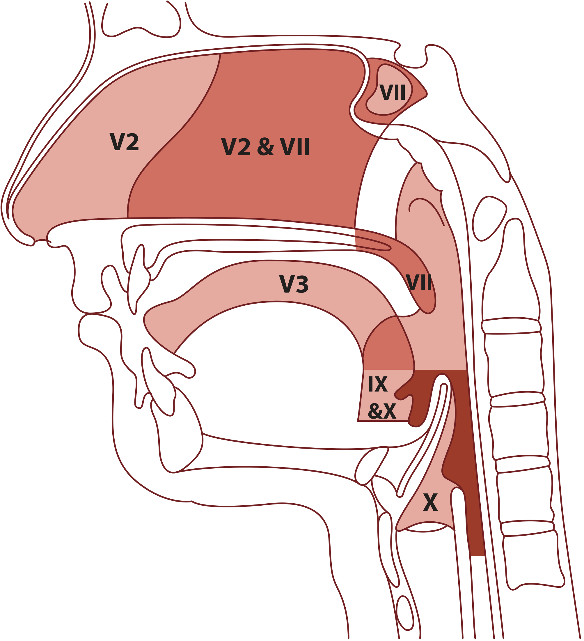
Figure: Otalgia arising from head and neck sources - any pathology within the sensory net of CNs V, VII, IX, X, and C2/C3 can produce referred ear pain (Scott-Brown's)
Causes of Primary Otalgia
| Site | Causes |
|---|---|
| Pinna | Chondrodermatitis nodularis helicis, perichondritis, trauma/haematoma, cellulitis, herpes zoster oticus, malignancy |
| External canal | Otitis externa (acute, chronic, malignant/necrotising), furuncle, bullous myringitis, keratosis obturans, canal cholesteatoma, trauma, foreign body |
| Middle ear | Otitis media (acute, with complications), mastoiditis, cholesteatoma, malignancy |
| Inner ear | Noise discomfort |
Primary otalgia is frequently accompanied by hearing loss and otorrhoea. Their presence points strongly toward an otogenic cause. - Cummings Otolaryngology
Causes of Referred (Secondary) Otalgia
1. Dental/TMJ (Most Common)
Dental disorders are the most common cause of referred otalgia overall.
- Pulpitis from dental caries - poorly localised pain via unmyelinated C fibres
- Acute apical periodontitis/abscess - severe throbbing, often poorly localised
- Pericoronitis from impacted/partially erupted wisdom teeth
- TMJ dysfunction syndrome - otalgia reported in 64% of TMJ dysfunction patients; bruxism is a major aetiological factor (>50% of cases); sufferers are more likely female, with elevated psychological stress; masticatory muscle tenderness is the most reliable clinical finding, with lateral pterygoid tenderness in 85%
2. Malignancy (Must Not Be Missed)
- Isolated unilateral otalgia with a normal ear exam should raise suspicion of occult aerodigestive tract malignancy
- Upper aerodigestive tract tumours (oropharynx, tongue base, hypopharynx, larynx) - referred via CN IX and X
- Skull base/petrous temporal bone tumours (adenoid cystic carcinoma most common; also adenocarcinoma, SCC)
- Temporal bone metastases - commonest primary sites: breast, lung, kidney, stomach, prostate; triad of otalgia + periauricular swelling + facial nerve weakness is highly suspicious
- Parotid malignancy, cervical lymph node malignancy, thyroid disease
3. Pharyngeal and Laryngeal Disease
- Tonsillitis, peritonsillar abscess, adenoiditis (via CN IX)
- Carcinoma of tongue base, tonsil, hypopharynx, larynx
- Pharyngitis, laryngitis
- Foreign bodies in throat
- Inflammatory parapharyngeal space lesions
4. Neuralgias (Rare but Important)
- Glossopharyngeal neuralgia - paroxysmal severe pain in throat/tongue base radiating to ear, preauricular area, neck, or EAC; lasts seconds to minutes; vascular compression (usually PICA) at root entry zone is the main cause; first-line treatment is carbamazepine; surgery (microvascular decompression) achieves complete relief in 64-75%
- Geniculate neuralgia (Ramsay Hunt syndrome) - herpes zoster of CN VII; vesicles in EAC/pinna, severe otalgia, facial palsy, sensorineural hearing loss
- Great auricular neuralgia - pain along the cervical plexus territory
5. Cervical Spine Disease
Cervical spine and cervical muscle pathology can refer pain to the ear via C2/C3 (greater auricular and lesser occipital nerves).
6. Eagle's Syndrome (Stylohyoid Syndrome)
Elongated styloid process (>4 cm, present in 4% of population, symptomatic in only 4% of those). Pain from compression of adjacent structures or carotid vessels. Occurs classically after tonsillectomy but not exclusively.
7. Laryngopharyngeal Reflux (LPR)
LPR has been implicated as a cause of otalgia in both children and adults, due to the sensitivity of upper airway mucosa to acid, even without typical GORD symptoms.
8. Cardiac Pain
Cardiac disease can, rarely, present as otalgia via the vagus nerve (Arnold's nerve). - Scott-Brown's; Cummings
Diagnostic Approach
Key Clinical Clue
Otalgia in the absence of otorrhoea, hearing loss, or otoscopic abnormality while the patient is symptomatic should strongly raise suspicion of secondary (referred) otalgia. - Scott-Brown's
History
- Character, location, radiation, duration, triggers
- Associated otological symptoms: hearing loss, discharge, tinnitus, vertigo
- Associated non-otological symptoms: sore throat, dysphagia, odynophagia, voice change, weight loss, dental pain, jaw click/trismus
- Relevant social history: tobacco and alcohol use (red flags for aerodigestive malignancy)
- Chronic pain history, psychological stressors
Examination
- Full ear examination - otoscopy, TM inspection
- Oral cavity and oropharynx - including tongue base, tonsillar fossae
- Neck - lymphadenopathy, thyroid
- TMJ and masticatory muscles - palpate lateral and medial pterygoids intraorally
- Dental inspection - occlusal wear, buccal mucosa scarring (bruxism)
- Cranial nerve assessment - especially CNs V, VII, IX, X
Investigations
- Dental X-rays, panoramic radiograph if dental/TMJ cause suspected
- Flexible nasendoscopy - to visualise nasopharynx, oropharynx, hypopharynx, larynx
- MRI with gadolinium - for skull base, perineural spread, parapharyngeal lesions
- CT - temporal bone, cervical spine pathology
- If neuralgia suspected: neurology referral for guidance on medical management
Red Flags in Otalgia
| Red Flag | Concern |
|---|---|
| Unilateral otalgia + normal otoscopy | Occult malignancy - aerodigestive tract |
| Tobacco/alcohol history | Head and neck cancer |
| Dysphagia, odynophagia, voice change, weight loss | Pharyngeal/laryngeal malignancy |
| Otalgia + periauricular swelling + facial weakness | Temporal bone malignancy |
| Vesicles in EAC + facial palsy | Ramsay Hunt syndrome |
| Pulsatile tinnitus + otalgia | Paraganglioma, vascular lesion |
Management Principles
- Treat the underlying cause
- For TMJ dysfunction: conservative first - splints, physiotherapy, NSAIDs, dietary modification; psychological assessment for stress/bruxism
- For neuralgias: first-line is carbamazepine; surgical microvascular decompression for refractory cases
- For otitis externa/media: topical/systemic antibiotics as appropriate
- Unexplained otalgia persisting despite normal ENT examination mandates thorough investigation to exclude malignancy
Sources: Scott-Brown's Otorhinolaryngology Head & Neck Surgery Vol. 2 (Ch. 92); Cummings Otolaryngology Head and Neck Surgery (Ch. 137); Tintinalli's Emergency Medicine (Ch. 242); Textbook of Family Medicine 9e
Tinnitus
Searching sources...
Finding Sources
Searching PubMed
tinnitus management treatment
Excellent - Scott-Brown's block 8 is a dedicated tinnitus chapter. Let me read that alongside Tintinalli and the key PubMed abstracts in parallel.
Reading File
Reading File
Reading Article
PMID: 40111327
Reading File
Reading Article
PMID: 40441764
Reading File
Reading File
Reading File
Reading File
Reading File
Now let me read the pulsatile tinnitus section and the causes/assessment sections I haven't covered yet.
Reading File
I now have comprehensive material. Let me compile the full answer.
Tinnitus
Tinnitus is the perception of sound in the absence of an external acoustic stimulus. It is one of the most common ear complaints, affecting approximately 10.1% of adults with persistent spontaneous tinnitus; 5% find it moderately or severely annoying, and 0.5% report a severe effect on daily life. It is most prevalent between ages 40 and 70. - Scott-Brown's Otorhinolaryngology, Vol. 2; Tintinalli's Emergency Medicine
Classification
By Audibility
| Type | Description |
|---|---|
| Subjective | Heard only by the patient - by far the most common type |
| Objective | Can be heard by the examiner (with stethoscope or microphone); always indicates a real sound source |
By Temporal Pattern
| Type | Description |
|---|---|
| Non-pulsatile | Constant or intermittent, not synchronous with heartbeat - most common; typically hissing, ringing, whistling, humming |
| Pulsatile - synchronous | Beats in time with the arterial pulse - implies a vascular source |
| Pulsatile - non-synchronous | Rhythmic but not in time with the pulse - e.g., myoclonus of stapedius, tensor tympani, or palatal muscles |
Subjective idiopathic tinnitus - non-pulsatile, associated with age-related hearing loss, no specific pathology - is the most prevalent form. More complex sounds (voices, music) suggest psychiatric rather than otological pathology.
Epidemiology and Risk Factors
- Noise exposure - the most important modifiable risk factor; noise-induced hearing loss significantly raises tinnitus risk
- Age-related hearing loss (presbycusis) - the most common associated pathology
- Drugs (ototoxic):
- Salicylates (aspirin) - often dose-dependent and reversible
- Quinine
- Aminoglycoside antibiotics
- Platinum-based antineoplastics (cisplatin)
- Specific ear diseases: Meniere's disease, otosclerosis, vestibular schwannoma ("syndromic tinnitus")
- Comorbidities: depression, anxiety (40% comorbidity - cause and effect are bidirectional), hyperacusis (40% of tinnitus patients have some degree of hyperacusis; conversely, 86% of patients with hyperacusis also have tinnitus), TMJ dysfunction
Pathophysiology
Tinnitus arises through both peripheral and central mechanisms. A key conceptual framework distinguishes:
- Ignition site - where the initial aberrant signal is generated (often peripheral - cochlea/auditory nerve)
- Promotion - central auditory processing that transforms the signal into a perceptible, potentially distressing sound
The important insight is that even if the peripheral auditory system is completely destroyed (e.g., after vestibular schwannoma surgery), tinnitus can persist - indicating that long-term tinnitus is maintained centrally.
Peripheral Mechanisms
- Discordant hair cell damage - outer hair cells (more vulnerable to noise, aminoglycosides) are lost while inner hair cells remain; the unsupported tectorial membrane sags and depolarises inner hair cells continuously
- Calcium channel dysfunction - salicylates and quinine alter intracellular calcium in cochlear hair cells
- Glutamate excitotoxicity - excess glutamate activates AMPA and NMDA receptors at inner hair cells; massive NMDA receptor activation is toxic to auditory nerve fibres
Central Mechanisms
- Loss of inhibition - cochlear damage reduces afferent activity, which downregulates inhibitory processes in higher auditory centres, increasing spontaneous cortical activity
- Increased neural synchrony - peripheral damage causes random spontaneous cortical activity to become synchronized, mimicking a sound signal
- Cortical map reorganisation - tonotopic reorganisation leads to over-representation of frequencies adjacent to damaged cochlear areas, generating increased neural activity at those frequencies
Neurophysiological Model (Jastreboff, 1990)
Tinnitus involves not only the classical auditory system but also altered activity in the limbic system, reticular system, and autonomic nervous system. High autonomic arousal prevents habituation to the tinnitus signal, making it intrusive. This model underpins Tinnitus Retraining Therapy (TRT). - Scott-Brown's
Causes
Non-Pulsatile Tinnitus
| Category | Examples |
|---|---|
| Cochlear/peripheral | Noise-induced hearing loss, presbycusis, Meniere's disease, otosclerosis, sudden SNHL, labyrinthitis |
| Retrocochlear | Vestibular schwannoma (acoustic neuroma), CPA tumours |
| Middle/external ear | Otitis media, otosclerosis, cerumen impaction, eustachian tube dysfunction |
| Drugs/ototoxic | Aspirin, quinine, aminoglycosides, cisplatin, loop diuretics |
| Systemic | Anaemia, hypothyroidism, hypertension |
| Neurological | Multiple sclerosis (involving auditory pathways) |
Pulsatile Tinnitus
| Subtype | Causes |
|---|---|
| Synchronous (vascular) | Arteriovenous malformation, arteriovenous fistula (esp. dural), glomus tumour (paraganglioma), carotid artery stenosis/dissection, high jugular bulb, intracranial hypertension (benign intracranial hypertension - "venous hum"), anaemia, thyrotoxicosis |
| Non-synchronous (muscular) | Myoclonus of stapedius, tensor tympani, or tensor veli palatini (palatal myoclonus); clicking quality |
Pulsatile tinnitus always warrants investigation - it may indicate a treatable or dangerous vascular lesion.
Assessment
History
- Character: pitch, quality (ringing, hissing, clicking, pulsatile), unilateral or bilateral
- Duration, onset (sudden vs. gradual), fluctuating or constant
- Associated symptoms: hearing loss, vertigo, aural fullness, discharge
- Noise exposure history, drug history
- Psychological impact: sleep disturbance, anxiety, depression
- Impact on daily life - use validated tools:
- Tinnitus Handicap Inventory (THI) - most widely used outcome measure
- Hospital Anxiety and Depression Scale (HADS)
- Insomnia Severity Index
Examination
- Otoscopy - middle/external ear disease
- Auscultation over ear, neck, orbit - for objective/vascular tinnitus
- Cranial nerve assessment
- Neck examination
- Somatic manoeuvres - ask if tinnitus changes with jaw movement, neck position (somatic modulation suggests central involvement)
Investigations
- Pure tone audiogram - mandatory; most patients have hearing loss even if subtle; high-frequency testing may detect loss at >8 kHz
- Tympanometry - middle ear pathology
- MRI with gadolinium - indicated for unilateral tinnitus with or without SNHL to exclude vestibular schwannoma
- MRA/CT angiography - for pulsatile tinnitus to exclude vascular lesions
- Blood tests - FBC, TFTs, lipids if systemic cause suspected
Management
1. Explanation and Reassurance
The first and most important step. Many patients have been incorrectly told "nothing can be done" - this negative counselling is harmful and should be avoided. Multiple RCTs support education and information-giving as a standalone treatment modality. - Scott-Brown's
2. Hearing Aids
- When tinnitus is associated with aidable hearing loss, hearing amplification is the primary intervention
- Amplifying external sounds masks tinnitus and reduces the central gain that drives it
- Indirect benefits: improved communication reduces stress and anxiety that exacerbate tinnitus
3. Sound Therapy / Masking
- Broadband noise generators worn in the ear ("white noise generators") or environmental sound enrichment
- Aims to reduce the contrast between tinnitus and background sound
- Classical tinnitus maskers (designed to completely mask tinnitus) have largely been superseded by partial maskers and sound enrichment
4. Cognitive Behavioural Therapy (CBT)
- The best-evidenced psychological treatment for tinnitus distress
- Does not eliminate the tinnitus perception but changes the patient's emotional and attentional response to it
- Addresses catastrophic thinking, avoidance behaviours, sleep hygiene
- Cochrane reviews support CBT for improving quality of life and reducing tinnitus distress
5. Tinnitus Retraining Therapy (TRT)
- Combines directive counselling (demystifying tinnitus) with long-term sound therapy
- Based on Jastreboff's neurophysiological model - promotes habituation at limbic and autonomic levels
- Evidence: superior to tinnitus masking alone in one RCT (Cochrane review); widely used but requires a rigorous, time-intensive protocol
6. Drug Treatments
Evidence for systemic drugs is generally poor due to underpowered trials. Key points:
- Antidepressants (tricyclics/SSRIs) - may help patients tolerate tinnitus and treat comorbid anxiety/depression but do not directly alter tinnitus perception
- Benzodiazepines - reduce tinnitus distress short-term but addiction risk limits use
- Betahistine - used in Meniere's-associated tinnitus; limited evidence for idiopathic tinnitus
- Carbamazepine - for rare cases of typewriter tinnitus (CN VIII irritation)
- Intratympanic steroids/glutamate antagonists - investigated for acute-onset tinnitus (e.g., following sudden SNHL); a small therapeutic window may exist; intratympanic dexamethasone showed no advantage over placebo in the only placebo-controlled RCT
- Esketamine (intratympanic NMDA antagonist) - under investigation in multinational trials at the time of publication
7. Electromagnetic/Neuromodulation Therapies
- Repetitive transcranial magnetic stimulation (rTMS) - Cochrane review finds very limited support for low-frequency rTMS
- Vagus nerve stimulation paired with sound - early-phase studies only; insufficient evidence
- Acoustic CR Neuromodulation - an RCT failed to show significant benefit
8. Treatment of Underlying Cause
- Otosclerosis: stapedectomy generally improves associated tinnitus
- Vestibular schwannoma: management per tumour protocol; tinnitus may persist post-surgery
- Meniere's disease: diuretics, low-salt diet, betahistine, intratympanic gentamicin/steroids
- Cerumen impaction: microsuction/syringing
- Ototoxic drugs: withdraw or substitute where possible
- Pulsatile tinnitus: directed at vascular cause (embolisation, surgery, IIH treatment)
Recent Evidence Update
A 2025 VA/DoD Clinical Practice Guideline (PMID: 40111327) provides the most current evidence-based framework for managing bothersome tinnitus in adults, based on a systematic review of 20 key clinical questions. It highlights significant evidence gaps and recommends patient education and self-management as central pillars.
A 2025 network meta-analysis of 60 RCTs (PMID: 40441764) on pharmacotherapy found that Ginkgo biloba + vitamins ranked highest for tinnitus severity reduction, acamprosate for annoyance reduction, and fluoxetine for loudness reduction - but all with very low GRADE certainty. Results should be interpreted cautiously pending higher-quality trials.
Summary Table: Tinnitus at a Glance
| Feature | Key Points |
|---|---|
| Prevalence | ~10% of adults; 0.5% severely affected |
| Most common type | Subjective, non-pulsatile, bilateral, high-frequency |
| Most common cause | Noise-induced / presbycusis hearing loss |
| Red flag | Unilateral tinnitus → rule out vestibular schwannoma (MRI) |
| Red flag | Pulsatile tinnitus → vascular imaging mandatory |
| Best-evidenced treatment | CBT for distress; hearing aids when hearing loss present |
| First step | Explanation and reassurance - never say "nothing can be done" |
Sources: Scott-Brown's Otorhinolaryngology Head & Neck Surgery Vol. 2 (Ch. on Tinnitus); Cummings Otolaryngology (Ch. 137); Tintinalli's Emergency Medicine (Ch. 242)
Allergic rhinitis
Searching sources...
Finding Sources
Searching PubMed
allergic rhinitis management guidelines
Excellent - fresh ARIA-EAACI 2024-2025 guidelines just published. Let me read the key textbook sections and the guideline abstracts in parallel.
Reading File
Reading File
Reading Article
PMID: 41324154
Reading File
Reading File
Reading Article
PMID: 41877472
Excellent content. Now let me read the treatment and immunotherapy sections from Cummings.
Reading File
Reading File
I have rich content from multiple sources and the fresh ARIA-EAACI 2024-2025 guidelines. Let me now fetch the pathophysiology diagram and compile the full answer.
Loading Image
Reading File
I have comprehensive, high-quality content from multiple authoritative sources plus the fresh 2024-2025 ARIA-EAACI guidelines. I can now compile a thorough answer.
Allergic Rhinitis (AR)
Allergic rhinitis is an IgE-mediated inflammatory condition of the nasal mucosa triggered by allergen exposure in a sensitised individual. It is among the most prevalent chronic conditions globally, affecting 10-30% of adults in North America (peak prevalence >30% in the fifth decade), with prevalence rising over the past two decades. It ranks 5th among all chronic diseases in the US in economic impact, with estimated direct costs of up to $5 billion annually and 3.5 million lost workdays per year. - Harrison's 22E; Cummings Otolaryngology
Classification
By Temporal Pattern
| Type | Definition | Common Allergens |
|---|---|---|
| Seasonal | Symptoms during specific pollen seasons | Tree pollen (spring), grass (early summer), weeds/ragweed (late summer-fall), outdoor moulds |
| Perennial | Symptoms year-round | House dust mites (D. farinae, D. pteronyssinus), animal dander, cockroach proteins, indoor moulds |
| Episodic | Intermittent upon specific exposure | Occupational or pet-related allergens |
ARIA Classification (Clinically Preferred)
| Intermittent | Persistent |
|---|---|
| Symptoms <4 days/week OR <4 weeks/year | Symptoms >4 days/week AND >4 consecutive weeks |
| Mild - no impairment of sleep/work/daily activities | Moderate-Severe - one or more: disturbed sleep, impaired work/daily activities, troublesome symptoms |
Local Allergic Rhinitis (LAR)
A recently defined phenotype where patients have classic AR symptoms but negative skin prick tests and serum-specific IgE. Local nasal IgE production occurs without systemic atopy. Prevalence may be 47-62.5% of perennial/seasonal rhinitis patients with negative systemic allergy testing. Common allergens: house dust mite, grass, olive pollen. Responds to oral antihistamines and intranasal corticosteroids. Confirmed by nasal provocation testing or nasal-specific IgE. - Cummings Otolaryngology
Predisposing Factors
- Atopy - AR occurs in atopic individuals; strongly associated with asthma (up to 50% of AR patients have asthma; 70-80% of asthmatics have AR), atopic dermatitis, food allergy, urticaria
- Family history of allergic disease
- Female sex, particulate air pollution, maternal tobacco smoking
- "Allergic march" - AR often precedes asthma development; allergen immunotherapy reduces asthma risk by ~40%
- Climate change - earlier tree pollination, prolonged ragweed season (delayed first frost); elevated CO₂ increases pollen production
Pathophysiology
The disease involves two phases - sensitisation and clinical disease - and two inflammatory responses within the clinical phase.
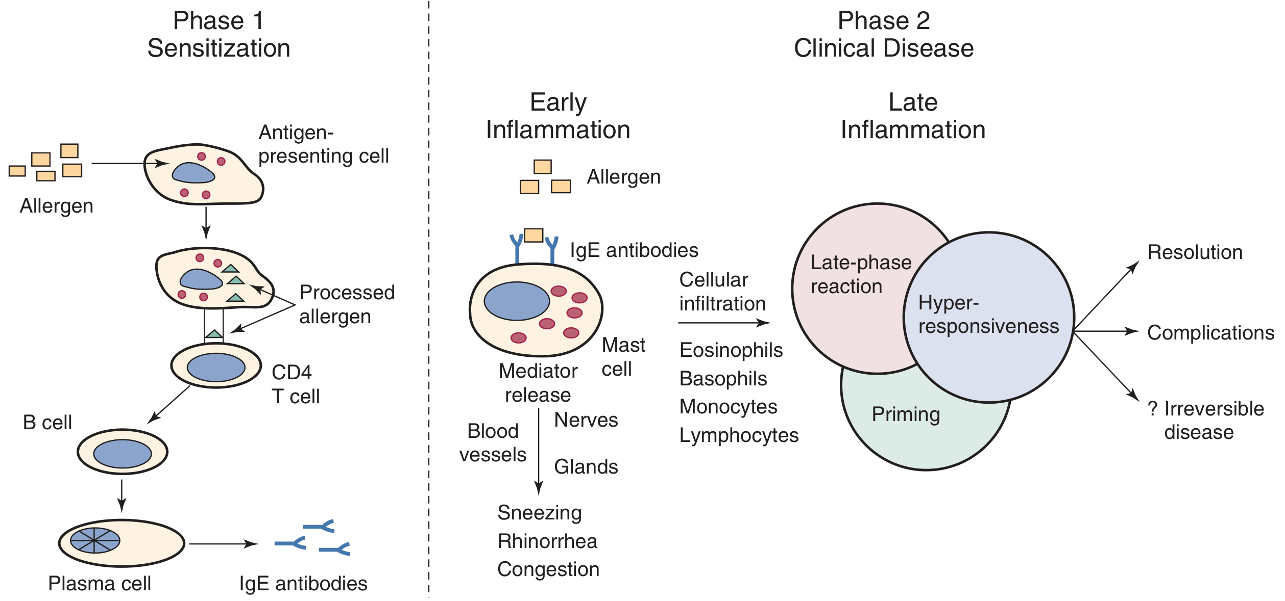
Fig. Pathophysiology of allergic rhinitis (Cummings Otolaryngology)
Phase 1 - Sensitisation
- Inhaled allergen deposited on nasal mucosa; mucosal enzymes digest outer coat releasing protein allergens
- Mucosal epithelial cells secrete TSLP → maturation of dendritic cells into TH2-promoting subtypes
- Antigen-presenting cells (macrophages, dendritic cells, Langerhans cells) process and present allergen to CD4+ T cells → TH2 polarisation
- TH2 cells secrete IL-4, IL-13 → B cell class switching → plasma cells produce allergen-specific IgE
- IgE binds high-affinity FcεRI receptors on mast cells and basophils → sensitisation complete
Phase 2a - Early-Phase Response (within 5-15 minutes of re-exposure)
- Cross-linking of IgE on mast cells triggers degranulation
- Released preformed mediators: histamine, heparin, tryptase
- Newly synthesised mediators: PGD₂, leukotrienes LTC₄, LTD₄, LTE₄, platelet-activating factor (PAF)
- Symptoms: sneezing, rhinorrhoea, nasal pruritus, conjunctival itching (histamine-dominant)
- PGD₂ attracts eosinophils and basophils; leukotrienes cause nasal congestion and increased vascular permeability
Phase 2b - Late-Phase Response (4-8 hours later)
- Recruited inflammatory cells: eosinophils, basophils, neutrophils, monocytes, CD4+ T lymphocytes
- Cytokine amplification sustains and prolongs inflammation
- Symptoms: predominantly nasal blockage, mucus hypersecretion, nasal hyperresponsiveness
- Priming effect: repeated allergen exposure lowers the threshold for subsequent responses ("priming phenomenon")
- Chronic inflammation → nasal hyperresponsiveness to non-specific stimuli (cold air, smoke, strong smells)
Consequences of Chronic Inflammation
- Swelling of turbinates obstructs sinus ostia → secondary sinusitis
- Obstruction of Eustachian tube → middle ear dysfunction, otitis media with effusion
- Olfactory cleft inflammation → hyposmia/anosmia
- Systemic effects: fatigue, malaise, sleep disturbance, impaired cognition
Clinical Features
Symptoms
- Cardinal nasal symptoms: sneezing (often paroxysmal), rhinorrhoea (clear, watery), nasal obstruction, nasal pruritus (most specific for allergic aetiology)
- Ocular symptoms: pruritus, lacrimation, conjunctival injection and oedema (allergic conjunctivitis)
- Palatal/pharyngeal pruritus ("itchy palate" - very characteristic)
- Ear symptoms: eustachian tube dysfunction (popping, clicking), otalgia
- Postnasal drip → throat clearing, snorting, cough
- Systemic: malaise, fatigue, irritability, sleep disturbance, snoring
- Pollen-food allergy syndrome (oral allergy syndrome): oropharyngeal pruritus after ingesting plant-based foods cross-reactive with pollen allergens
Signs
- Pale, boggy nasal mucosa with oedematous turbinates
- Conjunctival congestion and oedema
- Allergic shiners - dark infraorbital circles from venous stasis
- Allergic salute - transverse nasal crease from habitual upward nose rubbing
- Pharynx generally unremarkable
Diagnosis
Clinical Diagnosis
A clinical history of rhinitis symptoms in temporal relationship to allergen exposure + documentation of sensitisation to an environmental allergen.
Red flags requiring further investigation: unilateral obstruction, epistaxis, anosmia, purulent discharge, facial pain (consider sinusitis, sinonasal tumour, polyps), fixed constant obstruction (consider septal deviation or polyps).
Allergy Testing
| Test | Details | Advantages | Limitations |
|---|---|---|---|
| Skin prick test (SPT) | Gold standard; allergen applied to skin, IgE-sensitised mast cells react; wheal >3 mm positive | Immediate results, cheap, high sensitivity | Requires stopping antihistamines; contraindicated with severe eczema/dermatographism; anaphylaxis risk |
| Serum antigen-specific IgE (radioallergosorbent/ImmunoCAP) | Measures circulating allergen-specific IgE | No anaphylaxis risk; no medication stoppage; usable with skin disease | Higher cost; slower results; comparable sensitivity to SPT |
| Serum total IgE | Limited diagnostic utility alone; markedly elevated levels support atopy | Useful to identify atopic individuals | Low specificity |
| Nasal provocation test | Allergen applied intranasally; monitors symptoms ± nasal airflow | Confirms LAR; research standard | Time-consuming; not routine |
| Basophil activation test (BAT) | Flow cytometry assay (CD63, CD203c); useful when SPT and serum IgE discordant | Second-line for LAR diagnosis | Not widely available |
| Component resolved diagnosis (CRD) | Identifies specific allergen components; guides immunotherapy candidacy | Distinguishes true sensitisation from cross-reactivity | Specialised |
Important: A positive allergy test alone does NOT confirm AR - clinical correlation is mandatory. SPT positivity prevalence exceeds 50% in the US population, far exceeding clinical allergy prevalence.
Management
1. Allergen Avoidance
- Identify and reduce exposure to specific triggers
- Dust mite measures: impermeable mattress/pillow covers, hot washing, HEPA vacuuming, reduced humidity
- Pollen: close windows during peak season, sunglasses outdoors, shower after outdoor exposure
- Pet avoidance: most effective measure, though dander persists months after pet removal
- Cockroach: pest control, food storage, fixing water leaks
2. Pharmacotherapy
Updated ARIA-EAACI 2024-2025 Recommendations (Evidence Hierarchy)
Intranasal treatments (Part I - PMID 41324154):
- INCS + INAH combination > INAH or INCS alone (strongest recommendation)
- INCS > INAH alone (recommended)
- Intranasal decongestants: short-term use only (rhinitis medicamentosa risk with >5-7 days use)
Oral/ocular treatments (Part II - PMID 41877472):
- INCS > oral antihistamine (OAH) for nasal symptoms
- OAH > leukotriene receptor antagonist (LTRA) for AR
- OAH > ocular antihistamine (OcAH) for ocular symptoms
- Do NOT add LTRA to OAH (no added benefit)
Drug Classes in Detail
| Drug Class | Examples | Mechanism | Clinical Notes |
|---|---|---|---|
| Intranasal corticosteroids (INCS) | Mometasone, fluticasone, budesonide, beclometasone | Reduce all phases of inflammation; suppress eosinophil recruitment, cytokine production | Most effective single agent for nasal symptoms; onset 1-2 weeks; minimal systemic absorption with modern agents |
| Intranasal antihistamines (INAH) | Azelastine, olopatadine | H1 receptor antagonists; topical | Faster onset than INCS; effective for sneezing, rhinorrhoea; available as fixed-dose combination with INCS |
| Fixed combination INAH+INCS | Azelastine/fluticasone (Dymista) | Dual mechanism | Superior to either alone per ARIA 2024-2025; preferred first-line for moderate-severe AR |
| Oral antihistamines (OAH) | Cetirizine, loratadine, fexofenadine (2nd gen); chlorphenamine (1st gen) | H1 antagonism, systemically | 2nd generation preferred (non-sedating); better for ocular and systemic symptoms; less effective than INCS for nasal congestion |
| Oral decongestants | Pseudoephedrine | α-adrenergic agonist → vasoconstriction | Short-term only; caution in hypertension, cardiovascular disease, hyperthyroidism |
| Leukotriene receptor antagonists (LTRA) | Montelukast | Block LTC₄/LTD₄/LTE₄ receptors | Less effective than INCS and OAH; particularly useful with concomitant asthma; FDA black-box warning for neuropsychiatric events |
| Intranasal sodium cromoglicate | Cromoglicate spray | Mast cell stabiliser | Mild, short duration, requires frequent dosing; good safety profile; suitable in pregnancy/children |
| Intranasal anticholinergics | Ipratropium | Reduces rhinorrhoea only (no effect on congestion, sneezing, pruritus) | Useful for predominantly watery rhinorrhoea |
| Oral corticosteroids | Prednisolone (short course) | Potent anti-inflammatory | Reserved for severe refractory cases; short course only; avoid depot injections |
| Anti-IgE biologic | Omalizumab | Binds free IgE; reduces mast cell/basophil sensitisation | Licensed for severe persistent AR not controlled by standard therapy; expensive |
3. Allergen-Specific Immunotherapy (AIT) - Disease-Modifying Treatment
The only treatment that modifies the natural course of AR, with continued benefit after cessation. Reduces risk of asthma development by ~40% and prevents new sensitisations in monosensitised patients.
Routes:
- Subcutaneous immunotherapy (SCIT): injections of escalating allergen doses over 3-5 years; requires clinic visits; risk of systemic reactions
- Sublingual immunotherapy (SLIT): tablets or drops taken daily at home; lower systemic reaction risk; growing evidence base
Immunological mechanisms:
- Desensitisation of mast cells/basophils (upregulation of H2 receptor)
- Shift from TH2 to Treg dominance (IL-10, TGF-β mediated peripheral tolerance)
- Increase in allergen-specific IgG4 (blocking antibody) and gradual decrease in specific IgE
- Induction of FoxP3+/CD25+ Treg cells
Indications for AIT:
- Moderate-severe AR inadequately controlled by pharmacotherapy
- Patient preference to reduce long-term medication
- Prevention of asthma development
- Monosensitisation or clearly dominant allergen
4. Biologic Therapies
- Dupilumab (anti-IL-4Rα): targets IL-4/IL-13 signalling; licensed for chronic rhinosinusitis with nasal polyps; growing evidence in AR
- Omalizumab (anti-IgE): for severe persistent AR
Complications
| Complication | Mechanism |
|---|---|
| Rhinosinusitis | Obstruction of sinus ostia by turbinate oedema |
| Otitis media with effusion | Eustachian tube dysfunction |
| Asthma ("united airway disease") | Shared TH2 inflammatory pathway; AR directly worsens asthma control |
| Nasal polyps | Chronic eosinophilic inflammation |
| Sleep-disordered breathing | Nasal obstruction → mouth breathing, snoring, OSAS |
| Hyposmia/anosmia | Olfactory cleft obstruction/inflammation |
| Impaired quality of life | Sleep disturbance, cognitive impairment, school/work productivity loss |
Latest Guidelines Highlight
The ARIA-EAACI 2024-2025 Guidelines (published 2026) represent the most current evidence-based framework. The Part I intranasal guidelines and Part II oral/ocular guidelines both used the GRADE framework and notably incorporated mHealth real-world data alongside RCTs. The key departure from older guidelines is the elevation of the fixed-dose INCS+INAH combination as the preferred first-line intranasal treatment for moderate-severe AR, superseding INCS monotherapy as the automatic default.
Sources: Harrison's Principles of Internal Medicine 22E (2025); Cummings Otolaryngology Head & Neck Surgery (Ch. 35); Scott-Brown's Otorhinolaryngology Vol. 2; K.J. Lee's Essential Otolaryngology; ARIA-EAACI 2024-2025 Guidelines (PMIDs 41324154, 41877472)
Search for recent research on sublingual immunotherapy efficacy
This is a shared conversation. Sign in to Orris to start your own chat.