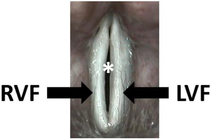
Contact laryngoscopy and virtual tracheobronchoscopy in detail in simplified manner according to standard ENT textbook
contact endoscopy larynx vocal cord microvasculature
virtual bronchoscopy CT airway 3D reconstruction
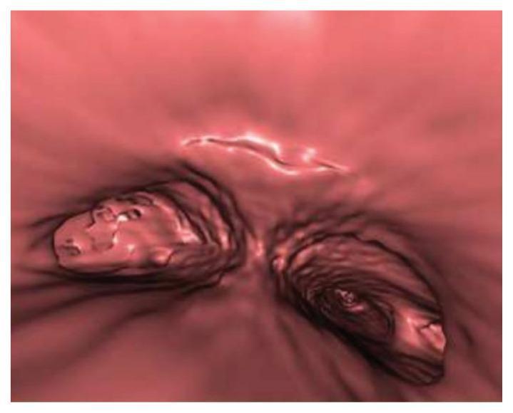
This diagnostic image is a virtual bronchoscopy reconstruction of the tracheobronchial tree, specifically focusing on the bifurcation and the left main bronchus. The reconstruction visualizes the internal luminal surface in a 3D perspective. In the left main bronchus, linear, thin, fenestrated membranes are visible, creating a web-like structure that partially occludes the lumen. Distal to these membranes, there is an area of soft tissue density, which contextually represents the left bronchial stump following a prior pneumonectomy. This density consists of collapsed lung parenchyma or scar tissue and possible suture material. Crucially, the virtual reconstruction demonstrates an intact bronchial stump with no evidence of a bronchopleural fistula (BPF). The image serves as an educational example of post-surgical anatomical changes and the use of non-invasive virtual bronchoscopy (derived from high-resolution CT) to evaluate airway patency and rule out complications like BPF in post-pneumonectomy patients.
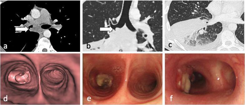
This composite figure illustrates a case of tracheobronchial tuberculosis involving lymph node invasion into the airway. (a) Axial CT in a mediastinal window demonstrates a 29 mm subcarinal lymph node (arrow) exhibiting central low attenuation, suggestive of necrosis. (b) Coronal CT in a lung window shows the node invading the wall of the right main bronchus. (c) Axial CT in a lung window reveals heterogeneous consolidation in the right lower lobe and a small right pleural effusion, likely representing post-obstructive changes. (d) CT virtual bronchoscopy provides a 3D reconstruction of the subcarinal mass protruding into the right main bronchial lumen. (e) Fiberoptic bronchoscopy confirms a yellowish/tan-colored, irregular necrotic nodule partially obstructing the right main bronchus. (f) Post-procedural bronchoscopy shows a patent airway lumen following successful cryoprobe removal of the necrotic lymph node tissue. The images collectively demonstrate the complications of tuberculous lymphadenopathy, including bronchial perforation and distal lung consolidation.
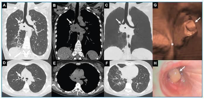
This composite figure presents multi-modal thoracic imaging and endoscopy. Panels A-F show chest CT scans in coronal (A-C) and axial (D-F) planes, utilizing both lung and mediastinal windows. A large, heterogeneous, hypodense subcarinal mediastinal mass/lymph node conglomerate is visible (asterisk), characteristic of necrotic lymphadenopathy. There is an associated endobronchial component (arrow) extending into the right main and intermediate bronchi. Lung window images (A, D, F) demonstrate secondary parenchymal changes in the middle and right lower lobes, including consolidation, subsegmental atelectasis, and centrilobular nodules in a 'tree-in-bud' pattern, suggesting infectious bronchiolitis or endobronchial dissemination. Panel G provides a 3D virtual bronchoscopy reconstruction, while Panel H shows a fiberoptic bronchoscopy view, both highlighting a smooth, polypoid endobronchial lesion (arrow) partially obstructing the right-sided airway. The findings are clinically significant for evaluating pulmonary cryptococcosis or other granulomatous infections causing mediastinal lymphadenitis and airway involvement.
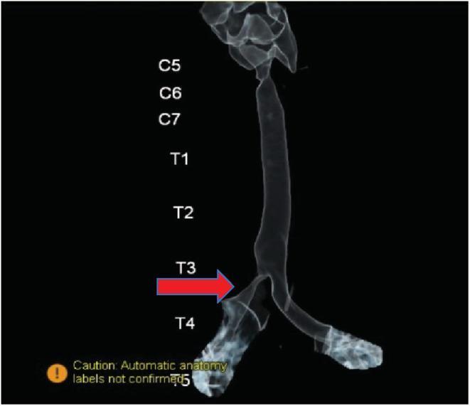
This diagnostic image is a 3D reconstructed chest CT scan, also known as virtual bronchoscopy, showcasing the tracheobronchial tree. The visualization tracks the airway from the cervical region down to the main bronchi. Labels indicate vertebral levels from C5 to T5 to provide anatomical context. A prominent red arrow points to the level of the third thoracic vertebra (T3), highlighting a focal, significant partial narrowing or stenosis of the right main bronchus. In contrast, the left main bronchus maintains a normal caliber and patency. This image demonstrates the clinical application of 3D reconstruction in identifying structural airway anomalies, such as a bronchial web, by mapping internal lumen dimensions relative to bony landmarks. The image is used in medical education to teach diagnostic imaging techniques for respiratory obstructions and the anatomical relationship between the tracheobronchial tree and the spinal column.
"virtual bronchoscopy" airway foreign body
1. Contact Laryngoscopy (Contact Endoscopy)
What it is
History (brief)
- First described by Desormaux (1865) for bladder mucosa.
- Refined into microcolpohysteroscopy in gynaecology (Hamou, 1983).
- The dedicated contact microlaryngoscope was developed in 1995, extending the technique to the larynx and later to the nose, nasopharynx, oral cavity, oropharynx, hypopharynx, and trachea.
- In 2007, combined with Narrow Band Imaging (NBI), which uses a specific light wavelength selectively absorbed by haemoglobin - this sharpens contrast of blood vessels against surrounding tissue and reveals deeper vessels.
Equipment
- Karl Storz contact microlaryngoscope: 5.8 mm diameter, 24 cm length (a 30-degree angled version also exists for difficult angles).
- Shorter, narrower scopes (4 mm, 18 cm) exist for the nose, nasopharynx, and oral cavity.
Technique
- Performed under general anaesthesia with endotracheal intubation for the larynx/hypopharynx; nasal, oral, and nasopharyngeal sites can often be examined without anaesthesia.
- Vascular pattern: the scope is applied directly to the mucosa with no stain, using standard or NBI illumination.
- Cellular pattern: the mucosa is first stained with methylene blue (stain lasts 4-5 minutes), then the scope is applied; a control ring on the eyepiece adjusts magnification (60x-150x) and focus.
- The scope is slid over the mucosa to "map" the lesion and adjoining areas.
- Always recorded on video (ideally high-definition) so the recording can be reviewed later with a pathologist/cytologist.
What you see
- Normal squamous epithelium: polyhedral cells in continuity, round dark nuclei, light-blue cytoplasm, regular nucleus:cytoplasm ratio, homogeneous pattern.
- Ciliated epithelium: round dark nuclei packed closely together (higher nuclear density per field), indistinct cytoplasmic borders.
- Dysplasia/malignancy: irregular and heterogeneous cells; nuclei vary in size, shape, and colour with an irregular nucleus:cytoplasm ratio; nuclear inclusions, prominent nucleoli, and mitoses may be seen. The vascular pattern (angioarchitecture) becomes atypical too - irregular calibre vessels, ectasias, haemorrhages, and areas of reduced flow/thrombosis.
- Papilloma: contact endoscopy helps define the true extent of the lesion.
Clinical uses
- Non-invasive, real-time evaluation of mucosal cells and microvasculature.
- Detects subtle cellular changes at transition zones (e.g., margin between tumour and normal mucosa).
- Helps map disease extent, guide biopsy site selection, guide cytology sampling, and define safe surgical margins during microlaryngeal surgery.
- Useful in both the outpatient clinic and the operating theatre.
Key limitation
Key points to remember (as per the textbook)
- Allows non-invasive evaluation of microvasculature and cells.
- Allows appreciation of cellular differences at transition zones.
- Most upper aerodigestive mucosa has vessels running parallel to the surface.
- Assists in identifying safe surgical margins.
2. Virtual Tracheobronchoscopy (Virtual Bronchoscopy)
What it is
How it works
- CT data is usually obtained in a single breath-hold at end-inspiration.
- Internal rendering produces a "virtual luminal view" that imitates what a bronchoscopist would see through a real scope.
- External rendering produces "CT bronchography," which shows the airway's outer dimensions and its relationship to surrounding structures (vessels, mediastinum, tumours).
Key advantage
Clinical applications
- Airway foreign bodies (especially in children): highly sensitive, with reported positive predictive value as high as ~98% for detecting tracheobronchial foreign bodies. Many children's hospital emergency departments now use low-dose "virtual bronchoscopy" CT protocols for this.
- Tracheobronchial stenosis: good accuracy in grading the degree, site, and length of narrowing (e.g., post-traumatic or post-intubation strictures).
- Tumour evaluation: shows an endobronchial mass and its relation to the airway wall and adjacent mediastinal structures, useful for surgical planning.
- Congenital tracheal anomalies (agenesis, stenosis, webs) and vascular ring evaluation in children, often combined with MRI/echocardiography for associated cardiac anomalies.
Limitations (important - standard exam point)
- It is less reliable for tracheobronchomalacia (dynamic airway collapse) since it is usually a static, single-phase acquisition.
- It cannot obtain tissue for histology/cytology and cannot replace therapeutic bronchoscopy (e.g., foreign body removal still needs real rigid bronchoscopy).
- If a patient needs a general anaesthetic anyway, most surgeons will proceed straight to a real rigid bronchoscopy rather than doing a CT first, since the airway can decompensate suddenly under anaesthesia.
- Best regarded as complementary to, not a substitute for, actual endoscopy - a "radiological survey" that helps plan the real procedure.
Quick Comparison Table
| Feature | Contact Laryngoscopy | Virtual Tracheobronchoscopy |
|---|---|---|
| Principle | Optical magnification (60-150x) of mucosa in direct contact | CT data post-processed into a simulated 3D endoluminal view |
| Invasiveness | Requires endoscope insertion (often under GA for larynx) | Fully non-invasive (just a CT scan) |
| What it shows | Individual cells and superficial microvasculature | Airway lumen, calibre, stenosis, mass, relation to surrounding structures |
| Main use | Early detection/mapping of laryngeal dysplasia, cancer margins, papilloma | Foreign body detection, stenosis grading, pre-op planning, paediatric airway anomalies |
| Tissue diagnosis | No (adjunct to biopsy/cytology, not a replacement) | No (purely anatomical/radiological) |
| Can go beyond a tight stricture? | No | Yes - key advantage |
| Dynamic assessment (malacia) | Not applicable | Poor (needs cine CT/MRI or real endoscopy) |
Stroboscopy in detail according to standard ENT textbook in simplified manner
1. Why we need stroboscopy at all
- The vocal folds vibrate at 75 to 1000 cycles per second.
- The human retina can only resolve about 5 images per second (Talbot's law). Any image lasting less than 0.2 seconds gets "fused" by the brain into apparent continuous motion.
- So with ordinary continuous light, the rapidly vibrating vocal folds simply look like a blurred, motionless (or smoothly moving) edge - the true vibratory pattern is invisible.
- Stroboscopy solves this by creating an illusion of slow motion, letting the examiner see (indirectly) how the vocal fold surface moves during phonation.
2. History
3. How it works (the principle)
- A xenon light source is used because xenon can produce very rapid on-off flashes (as brief as 1/1000 second).
- A microphone picks up the patient's voice and detects the fundamental frequency (F0) of vocal fold vibration.
- This frequency is used to control the timing of the xenon flashes.
- The light samples the vocal folds at different, slightly-shifted points across many successive vibratory cycles - never during one single cycle.
- These brief snapshots, gathered from many different cycles, are strung together by the brain into a smooth, apparent slow-motion movie of the vocal fold vibrating.
- Out-of-phase sampling -> the vocal folds appear to move in slow motion (this is how you study the vibratory cycle).
- In-phase (locked/stop) sampling, where the flash rate exactly matches the vibration rate -> the vocal folds appear to freeze/stand still (useful for detailed still-frame inspection of a lesion at one point in the cycle).
4. What normal vocal fold vibration looks like
- Lateral displacement and return to midline - cyclically opens and closes the glottis.
- Vertical phase difference - the inferior edge of the fold leads the superior edge in moving away from, and back to, midline. This timing difference (together with the inertia of the air column above the glottis) is what actually helps start and sustain vibration.
5. What stroboscopy allows you to assess (the key parameters)
| Parameter | What it means |
|---|---|
| Symmetry | Are the two vocal folds mirror images of each other during vibration? Differences in mass, tension, or mucosal pliability cause asymmetry, which can cause a rough or hoarse voice (dysphonia). |
| Amplitude | The sideways excursion of the mid-membranous vocal fold - normally 1/3 to 1/2 the width of the visible fold. Reduced by lesions that stiffen the fold, or by higher pitch (stiffer, thinner fold); increased with louder phonation. |
| Periodicity | Regularity of successive cycles. Can be completely periodic, mostly periodic, mostly aperiodic, or completely aperiodic. |
| Mucosal wave | A ripple of light travelling across the vocal fold surface as the mucosa vibrates - like a ripple spreading across a pond. Has a vertical phase (upper/lower "lip" formation of the vocal fold cover) and a horizontal phase (the visible light ripple). Reduced or absent mucosal wave suggests scarring, stiffness, or a lesion infiltrating deep into the lamina propria. |
| Glottic closure | Pattern of closure during vibration - described as complete, small/large posterior gap, slit, elliptical, hourglass, or asymmetric hourglass. Men usually show complete closure; up to 70% of women normally show a small posterior gap (from the cartilaginous glottis, which is not functionally important unless it extends into the membranous fold). |
6. Clinical value
- Detects small vocal fold lesions, submucosal scarring/stiffness, or asymmetric mass/tension that continuous-light endoscopy would miss.
- Very useful for monitoring healing after phonosurgery.
- Studies show stroboscopy changes the diagnosis in 30-47% of patients initially assessed only with flexible (continuous-light) endoscopy, and adds important detail to the diagnosis in another ~32% of cases.
- Ideally, a baseline stroboscopic exam should be recorded in every professional voice user while healthy, so it can later be compared against exams done during a dysphonic episode.
- Recorded exams allow interval/serial follow-up to track the effect of voice therapy, medical treatment, or surgery over days to years.
7. Equipment (a stroboscopy system typically includes)
- Bright light source capable of stroboscopic flashing (xenon)
- Rigid (70- or 90-degree) and/or flexible endoscopes
- Camera/lens and monitor
- A contact microphone or electroglottograph (to sense F0 and time the flashes)
- Computer for digital recording and a printer
8. Important limitation
- Stroboscopy is not designed to assess pharyngeal motion, gross vocal fold abduction/adduction, or to characterize pharyngeal/laryngeal lesions structurally - continuous light endoscopy is used for that.
- It has limited value in severe aperiodic dysphonia (very irregular vibration), because the strobe algorithm depends on being able to lock onto a fundamental frequency. In such cases, high-speed digital videoendoscopy (currently more of a research tool) is a promising alternative that does not depend on periodicity.
Simplified example - what an abnormal stroboscopy image looks like