
So as you know i have to present seminar with ppt on topic called diffuse sclerotic diseases so give me ultimate game plan in most professional way and as i say seminar every body should understand easily in a way that matter should come beside images of each disease if u want we can do in sessions wise but i want each and every thing in depth I should be top of the seminar
What is your field/speciality? This shapes which diseases are included and how deep to go. · Who is your audience? · How do you want to build this?
Which organ system are you focusing on for 'diffuse sclerotic diseases'?
diffuse sclerotic bone disease radiology
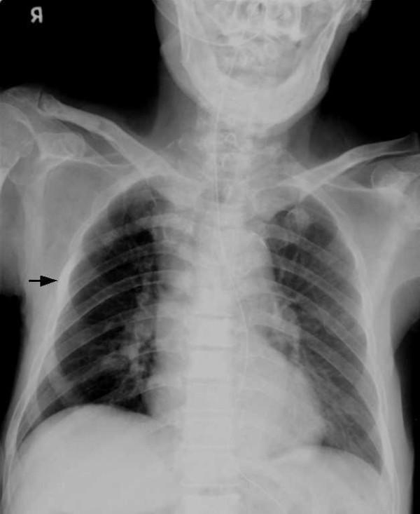
This diagnostic image is an anteroposterior (AP) chest radiograph demonstrating diffuse, multifocal osteoblastic bone lesions. The skeletal structures, including the thoracic spine, bilateral ribs, scapulae, and clavicles, exhibit widespread increased bone density appearing as irregular, patchy regions of heightened radiopacity (sclerosis). A black arrow highlights a particularly conspicuous focal osteoblastic lesion on the lateral aspect of the right fourth rib, appearing markedly whiter and more opaque than the surrounding bone. These radiographic findings are characteristic of sclerotic/osteoblastic metastases, frequently associated with advanced prostate cancer in older male patients. The underlying lung fields and cardiac silhouette appear within normal limits relative to the significant skeletal pathology. This visual material is ideal for teaching radiological manifestations of systemic metastatic disease and differentiating between lytic and blastic bone lesions in a clinical oncology or radiology context.
This diagnostic image is a lateral radiograph of a human skull, demonstrating significant skeletal abnormalities. The primary finding is diffuse hyperostosis of the calvaria, characterized by a marked thickening of the cranial vault and increased radiodensity, resulting in a sclerotic appearance. A black arrow specifically points to the skull base, highlighting localized sclerosis and increased bone density in the sphenoid and temporal regions. The normal trabecular pattern and clear differentiation of the cortical margins are obscured by the pathological bone deposition. Additionally, there is evidence of mandibular enlargement and facial bone thickening. These features are educationally significant for identifying systemic bone disorders, such as sclerosing bone dysplasias or metabolic bone diseases. The image is an essential resource for medical students and residents studying radiology and musculoskeletal pathology.
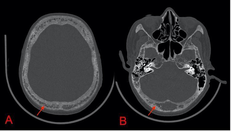
This diagnostic image consists of two axial non-contrast CT scans of the head in the bone window, highlighting the calvaria (A) and the skull base (B). The images demonstrate diffuse, spotty osteopenia throughout the cranial vault and base. There is a characteristic 'salt and pepper' morphology, signifying a loss of normal trabecular pattern and the presence of multiple tiny, interspersed radiolucent and sclerotic foci. Red arrows in both panels point to specific areas of bone remodeling and decreased density. These radiographic findings are classic manifestations of hyperparathyroidism, such as that seen in tertiary hyperparathyroidism secondary to chronic kidney disease (CKD), where excessive parathyroid hormone leads to significant bone resorption. The image serves as an educational reference for identifying 'salt and pepper skull' and understanding the skeletal complications of metabolic bone disease within the field of radiology and endocrinology.
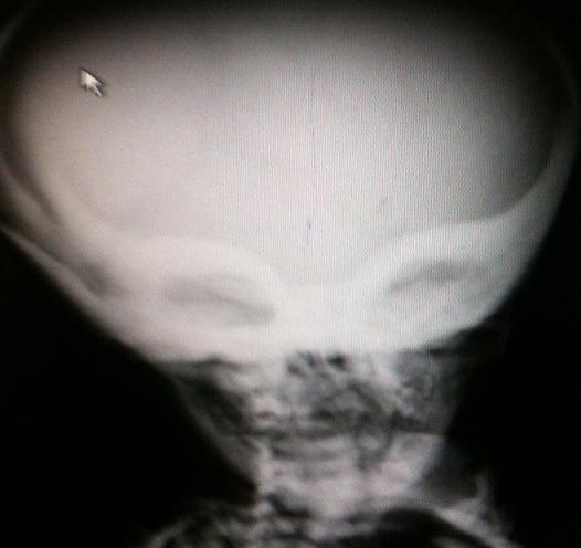
Anteroposterior facial skull radiograph demonstrating generalized osteosclerosis, a hallmark of osteopetrosis. The image reveals a significant increase in bone density throughout the craniofacial skeleton, with marked involvement of the skull base, sphenoid bones, and orbital margins. This dense sclerotic appearance of the orbits and sphenoid wings creates a characteristic 'Harlequin mask' sign. The cervical vertebrae are partially visible at the bottom of the frame, showing increased radiopacity consistent with 'bone-in-bone' appearance. This diagnostic image illustrates key radiographic manifestations of marble bone disease, highlighting the pathological failure of osteoclast-mediated bone resorption. Such findings are critical for diagnosing metabolic bone disorders in pediatric and adult radiology, particularly where diffuse skeletal hyperostosis is present.
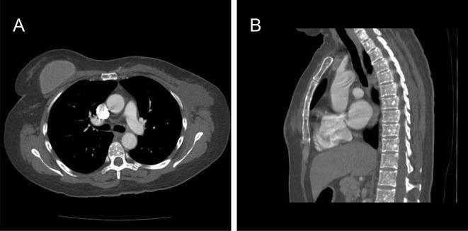
This diagnostic image consists of two Computed Tomography (CT) scans of the thorax in axial (A) and sagittal (B) planes, demonstrating diffuse skeletal findings. Image A, an axial section at the level of the T7 vertebra, reveals multiple, small, well-circumscribed, rounded sclerotic foci (osteosclerotic lesions) within the vertebral body. Image B, a sagittal reconstruction, highlights the extensive distribution of these high-density sclerotic nodules throughout the thoracic vertebral bodies and the sternum. The lesions are uniform in density and lack associated soft tissue masses or cortical destruction. In an oncology context, such as breast cancer, these findings necessitate differentiation between osteoblastic metastases and benign skeletal dysplasias like osteopoikilosis. The image illustrates a key diagnostic challenge in radiology: distinguishing widespread benign sclerotic bone islands from metastatic disease. Visible anatomical landmarks include the cardiac chambers, pulmonary trunk, descending aorta, and the thoracic spinal column.
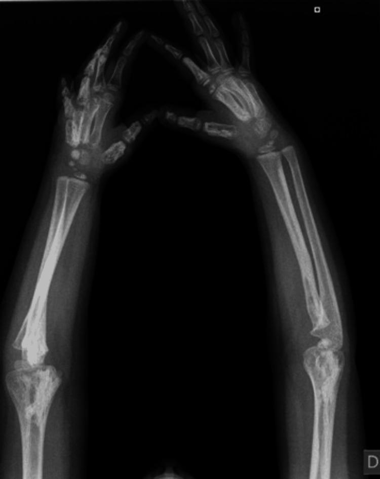
This diagnostic image is a plain radiograph (X-ray) of the bilateral forearms, wrists, and hands of a pediatric patient, demonstrated by the presence of open growth plates. The imaging reveals significant cortical hyperostosis and sclerosis affecting the diaphyses and metaphyses of the radius and ulna bilaterally. There is a marked increase in bone density (radiopacity) and thickening of the cortical layer, resulting in an irregular, undulating surface morphology. In the hands, diffuse hyperdensities are observed within the carpal bones, metacarpals, and phalanges, which appear abnormally sclerotic and irregularly shaped. The radiographic appearance is characteristic of a sclerosing bone dysplasia, such as melorheostosis or Camurati-Engelmann disease, where excessive bone formation alters normal skeletal architecture. These findings are clinically relevant for evaluating causes of limb stiffness, joint contractures, and skeletal deformities in pediatric orthopedics and radiology.
Paget disease bone X-ray radiograph cotton wool skull
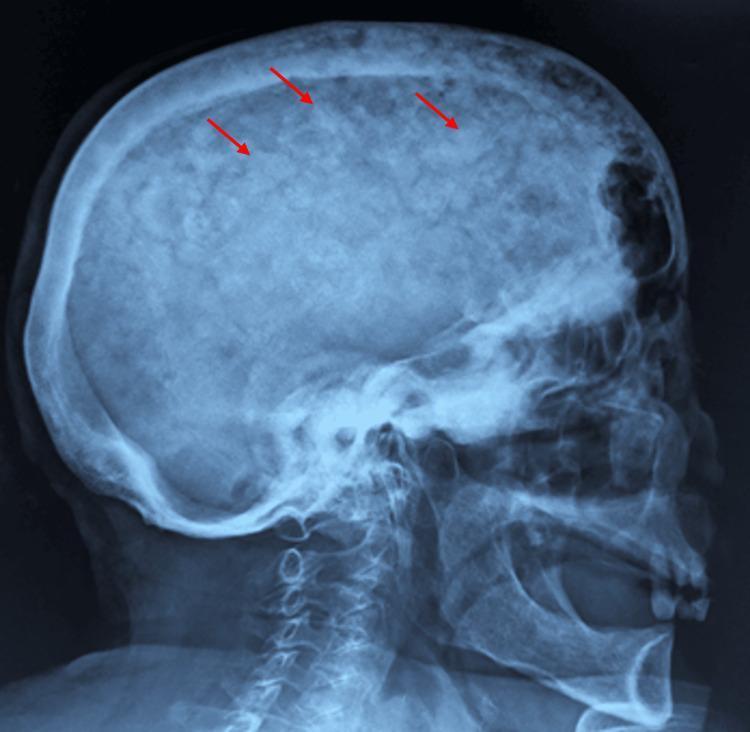
This lateral skull X-ray demonstrates a classic 'cotton wool' appearance of the cranium, a hallmark radiographic finding of Paget's disease of bone (osteitis deformans). The image shows diffuse thickening of the calvarium with a characteristic mottled texture. Multiple, ill-defined sclerotic patches are visible throughout the skull (highlighted by red arrows), representing areas of disordered, high-density bone formation interspersed with radiolucent areas of osteolysis. There is a noticeable widening of the diploic space and a loss of distinction between the inner and outer tables of the skull. The irregular mineralization replaces the normally smooth, homogeneous trabecular pattern of the cranial vault. These visual features are clinically significant as they reflect the late sclerotic phase of Paget's disease, where excessive and disorganized bone remodeling leads to structural deformities and potential neurological complications due to foraminal narrowing or cranial vault thickening.
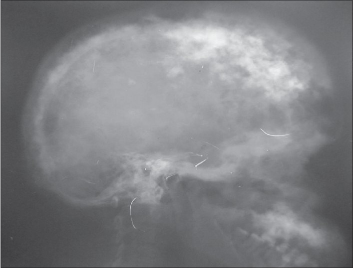
**Imaging Modality:** Lateral plain film radiograph (X-ray). **Anatomical Region:** Skull (cranium). **Observed Pathology:** Advanced Paget's disease of bone (osteitis deformans). **Characteristic Visual Features:** The image demonstrates a classic "cotton wool appearance" characterized by multifocal, ill-defined sclerotic patches interspersed with radiolucent areas throughout the calvarium. There is marked thickening of the diploic space and expansion of the outer table of the skull. The normal cortical margins of the inner and outer tables are blurred due to disorganized bone remodeling. **Key Diagnostic Features:** - **Cotton wool spots:** Focal areas of opacification representing the sclerotic phase of Paget's disease. - **Diploic thickening:** Significant widening of the cranial vault. - **Bone expansion:** Generalized enlargement of the skull landmarks compared to normal anatomical proportions. **Clinical Significance:** These radiographic findings are pathognomonic for the mixed lytic and sclerotic stages of Paget’s disease involving the axial skeleton, specifically the calvarium.
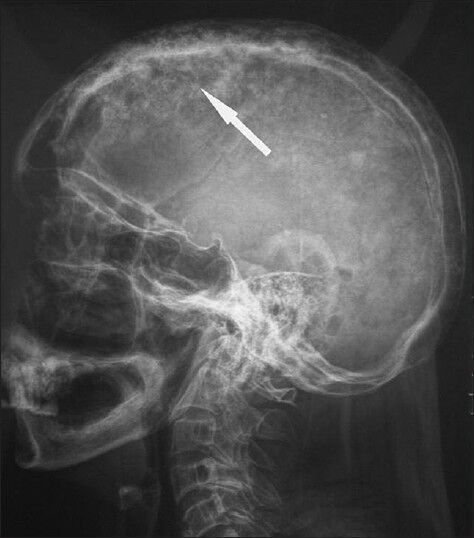
**Imaging Modality:** Lateral plain film radiograph (X-ray). **Anatomical Region:** Lateral view of the skull (calvaria) and upper cervical spine. **Observed Pathology:** The image demonstrates classic radiologic features of Paget's disease of bone (osteitis deformans) affecting the skull. **Characteristic Visual Features:** * **Cotton-Wool Appearance:** The calvaria exhibits generalized, patchy, mixed radiolucent and radiopaque areas, creating a characteristic "cotton-wool" appearance. * **Cortical Thickening:** Significant thickening of the inner and outer tables of the skull is evident, particularly in the frontal and parietal regions. * **Diploic Space Expansion:** There is a marked widening of the diploic space with loss of the normal distinct margins between the tables. * **Distribution:** Findings are diffuse throughout the vault of the skull. **Key Diagnostic Features:** The combination of calvarial thickening and the multifocal sclerotic patches (cotton-wool spots) is highly suggestive of the sclerotic (late) phase of Paget’s disease, distinguishing it from metastatic disease or primary bone malignancies. White arrow indicates a prominent area of mixed bone density within the parietal bone.
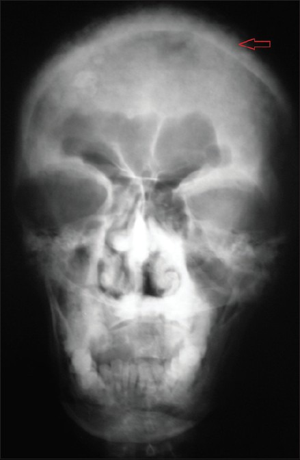
**Imaging Modality:** Conventional Radiography (X-ray) **Anatomical Region:** Skull, Postero-anterior (PA) view **Observed Pathology:** The radiograph demonstrates diffuse, multi-focal osseous changes throughout the calvarium. There is a characteristic "cotton wool" appearance, defined by irregular, sclerotic radiopaque flecks and patches interspersed with areas of lucency. **Characteristic Visual Features:** - **Calvarial Thickening:** Marked expansion and thickening of the inner and outer tables of the skull. - **Sclerotic Pattern:** Generalized distribution of patchy, ill-defined radiopacities involving the frontal, parietal, and facial bones. - **Sinus Involvement:** Opacification of the bilateral maxillary antra, consistent with bone remodeling or secondary sinus obstruction. - **Structural Integrity:** Loss of normal cortical-medullary differentiation within the cranial vault. **Relevant Clinical Context:** The radiographic presentation is highly characteristic of the sclerotic (late) phase of Paget’s disease of bone (osteitis deformans). **Key Diagnostic Features:** The combination of cortical thickening, skull expansion, and the "cotton wool" matrix of irregular radiopacities are pathognomonic findings for Pagetoid involvement of the craniofacial skeleton.
osteopetrosis marble bone disease X-ray sandwich vertebra
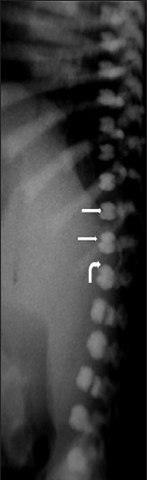
**Imaging Modality:** Lateral projection radiograph (X-ray). **Anatomical Region:** Thoracolumbar spine and posterior rib cage. **Observed Pathology:** Generalized osteosclerosis consistent with osteopetrosis (Albers-Schönberg disease). **Characteristic Visual Features:** * **Sandwich Vertebra Sign:** The vertebral bodies exhibit dense, sclerotic bands at the superior and inferior endplates (curved arrow) with relative central radiolucency (straight arrows), creating a distinct layered or "sandwich" appearance. * **Diffuse Sclerosis:** Increased radiodensity is visible across the visualized vertebrae and posterior ribs, indicating a systemic failure of osteoclast-mediated bone resorption. * **Bone-within-bone Appearance:** The morphology of the vertebral bodies suggests an endosteal thickening pattern typical of metabolic bone disorders. **Key Diagnostic Features:** The "sandwich vertebra" sign is a pathognomonic radiologic finding for osteopetrosis, distinguishing it from other causes of vertebral sclerosis such as Paget’s disease (which typically presents with a "picture frame" vertebra) or renal osteodystrophy (rugger-jersey spine). This presentation in an infant is highly indicative of the autosomal recessive (malignant) form of the condition.
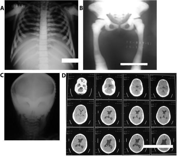
This composite of diagnostic images demonstrates pathognomonic radiological features of osteopetrosis (Albers-Schönberg disease). Panel A (chest/spine X-ray) shows generalized osteosclerosis with a characteristic 'sandwich vertebrae' sign, appearing as dense bands of sclerosis at the superior and inferior vertebral endplates. Panel B (pelvic and lower limb X-ray) exhibits diffuse thickening and a 'bone-within-bone' appearance in the iliac wings and proximal femurs. Panel C (skull X-ray) reveals a strikingly dense, homogenous sclerotic skull base and calvarium. Panel D (axial brain CT) demonstrates extreme hyperdensity of the skull, encroachment of the optic foramina due to bony overgrowth, and secondary mild dilatation of the lateral and third ventricles, indicating potential hydrocephalus or intracranial pressure changes. These features are hallmark signs of impaired osteoclast function leading to defective bone resorption and excessive bone accumulation, typically seen in autosomal dominant osteopetrosis type II (ADO-II).
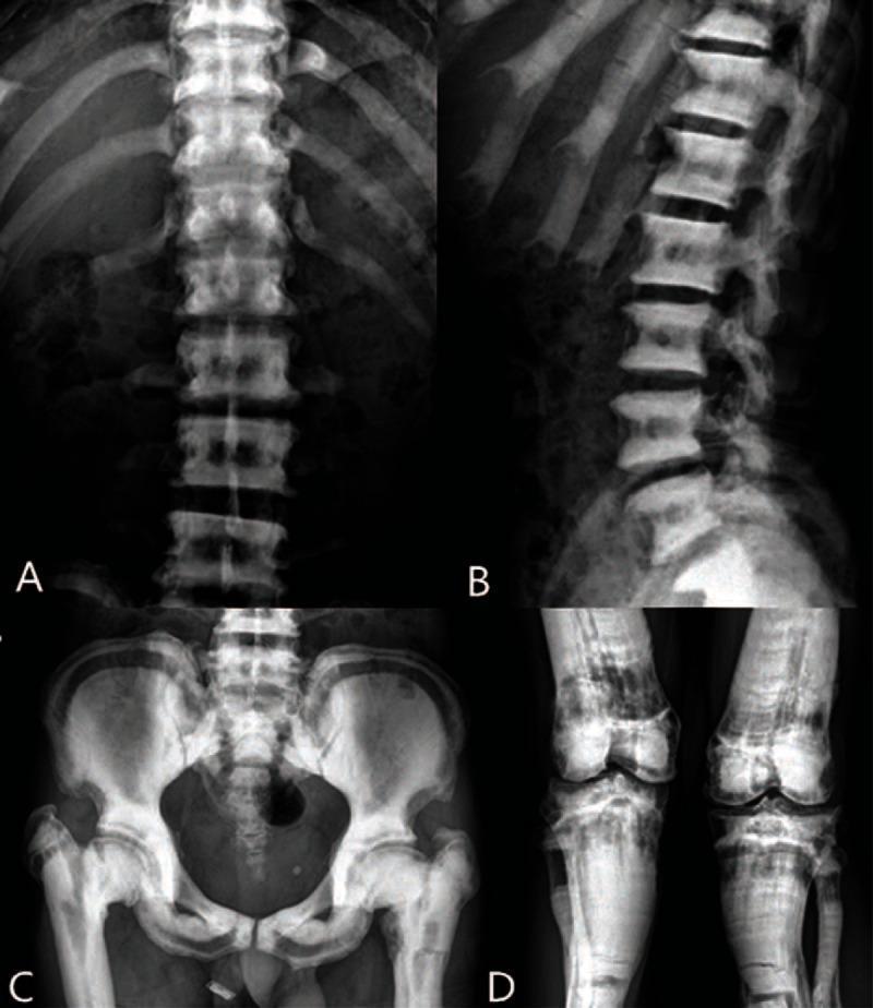
This composite of diagnostic X-ray radiographs (A–D) illustrates systemic bone sclerosis and metabolic bone disease, consistent with osteopetrosis. Panels A and B show the thoracolumbar spine and ribs, demonstrating diffusely increased bone density and a characteristic 'sandwich vertebrae' (or 'layer cake') appearance, where sclerotic bands involve the superior and inferior endplates of the vertebral bodies. Transverse fracture lines are visible in the ribs. Panel C presents a pelvic X-ray revealing marked sclerosis of the ilia, pubis, and ischium, with visible low-density zones. Significant structural abnormalities include shortening of the bilateral femoral necks, superior displacement of the femoral shafts, and transverse fractures of the proximal femurs. Panel D displays the bilateral knees, exhibiting intense bone mineral density and multiple transverse fracture lines across the proximal tibiae and fibulae. These findings represent a classic presentation of Albers-Schönberg disease, emphasizing the increased fracture risk despite increased radiodensity. The images serve as an educational resource for identifying metabolic bone disorders through hallmark radiological signs like 'bone-within-bone' and 'sandwich' vertebrae.
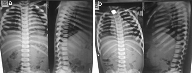
This diagnostic imaging set consists of two conventional X-ray panels (labeled 'a' and 'b') displaying anteroposterior (AP) and lateral views of the thoracolumbar spine and rib cage in two pediatric patients (aged 5 and 8 years). The primary radiological finding is marked sclerosis of the superior and inferior vertebral endplates throughout the spine. This selective thickening creates a classic 'sandwich vertebrae' appearance, where the opaque endplates contrast sharply with the relatively lucent center of the vertebral bodies. Additionally, there is a generalized, homogeneous increase in bone density (osteosclerosis) involving the ribs and visible pedicles, leading to a loss of the normal corticomedullary differentiation. These skeletal features are pathognomonic for osteopetrosis (marble bone disease), illustrating the progressive failure of osteoclast-mediated bone resorption. The images serve as an educational example of metabolic bone disorders and their characteristic radiographic manifestations in a clinical genetics or pediatric radiology context.
fluorosis bone skeletal fluorosis radiograph
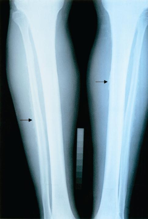
This diagnostic X-ray (radiograph) demonstrates the bilateral lower legs, specifically the tibiae and fibulae, of a patient with Stage II skeletal fluorosis. The image illustrates key osteosclerotic changes and cortical abnormalities characteristic of chronic fluoride toxicity. Visible findings include a general thinning of the diaphyseal regions and notable ill-defined or blurred cortical margins, highlighted by black arrows. Unlike the sharp demarcation seen in healthy bone, these margins appear irregular, suggesting periosteal reaction or subperiosteal bone formation. The educational focus is on identifying the skeletal manifestations of fluorosis, which often present with increased bone density (osteosclerosis), coarsening of the trabecular pattern, and potential calcification of interosseous membranes or ligaments. This radiographic study serves as a clinical example of occupational or environmental toxicological pathology affecting the appendicular skeleton.
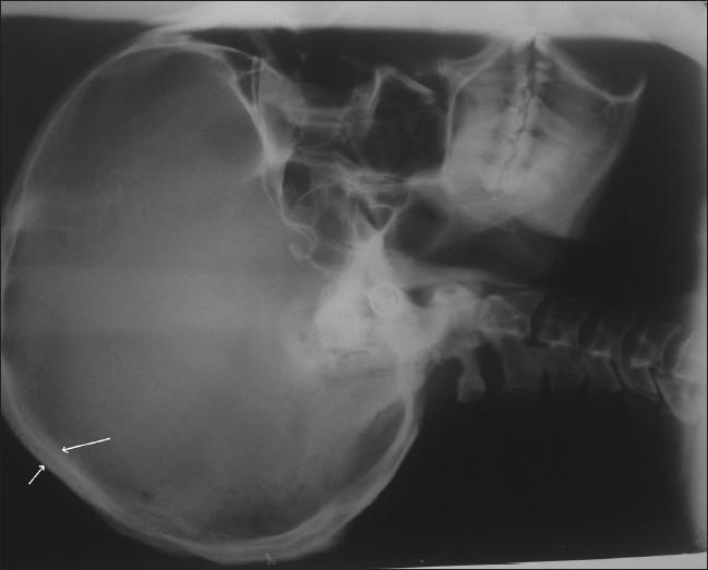
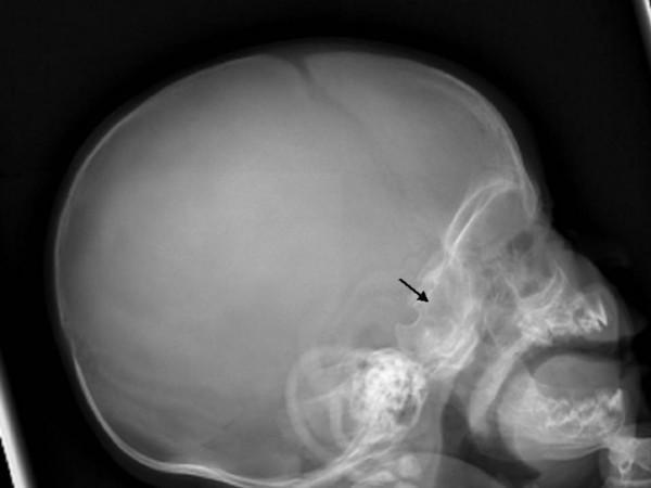
This diagnostic image is a lateral skull radiograph (X-ray) demonstrating significant skeletal manifestations often associated with chronic fluoride toxicity (skeletal fluorosis) or metabolic bone disorders. The image reveals marked thickening and increased radiopacity (sclerosis) of both the inner and outer tables of the skull. White arrows indicate areas of cortical thickening and expansion of the diploic space. There is a visible loss of the normal sharp demarcation between the tables and the intervening diploe, creating a blurred, dense bony architecture. Additional findings include prominent marrow spaces and altered density within the cervical vertebrae and mandibular structures. This visual represents a classic clinical example used to teach the radiological signs of generalized osteosclerosis and the structural bone changes resulting from prolonged ingestion of high fluoride levels. The image is relevant for medical students and radiology residents studying metabolic bone diseases and toxicology.
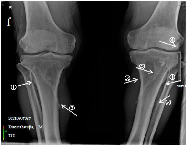
This diagnostic image is an anteroposterior (AP) X-ray of both lower limbs, specifically focusing on the knee joints and proximal shafts of the tibiae and fibulae. The radiograph illustrates severe manifestations of Skeletal Fluorosis (SF), characterized by extensive ectopic ossification. Label ① points to dense, irregular ossification of the tibiofibular interosseous membrane, exhibiting a characteristic 'colliculus' or nodular appearance between the bones. Label ③ highlights multiple 'ossification shadows' or opaque bony outgrowths along the shafts of the tibia and fibula. Label ④ identifies significant ossification of the knee joint capsule on the right limb, appearing as a radiopaque mass near the joint line. Label ⑤ indicates ossification of the soleus tendon, visible as a distinct bony density posterior to the proximal tibia. These findings represent advanced skeletal changes including periosteal new bone formation and ligamentous/tendinous calcification, relevant for diagnosing and staging chronic fluoride toxicity in clinical radiology.
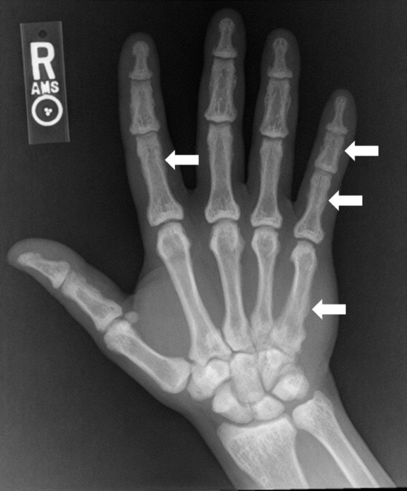
This anteroposterior (AP) radiograph of the right hand and wrist demonstrates diffuse skeletal sclerosis, characterized by a generalized increase in bone density across the metacarpals, phalanges, and carpal bones. The osseous structures appear hyper-opaque with a significant loss of normal corticomedullary differentiation; the medullary cavities are nearly obliterated by dense trabecular bone. Several white arrows highlight focal abnormalities along the diaphyses, including the second proximal phalanx, the fifth metacarpal, and the fifth proximal and intermediate phalanges. These areas exhibit irregular, indistinct cortical margins and subtle periosteal reactions, giving the bone surfaces a smudged appearance. Such widespread osteosclerosis in the small bones of the hand is a key radiological finding for systemic metabolic or toxic bone conditions, such as skeletal fluorosis. The image serves as a clinical example of secondary osteosclerosis and its impact on peripheral skeletal architecture.
renal osteodystrophy rugger jersey spine radiograph
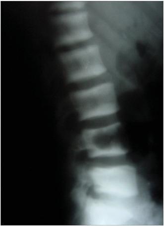
A lateral view X-ray of the lumbar spine illustrating the 'rugger jersey spine' sign. The radiograph reveals a characteristic pattern of alternating radiopacity within the vertebral bodies. Specifically, there are prominent horizontal bands of increased bone density (osteosclerosis) located at the superior and inferior endplates of each vertebra. These sclerotic bands contrast with the relatively radiolucent (normal to osteopenic) central portion of the vertebral bodies, creating a striped appearance reminiscent of a rugby jersey. This finding is highly indicative of renal osteodystrophy secondary to chronic kidney disease or hyperparathyroidism. The image demonstrates the structural skeletal manifestations of systemic metabolic bone disease, focusing on the lumbar region's vertebral architecture and density distribution.
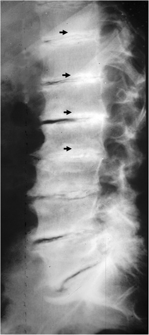
**Imaging Modality:** Lateral plain radiograph (X-ray). **Anatomical Region:** Thoracolumbar spine. **Observed Pathology:** Diffuse osteosclerosis with a characteristic "rugger-jersey spine" appearance, diagnostic of renal osteodystrophy (secondary hyperparathyroidism). **Characteristic Visual Features:** * **Distribution:** Multiple contiguous vertebral levels are affected. * **Sclerotic Bands:** Prominent, horizontal radiopaque (dense) bands are visible along the superior and inferior endplates of the vertebral bodies. * **Central Lucency:** The mid-portion of each vertebral body remains relatively radiolucent compared to the thickened endplates, creating a striped or "sandwich" appearance. * **Associated Findings:** There is a generalized increase in bone density (osteosclerosis) throughout the visualized axial skeleton. Black arrows highlight the specific sub-endplate densities. **Clinical Context:** This manifestation is typically seen in patients with chronic kidney disease (CKD) and end-stage renal disease undergoing long-term hemodialysis. **Key Diagnostic Features:** The alternating pattern of dense sclerotic endplates and lucent vertebral centers is the pathognomonic sign of the rugger-jersey spine, distinguishing it from other causes of diffuse osteosclerosis like osteopetrosis or Paget's disease.
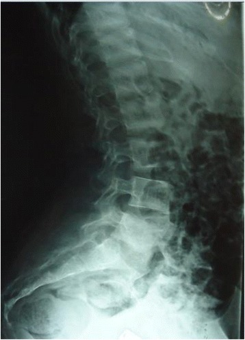
**Modality:** Lateral projection conventional radiograph (X-ray). **Anatomical Region:** Lumbar spine and sacrum. **Observed Pathology:** The image demonstrates classic "rugger jersey spine" appearance, a hallmark radiologic sign of renal osteodystrophy (secondary hyperparathyroidism). **Characteristic Visual Features:** - **Sclerotic Bands:** Prominent horizontal bands of increased radiodensity are visible at the superior and inferior endplates of multiple vertebral bodies. - **Central Lucency:** The mid-portion of the vertebral bodies demonstrates relative osteopenia or decreased radiodensity compared to the dense endplates. - **Pattern:** This alternating radiopaque-radiolucent-radiopaque pattern creates a striped appearance across the longitudinal axis of the spine. - **Bone Quality:** Generalized alteration in trabecular pattern and mineralization is noted throughout the visualized lumbar vertebrae. **Clinical Context/Differentiating Features:** This pattern is highly specific for chronic kidney disease (CKD) induced metabolic bone disease. The distribution of sclerosis at the endplates distinguishes it from other causes of osteosclerosis, such as osteopetrosis (which typically presents with more diffuse "bone-within-bone" appearance) or Paget's disease (which involves cortical thickening and coarser trabeculae).
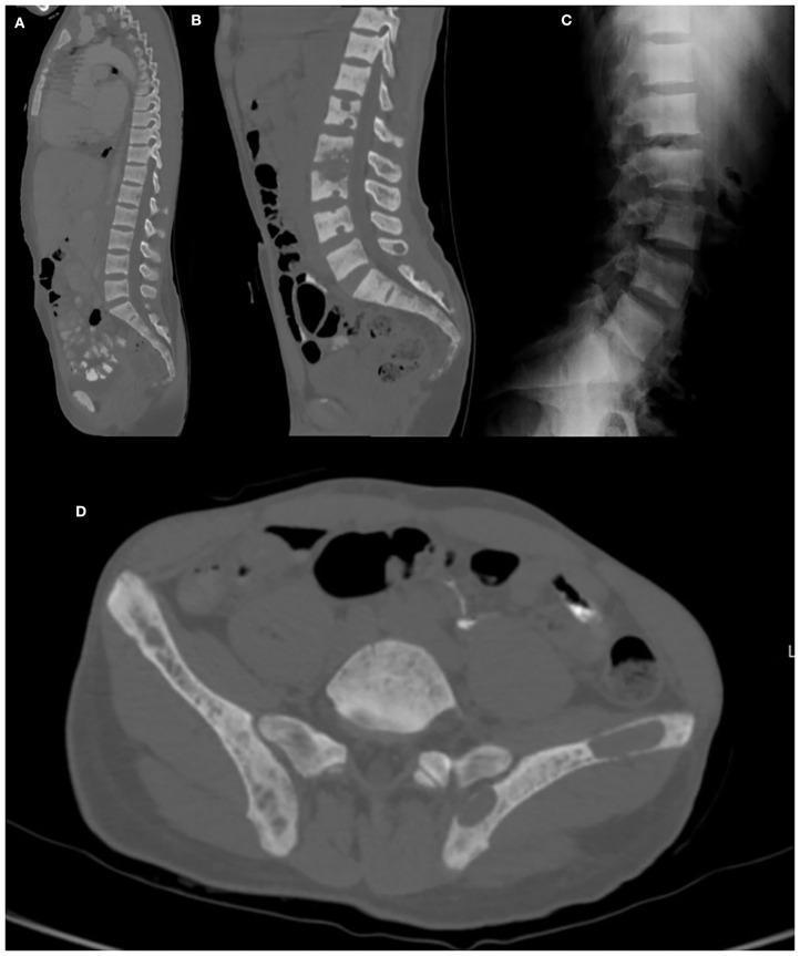
This composite of diagnostic images illustrates musculoskeletal manifestations of renal osteodystrophy and hyperparathyroidism. Images A and B are CT sagittal reconstructions of the thoracic and lumbar spine, demonstrating diffuse osteosclerosis and multiple areas of subperiosteal resorption, reflecting abnormal bone turnover. Image C is a lateral lumbar spine radiograph showing the classic 'rugger jersey spine' sign, characterized by dense sclerotic bands at the superior and inferior vertebral endplates with a more lucent central region, creating a striped horizontal appearance. Image D is an axial CT scan of the pelvis revealing a large, well-defined, expansive 'brown tumor' (osteoclastoma) within the left iliac bone. This lesion appears as a heterogeneous, cystic-like mass that disrupts normal cortical architecture. Collectively, these images provide a comprehensive visual overview of secondary hyperparathyroidism's impact on bone density, including generalized sclerosis, focal resorptive tumors, and characteristic vertebral mineralization patterns.
myelofibrosis bone marrow radiograph osteosclerosis
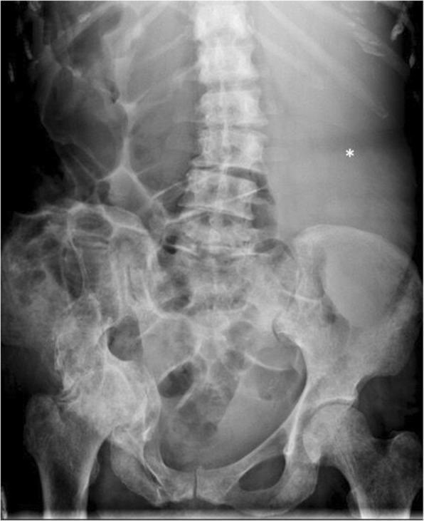
This abdominal radiograph (X-ray) illustrates the classical imaging triad of primary myelofibrosis. The axial and appendicular skeleton, including the lumbar vertebrae, pelvic bones, and proximal femurs, demonstrate diffuse, symmetric osteosclerosis, evidenced by an overall increased radiographic density (whiter appearance) and loss of normal trabecular clarity. In the soft tissues, a massive, homogeneous soft tissue opacity is visible in the left upper quadrant (marked with an asterisk), representing severe splenomegaly. This enlarged splenic mass exerts significant mass effect, resulting in the marked displacement of gas-filled small and large bowel loops toward the right side of the abdomen. These findings reflect the systemic bone marrow remodeling and extramedullary hematopoiesis characteristic of myeloproliferative neoplasms. The image serves as a key diagnostic example of how hematological disorders can manifest through both skeletal density changes and profound organomegaly on plain film imaging.
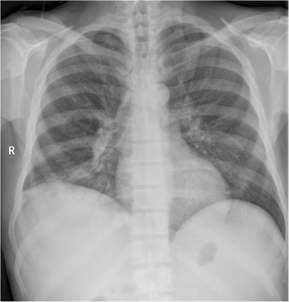
This diagnostic image is a posterior-anterior (PA) chest radiograph illustrating diffuse osteosclerosis secondary to primary myelofibrosis. The primary visual finding is a generalized, symmetrical increase in radiographic density (bone whitening) across all visible skeletal elements, including the ribs, clavicles, and vertebral bodies. This sclerotic change is characterized by a loss of the normal trabecular pattern and a markedly opaque appearance of the bone compared to surrounding soft tissues. Despite the axial skeleton involvement, the lung parenchyma remains relatively clear with preserved vascular markings and no evidence of consolidation, pulmonary edema, or pleural effusion. The cardiac and mediastinal contours are within normal limits, and the hila are not enlarged. This presentation is a classic imaging hallmark of myeloproliferative neoplasms (MPN), where bone marrow fibrosis leads to increased bone formation. It serves as a key educational example of how systemic hematologic disorders can manifest incidentally as metabolic bone disease on routine imaging.
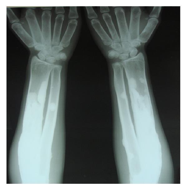
This diagnostic image is an anteroposterior (AP) radiograph of both forearms and hands, illustrating hallmark signs of generalized osteosclerosis, consistent with a metabolic or genetic bone disorder such as osteopetrosis. The imaging shows a marked increase in radiodensity (sclerosis) throughout the radius, ulna, and metacarpals bilaterally. There is a notable loss of the normal corticomedullary differentiation, with the marrow spaces appearing obliterated by dense bone. The surfaces of the long bones exhibit a characteristic smoothening. The metacarpals display an atypical cylindrical morphology, losing their usual mid-shaft tapering. These features indicate significant bone modeling and remodeling defects. The clinical significance of these findings lies in the demonstration of pathologic bone thickening, which often leads to skeletal fragility and hematological complications due to marrow space compression. This visual material is suitable for advanced medical education in radiology and orthopedics, specifically for teaching bone density pathologies.
mastocytosis diffuse sclerotic bone radiograph
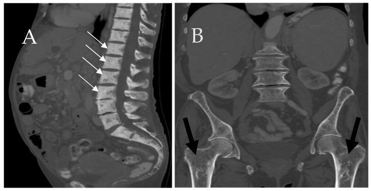
This dual-panel figure displays computed tomography (CT) scans of the skeletal system in a patient with systemic mastocytosis. Image A is a sagittal CT of the lumbar and sacral spine, demonstrating diffuse osteosclerosis of the axial skeleton. White arrows indicate multiple vertebral bodies showing pathologically increased bone density with a patchy, sclerotic appearance. Image B is a coronal CT of the pelvis and proximal femurs, where black arrows highlight a diffuse loss of normal trabecular architecture. The proximal femurs exhibit a heterogeneous, osteolytic, and somewhat 'mottled' internal structure, indicative of bone marrow infiltration. The images collectively illustrate the varied osseous manifestations of mast cell infiltration, ranging from lytic-appearing trabecular loss to profound focal or diffuse sclerosis. These findings are clinically significant for the radiological evaluation of hematologic malignancies and metabolic bone diseases.
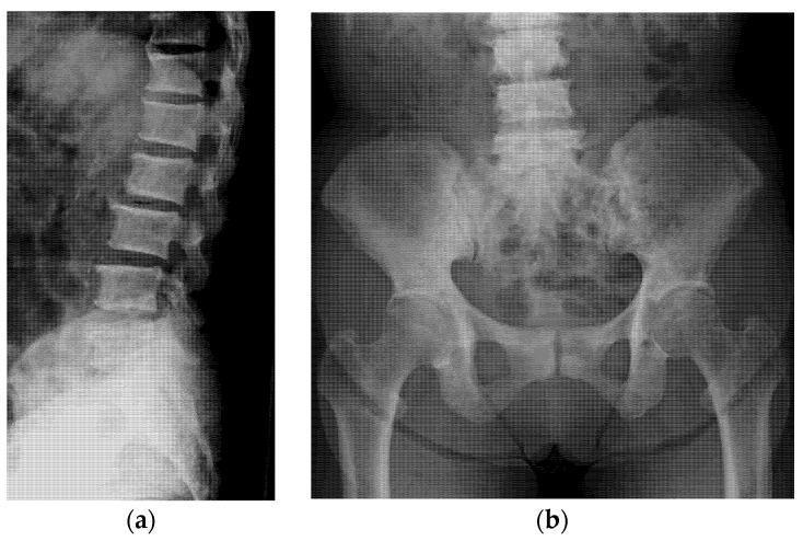
Two-panel plain film radiograph of the axial skeleton illustrating diffuse osteoblastic activity. Image (a) is a lateral projection of the thoracolumbar spine showing generalized, marked increase in radiodensity (sclerosis) within the vertebral bodies, resulting in a 'chalky' or 'ivory' appearance of the vertebrae. The intervertebral disc spaces appear relatively preserved. Image (b) is an anteroposterior (AP) view of the pelvis and lumbar spine, demonstrating widespread sclerotic changes throughout the sacrum and bilateral iliac bones. Notably, the proximal femurs maintain normal cortical and medullary radiodensity, highlighting the axial distribution of the pathology. These radiographic findings are characteristic of osteoblastic metastases, commonly seen in secondary malignancies such as prostate or gastric cancer. The educational focus is on the recognition of diffuse bone sclerosis as a manifestation of systemic disease and the clinical utility of plain radiographs in identifying metastatic bone lesions.
This diagnostic image is an anteroposterior (AP) chest radiograph demonstrating diffuse, multifocal osteoblastic bone lesions. The skeletal structures, including the thoracic spine, bilateral ribs, scapulae, and clavicles, exhibit widespread increased bone density appearing as irregular, patchy regions of heightened radiopacity (sclerosis). A black arrow highlights a particularly conspicuous focal osteoblastic lesion on the lateral aspect of the right fourth rib, appearing markedly whiter and more opaque than the surrounding bone. These radiographic findings are characteristic of sclerotic/osteoblastic metastases, frequently associated with advanced prostate cancer in older male patients. The underlying lung fields and cardiac silhouette appear within normal limits relative to the significant skeletal pathology. This visual material is ideal for teaching radiological manifestations of systemic metastatic disease and differentiating between lytic and blastic bone lesions in a clinical oncology or radiology context.
mkdir -p /tmp/workspace/diffuse-sclerotic-ppt && ls /tmp/workspace/diffuse-sclerotic-ppt/
~/diffuse-sclerotic-ppt/build_ppt.py
#!/usr/bin/env python3
"""
Diffuse Sclerotic Bone Diseases - Radiology Seminar
Professional PPT with images beside content for each disease.
"""

import json, subprocess, base64, textwrap
from io import BytesIO
from pptx import Presentation
from pptx.util import Inches, Pt, Emu
from pptx.dml.color import RGBColor
from pptx.enum.text import PP_ALIGN, MSO_ANCHOR
from pptx.util import Inches, Pt

# ─── COLOUR PALETTE ───────────────────────────────────────────────────────────
DARK_NAVY   = RGBColor(0x0D, 0x1B, 0x2A)   # deep navy - backgrounds
MID_BLUE    = RGBColor(0x1B, 0x44, 0x7A)   # section headers
ACCENT_TEAL = RGBColor(0x00, 0xB4, 0xD8)   # accent lines / highlights
WHITE       = RGBColor(0xFF, 0xFF, 0xFF)
LIGHT_GREY  = RGBColor(0xF0, 0xF4, 0xF8)
GOLD        = RGBColor(0xF7, 0xB7, 0x31)
TEXT_DARK   = RGBColor(0x1A, 0x1A, 0x2E)
SLIDE_W     = Inches(13.333)
SLIDE_H     = Inches(7.5)

# ─── IMAGE URLS ───────────────────────────────────────────────────────────────
IMAGES = {
    "paget_skull1":   "https://cdn.orris.care/cdss_images/roco_radiology_ROCO_14463_1766646866328.png",
    "paget_skull2":   "https://cdn.orris.care/cdss_images/pmc_clinical_VQA_7e4cde5dcc9978e5c8b2337ba5557825e80ffff7235c57d964c40be160010569.jpg",
    "paget_pelvis":   "https://cdn.orris.care/cdss_images/ca1d7ec0af4401e54b948c104fc2110d271be62498a34f17f81ed3ffba768b4c.png",
    "osteopetrosis1": "https://cdn.orris.care/cdss_images/roco_radiology_ROCO_78989_1766650467211.png",
    "osteopetrosis2": "https://cdn.orris.care/cdss_images/pmc_clinical_VQA_89154655305fa9df745e30668afd2153790bf1c4e3d8ff86fd86bc4e65a95dd7.jpg",
    "osteopetrosis3": "https://cdn.orris.care/cdss_images/031a0a10fbdd4f2e88d8642b8e4ea18966327a41ed95f78cd5e9dc0ede71cc47.png",
    "fluorosis1":     "https://cdn.orris.care/cdss_images/pmc_clinical_VQA_25c4822ce6516c08af23c2ac3e87411ae4d555daf257b46b4d85a3dd4546cc46.jpg",
    "fluorosis2":     "https://cdn.orris.care/cdss_images/pmc_clinical_VQA_bb872e370bc52fe92e90fcf22ed33d38acd80a066624f530918d6a62fe74b26d.jpg",
    "rugger_jersey":  "https://cdn.orris.care/cdss_images/roco_radiology_ROCO_65340_1766649702501.png",
    "rugger_jersey2": "https://cdn.orris.care/cdss_images/pmc_clinical_VQA_24f58b957a4c12b38f0bdf7bfba59d9c18e09d958c37d7857b8939bbf4ed7ccb.jpg",
    "myelofibrosis1": "https://cdn.orris.care/cdss_images/pmc_clinical_VQA_ca4c98ddaa0463096fd1e721809d724a88a5a7e3a0a5ced67e06150919ee4d40.jpg",
    "myelofibrosis2": "https://cdn.orris.care/cdss_images/pmc_clinical_VQA_d8db5303bc9d79e569e89cbde384801c053de98a88bc93e57a7c8d8b86e3ddec.jpg",
    "mastocytosis":   "https://cdn.orris.care/cdss_images/pmc_clinical_VQA_cb1bba97e79a3d8fb07c2a6af7aa5fe52cae5ecfee15cc05ca4c3be2b8be1760.jpg",
    "diff_sclerotic": "https://cdn.orris.care/cdss_images/pmc_clinical_VQA_fdb51f758bfbdfaa5b9135cafa582a5c1710d9d046c374cf407df0c28c58962c.jpg",
    "camurati":       "https://cdn.orris.care/cdss_images/pmc_clinical_VQA_60f92d6122c1085232f42a4217cbca1205d44bee83fb587dc61627b03fc3e094.jpg",
}

# ─── FETCH ALL IMAGES ─────────────────────────────────────────────────────────
print("Fetching images...")
urls = list(IMAGES.values())
result_raw = subprocess.check_output(
    ["python", "/tmp/skills/shared/scripts/fetch_images.py"] + urls,
    timeout=120
)
img_data = json.loads(result_raw)
img_map = {}
for item in img_data:
    for key, url in IMAGES.items():
        if item["url"] == url and item.get("base64"):
            raw = item["base64"]
            if "," in raw:
                raw = raw.split(",", 1)[1]
            img_map[key] = base64.b64decode(raw)

print(f"Fetched {len(img_map)}/{len(IMAGES)} images")

# ─── HELPERS ──────────────────────────────────────────────────────────────────
def make_prs():
    prs = Presentation()
    prs.slide_width  = SLIDE_W
    prs.slide_height = SLIDE_H
    return prs

def blank(prs):
    return prs.slides.add_slide(prs.slide_layouts[6])

def rect(slide, x, y, w, h, fill, alpha=None):
    shape = slide.shapes.add_shape(1, x, y, w, h)
    shape.line.fill.background()
    shape.fill.solid()
    shape.fill.fore_color.rgb = fill
    return shape

def textbox(slide, text, x, y, w, h, size=18, bold=False, color=WHITE,
            align=PP_ALIGN.LEFT, wrap=True, italic=False, anchor=MSO_ANCHOR.TOP):
    tb = slide.shapes.add_textbox(x, y, w, h)
    tf = tb.text_frame
    tf.word_wrap = wrap
    tf.vertical_anchor = anchor
    tf.margin_left  = Inches(0.05)
    tf.margin_right = Inches(0.05)
    tf.margin_top   = Pt(2)
    tf.margin_bottom= Pt(2)
    p  = tf.paragraphs[0]
    p.alignment = align
    r  = p.add_run()
    r.text = text
    r.font.size   = Pt(size)
    r.font.bold   = bold
    r.font.italic = italic
    r.font.color.rgb = color
    r.font.name = "Calibri"
    return tf

def multiline_textbox(slide, lines, x, y, w, h, size=15, color=WHITE, leading_bold=False):
    """lines = list of (text, bold, size_override_or_None)"""
    tb = slide.shapes.add_textbox(x, y, w, h)
    tf = tb.text_frame
    tf.word_wrap = True
    tf.vertical_anchor = MSO_ANCHOR.TOP
    tf.margin_left   = Inches(0.1)
    tf.margin_right  = Inches(0.05)
    tf.margin_top    = Pt(4)
    tf.margin_bottom = Pt(2)
    first = True
    for (txt, bld, sz) in lines:
        if first:
            p = tf.paragraphs[0]
            first = False
        else:
            p = tf.add_paragraph()
        p.space_before = Pt(2)
        r = p.add_run()
        r.text = txt
        r.font.size  = Pt(sz if sz else size)
        r.font.bold  = bld
        r.font.color.rgb = color
        r.font.name  = "Calibri"
    return tf

def add_image(slide, key, x, y, w, h):
    if key in img_map:
        img_bytes = BytesIO(img_map[key])
        slide.shapes.add_picture(img_bytes, x, y, w, h)
    else:
        # placeholder grey box
        r = rect(slide, x, y, w, h, RGBColor(0x88,0x88,0x88))
        textbox(slide, "[ Image ]", x, y+h//2-Inches(0.2), w, Inches(0.4),
                size=11, color=WHITE, align=PP_ALIGN.CENTER)

def accent_line(slide, x, y, w, thickness=Pt(3)):
    ln = slide.shapes.add_shape(1, x, y, w, int(thickness))
    ln.fill.solid(); ln.fill.fore_color.rgb = ACCENT_TEAL
    ln.line.fill.background()

def header_bar(slide, title, subtitle=""):
    # full-width dark top bar
    rect(slide, 0, 0, SLIDE_W, Inches(1.05), DARK_NAVY)
    accent_line(slide, 0, Inches(1.05), SLIDE_W, Pt(4))
    textbox(slide, title, Inches(0.4), Inches(0.08), Inches(10), Inches(0.55),
            size=30, bold=True, color=WHITE)
    if subtitle:
        textbox(slide, subtitle, Inches(0.4), Inches(0.6), Inches(10), Inches(0.4),
                size=14, color=ACCENT_TEAL, italic=True)

def footer(slide, page_num, total):
    rect(slide, 0, Inches(7.2), SLIDE_W, Inches(0.3), DARK_NAVY)
    textbox(slide, "Diffuse Sclerotic Bone Diseases  |  Radiology Seminar",
            Inches(0.3), Inches(7.2), Inches(10), Inches(0.3),
            size=9, color=RGBColor(0xAA,0xCC,0xEE))
    textbox(slide, f"{page_num} / {total}",
            Inches(12.2), Inches(7.2), Inches(1), Inches(0.3),
            size=9, color=ACCENT_TEAL, align=PP_ALIGN.RIGHT)

# ─── DISEASE SLIDE BUILDER ────────────────────────────────────────────────────
def disease_slide(prs, page_num, total, title, subtitle,
                  content_lines, img_key1, img_key2=None,
                  caption1="", caption2="", label_color=ACCENT_TEAL):
    """
    Layout: Left 55% = text content | Right 45% = image(s)
    content_lines: list of (text, bold, size)
    """
    slide = blank(prs)
    # Background
    rect(slide, 0, 0, SLIDE_W, SLIDE_H, LIGHT_GREY)
    rect(slide, 0, 0, SLIDE_W, SLIDE_H, RGBColor(0xF5,0xF8,0xFF))
    # Left panel background
    rect(slide, 0, Inches(1.09), Inches(7.3), Inches(6.11), MID_BLUE)
    # Right panel background
    rect(slide, Inches(7.35), Inches(1.09), Inches(5.98), Inches(6.11), DARK_NAVY)
    # Header
    header_bar(slide, title, subtitle)
    # Content
    multiline_textbox(slide, content_lines,
                      Inches(0.2), Inches(1.18), Inches(6.9), Inches(5.9),
                      size=14, color=WHITE)
    # Images
    if img_key2:
        add_image(slide, img_key1, Inches(7.4), Inches(1.15), Inches(5.85), Inches(2.95))
        add_image(slide, img_key2, Inches(7.4), Inches(4.2),  Inches(5.85), Inches(2.75))
        if caption1:
            textbox(slide, caption1, Inches(7.4), Inches(4.08), Inches(5.85), Inches(0.28),
                    size=8, color=ACCENT_TEAL, italic=True)
        if caption2:
            textbox(slide, caption2, Inches(7.4), Inches(6.88), Inches(5.85), Inches(0.28),
                    size=8, color=ACCENT_TEAL, italic=True)
    else:
        add_image(slide, img_key1, Inches(7.4), Inches(1.15), Inches(5.85), Inches(5.8))
        if caption1:
            textbox(slide, caption1, Inches(7.4), Inches(6.88), Inches(5.85), Inches(0.28),
                    size=8, color=ACCENT_TEAL, italic=True)
    footer(slide, page_num, total)
    return slide

# ─── SECTION DIVIDER ──────────────────────────────────────────────────────────
def section_divider(prs, page_num, total, number, title, subtitle):
    slide = blank(prs)
    rect(slide, 0, 0, SLIDE_W, SLIDE_H, DARK_NAVY)
    rect(slide, 0, 0, Inches(0.25), SLIDE_H, ACCENT_TEAL)
    textbox(slide, number, Inches(1), Inches(1.5), Inches(11), Inches(1.5),
            size=80, bold=True, color=RGBColor(0x1B,0x44,0x7A))
    textbox(slide, title, Inches(1), Inches(3.0), Inches(11), Inches(1.2),
            size=38, bold=True, color=WHITE)
    textbox(slide, subtitle, Inches(1), Inches(4.1), Inches(11), Inches(0.8),
            size=20, color=ACCENT_TEAL, italic=True)
    accent_line(slide, Inches(1), Inches(2.95), Inches(11), Pt(3))
    footer(slide, page_num, total)

# ─── BUILD ────────────────────────────────────────────────────────────────────
TOTAL = 30
prs = make_prs()

# ── SLIDE 1: TITLE ────────────────────────────────────────────────────────────
s = blank(prs)
rect(s, 0, 0, SLIDE_W, SLIDE_H, DARK_NAVY)
rect(s, 0, 0, Inches(0.35), SLIDE_H, ACCENT_TEAL)
rect(s, 0, Inches(5.5), SLIDE_W, Inches(2.0), MID_BLUE)
textbox(s, "DIFFUSE SCLEROTIC", Inches(1), Inches(1.0), Inches(12), Inches(1.4),
        size=54, bold=True, color=WHITE, align=PP_ALIGN.CENTER)
textbox(s, "BONE DISEASES", Inches(1), Inches(2.3), Inches(12), Inches(1.4),
        size=54, bold=True, color=ACCENT_TEAL, align=PP_ALIGN.CENTER)
accent_line(s, Inches(1.5), Inches(3.6), Inches(10.5), Pt(3))
textbox(s, "A Comprehensive Radiology Seminar", Inches(1), Inches(3.8), Inches(12), Inches(0.6),
        size=22, color=GOLD, align=PP_ALIGN.CENTER)
textbox(s, "Radiological Patterns  |  Pathophysiology  |  Differential Diagnosis  |  Clinical Correlates",
        Inches(1), Inches(4.5), Inches(12), Inches(0.5),
        size=14, color=RGBColor(0xAA,0xCC,0xEE), align=PP_ALIGN.CENTER)
textbox(s, "Department of Radiology  •  July 2026",
        Inches(1), Inches(5.8), Inches(12), Inches(0.5),
        size=14, color=GOLD, align=PP_ALIGN.CENTER)
footer(s, 1, TOTAL)

# ── SLIDE 2: OVERVIEW / INDEX ─────────────────────────────────────────────────
s = blank(prs)
rect(s, 0, 0, SLIDE_W, SLIDE_H, RGBColor(0xF5,0xF8,0xFF))
header_bar(s, "Session Overview", "Diseases Covered in This Seminar")
rect(s, Inches(0.3), Inches(1.2), Inches(6.1), Inches(5.9), MID_BLUE)
rect(s, Inches(6.6), Inches(1.2), Inches(6.4), Inches(5.9), DARK_NAVY)

diseases_left = [
    ("SESSION 1 - METABOLIC / SYSTEMIC", True, 13),
    ("", False, 8),
    ("01  Paget's Disease of Bone", False, 15),
    ("02  Renal Osteodystrophy", False, 15),
    ("03  Fluorosis", False, 15),
    ("", False, 8),
    ("SESSION 2 - GENETIC / DYSPLASTIC", True, 13),
    ("", False, 8),
    ("04  Osteopetrosis (Marble Bone)", False, 15),
    ("05  Camurati-Engelmann Disease", False, 15),
    ("06  Melorheostosis", False, 15),
]
diseases_right = [
    ("SESSION 3 - HEMATOLOGIC / INFILTRATIVE", True, 13),
    ("", False, 8),
    ("07  Myelofibrosis", False, 15),
    ("08  Systemic Mastocytosis", False, 15),
    ("09  Sclerotic Metastases", False, 15),
    ("", False, 8),
    ("SESSION 4 - DIFFERENTIAL & APPROACH", True, 13),
    ("", False, 8),
    ("10  Radiological Approach & Key Differentials", False, 15),
    ("11  Imaging Pearls & Pitfalls", False, 15),
    ("12  Case-Based Summary", False, 15),
]
multiline_textbox(s, diseases_left,  Inches(0.45), Inches(1.3), Inches(5.9), Inches(5.8), size=15, color=WHITE)
multiline_textbox(s, diseases_right, Inches(6.75), Inches(1.3), Inches(6.1), Inches(5.8), size=15, color=WHITE)
footer(s, 2, TOTAL)

# ── SLIDE 3: DEFINITION & CLASSIFICATION ──────────────────────────────────────
s = blank(prs)
rect(s, 0, 0, SLIDE_W, SLIDE_H, RGBColor(0xF5,0xF8,0xFF))
header_bar(s, "What Are Diffuse Sclerotic Bone Diseases?", "Definition, Mechanism & Classification")
rect(s, Inches(0.3), Inches(1.15), Inches(12.7), Inches(5.95), MID_BLUE)
lines = [
    ("DEFINITION", True, 16),
    ("Conditions characterized by diffuse or widespread increase in bone density (osteosclerosis)", False, 14),
    ("due to excessive bone formation, impaired resorption, or infiltration of marrow.", False, 14),
    ("", False, 8),
    ("MECHANISM", True, 16),
    ("Normal bone remodeling: Osteoclast resorption  →  Osteoblast formation (balanced)", False, 14),
    ("In sclerotic diseases: Balance tips toward FORMATION > RESORPTION", False, 14),
    ("", False, 8),
    ("CLASSIFICATION", True, 16),
    ("1.  METABOLIC / SYSTEMIC     Paget's disease, Fluorosis, Renal Osteodystrophy", False, 14),
    ("2.  GENETIC / DYSPLASTIC     Osteopetrosis, Camurati-Engelmann, Melorheostosis", False, 14),
    ("3.  HEMATOLOGIC              Myelofibrosis, Systemic Mastocytosis", False, 14),
    ("4.  NEOPLASTIC               Sclerotic metastases (Prostate, Breast)", False, 14),
    ("", False, 8),
    ("RADIOLOGICAL HALLMARK: Increased radiodensity, loss of corticomedullary differentiation", True, 14),
]
multiline_textbox(s, lines, Inches(0.5), Inches(1.2), Inches(12.3), Inches(5.9), color=WHITE)
footer(s, 3, TOTAL)

# ── SLIDE 4: SECTION DIVIDER - PAGET'S ────────────────────────────────────────
section_divider(prs, 4, TOTAL, "01", "PAGET'S DISEASE OF BONE",
                "Osteitis Deformans  |  Most Common Metabolic Bone Disease in Adults")

# ── SLIDE 5: PAGET'S - PATHOPHYSIOLOGY & CLINICAL ─────────────────────────────
disease_slide(
    prs, 5, TOTAL,
    "Paget's Disease - Pathophysiology & Clinical Features",
    "Uncontrolled osteoclastic resorption followed by chaotic osteoblastic repair",
    [
        ("PATHOPHYSIOLOGY", True, 15),
        ("Phase 1 (Lytic): Overactive osteoclasts cause massive resorption", False, 13),
        ("Phase 2 (Mixed): Compensatory osteoblastic activity - disordered bone", False, 13),
        ("Phase 3 (Sclerotic): Chaotic mosaic of woven + lamellar bone (cement lines)", False, 13),
        ("Gene: SQSTM1/p62 mutations in familial cases; RANK pathway dysregulation", False, 13),
        ("", False, 6),
        ("CLINICAL FEATURES", True, 15),
        ("Age: >40 yrs  |  Males > Females  |  Prevalence 3% >40 yrs at autopsy", False, 13),
        ("Sites: Pelvis (70%) > Lumbar spine > Skull > Femur > Tibia", False, 13),
        ("Symptoms: Bone pain, bowing of tibia/femur, skull enlargement, deafness", False, 13),
        ("Complications: Pathological fractures, secondary osteoarthritis, sarcoma (1%)", False, 13),
        ("", False, 6),
        ("BIOCHEMISTRY", True, 15),
        ("ALP markedly elevated (bone formation marker)", False, 13),
        ("Normal serum Ca²⁺, PO₄  |  Elevated PINP, NTX/CTX", False, 13),
        ("Bone scan: Intense focal uptake - most sensitive for extent of disease", False, 13),
    ],
    "paget_skull1", "paget_pelvis",
    "Cotton-wool skull: Paget's sclerotic phase",
    "Paget's right femur: Cortical thickening, joint narrowing"
)

# ── SLIDE 6: PAGET'S - RADIOLOGY ──────────────────────────────────────────────
disease_slide(
    prs, 6, TOTAL,
    "Paget's Disease - Radiological Findings",
    "Pathognomonic X-ray signs you must know",
    [
        ("SKULL", True, 15),
        ("'Cotton-wool' appearance: Patchy sclerosis + lucency intermixed", False, 13),
        ("Diploic space widening, thickened inner & outer tables", False, 13),
        ("'Osteoporosis circumscripta': Lytic phase - geographic lucency, frontal/occipital", False, 13),
        ("'Tam-o'-shanter' skull: Skull base invagination (platybasia)", False, 13),
        ("", False, 6),
        ("SPINE", True, 15),
        ("'Picture frame' vertebra: Cortical thickening of all 4 borders", False, 13),
        ("'Ivory vertebra': Dense homogeneous sclerosis of entire vertebral body", False, 13),
        ("Vertebral expansion distinguishes it from metastases", False, 13),
        ("", False, 6),
        ("LONG BONES / PELVIS", True, 15),
        ("Cortical thickening, coarsened trabeculae, bowing deformity", False, 13),
        ("'Blade of grass' / 'Candle flame' sign: Advancing lytic front in long bones", False, 13),
        ("Pelvic involvement: Iliopectineal line thickening, acetabular protrusion", False, 13),
        ("", False, 6),
        ("KEY DIFFERENTIALS: Sclerotic metastases, osteopetrosis, lymphoma", True, 13),
    ],
    "paget_skull2", None,
    "PA skull: Classic cotton-wool mixed lytic-sclerotic pattern"
)

# ── SLIDE 7: SECTION DIVIDER - RENAL OSTEODYSTROPHY ──────────────────────────
section_divider(prs, 7, TOTAL, "02", "RENAL OSTEODYSTROPHY",
                "Chronic Kidney Disease-Mineral Bone Disorder (CKD-MBD)  |  Rugger-Jersey Spine")

# ── SLIDE 8: RENAL OSTEODYSTROPHY ─────────────────────────────────────────────
disease_slide(
    prs, 8, TOTAL,
    "Renal Osteodystrophy - Radiology & Pathophysiology",
    "CKD-MBD: Secondary hyperparathyroidism drives sclerotic endplates",
    [
        ("PATHOPHYSIOLOGY", True, 15),
        ("CKD → Phosphate retention → Low Ca²⁺ → PTH surge", False, 13),
        ("PTH drives osteoclastic resorption PLUS paradoxical subendplate sclerosis", False, 13),
        ("Low 1,25-OH Vit D → Osteomalacia component co-exists", False, 13),
        ("", False, 6),
        ("KEY RADIOLOGICAL SIGNS", True, 15),
        ("'Rugger-Jersey' Spine: Dense sclerotic bands at superior + inferior endplates", False, 13),
        ("  of each vertebral body with a relatively lucent center (alternating stripes)", False, 13),
        ("Subperiosteal bone resorption: Radial side of middle phalanges (PATHOGNOMONIC)", False, 13),
        ("'Salt and pepper skull': Multiple tiny lucencies throughout calvarium", False, 13),
        ("'Brown tumors': Expansile lytic lesions (osteitis fibrosa cystica)", False, 13),
        ("Soft-tissue calcification: Vascular & periarticular (Ca x PO₄ product elevation)", False, 13),
        ("", False, 6),
        ("DISTINGUISH FROM:", True, 15),
        ("Osteopetrosis: Sandwich vertebra (more diffuse endplate sclerosis, no subperiosteal resorption)", False, 12),
        ("Paget's: Picture-frame vertebra + expanded vertebral body", False, 12),
    ],
    "rugger_jersey", "rugger_jersey2",
    "Rugger-jersey spine: CKD-MBD secondary hyperparathyroidism",
    "CT: Rugger-jersey + brown tumor in ilium (secondary HPT)"
)

# ── SLIDE 9: SECTION DIVIDER - FLUOROSIS ─────────────────────────────────────
section_divider(prs, 9, TOTAL, "03", "SKELETAL FLUOROSIS",
                "Endemic Fluorosis  |  Calcification of Ligaments & Interosseous Membranes")

# ── SLIDE 10: FLUOROSIS ───────────────────────────────────────────────────────
disease_slide(
    prs, 10, TOTAL,
    "Skeletal Fluorosis - Radiological Features",
    "Chronic fluoride excess → Dense cortical bone + ligamentous ossification",
    [
        ("EPIDEMIOLOGY & CAUSES", True, 15),
        ("Endemic: High fluoride groundwater (India, China, Africa) - >1.5 mg/L", False, 13),
        ("Industrial: Aluminum smelting, phosphate fertilizer workers", False, 13),
        ("Threshold: Skeletal fluorosis at >3-6 mg/day chronic intake", False, 13),
        ("", False, 6),
        ("STAGING (WHO / Franke Grading)", True, 15),
        ("Stage I: Increased bone density, early trabecular coarsening", False, 13),
        ("Stage II: Osteosclerosis + calcification of ligaments/interosseous membranes", False, 13),
        ("Stage III: Crippling fluorosis - severe kyphosis, fixed joints, myelopathy", False, 13),
        ("", False, 6),
        ("RADIOLOGICAL HALLMARKS", True, 15),
        ("Axial skeleton: Dense chalky vertebrae, calcified anterior longitudinal ligament", False, 13),
        ("Pelvis: Calcified sacrotuberous/sacrospinous ligaments ('snowflake' pattern)", False, 13),
        ("Long bones: Coarse trabeculae, periosteal new bone formation, blurred cortex", False, 13),
        ("PATHOGNOMONIC: Calcification of interosseous membrane (forearm, leg)", False, 13),
        ("", False, 6),
        ("DIFFERENTIALS: Hypoparathyroidism, DISH, diffuse idiopathic skeletal hyperostosis", True, 13),
    ],
    "fluorosis1", "fluorosis2",
    "Fluorosis: Interosseous membrane & periosteal ossification (lower leg)",
    "Fluorosis: Dense skull with outer table thickening"
)

# ── SLIDE 11: SECTION DIVIDER - OSTEOPETROSIS ────────────────────────────────
section_divider(prs, 11, TOTAL, "04", "OSTEOPETROSIS",
                "Marble Bone Disease  |  Albers-Schönberg Disease  |  Defective Osteoclast Function")

# ── SLIDE 12: OSTEOPETROSIS ───────────────────────────────────────────────────
disease_slide(
    prs, 12, TOTAL,
    "Osteopetrosis - Radiological Features",
    "Most dramatic generalized osteosclerosis; bones are dense but brittle",
    [
        ("TYPES", True, 15),
        ("AR Malignant (Infantile): CLCN7/TCIRG1 mutations, fatal in infancy", False, 13),
        ("AD Benign (Adult/Albers-Schonberg): Milder, incidental or fracture-related", False, 13),
        ("Carbonic Anhydrase II Deficiency: + Renal tubular acidosis + cerebral calcification", False, 13),
        ("", False, 6),
        ("PATHOPHYSIOLOGY", True, 15),
        ("Defective osteoclast proton pump (H⁺ ATPase) → Cannot acidify resorption lacuna", False, 13),
        ("Result: Primary spongiosa (calcified cartilage) NOT resorbed → Obliterates medullary canal", False, 13),
        ("Bone is sclerotic but BRITTLE (no normal lamellar remodeling)", False, 13),
        ("", False, 6),
        ("PATHOGNOMONIC RADIOLOGICAL SIGNS", True, 15),
        ("'Bone-within-bone' (endobone): Innermost ghost-bone appearance in all bones", False, 13),
        ("'Sandwich vertebra': Dense sclerotic bands at BOTH endplates - entire endplate involved", False, 13),
        ("'Erlenmeyer flask deformity': Failure of tubulation - widened metaphyses (distal femur)", False, 13),
        ("'Harlequin mask': Dense orbital rings on skull XR", False, 13),
        ("Total obliteration of medullary cavity → Extramedullary hematopoiesis", False, 13),
        ("Brittle bones: Horizontal transverse fractures in dense bone (chalk-stick fractures)", False, 13),
    ],
    "osteopetrosis1", "osteopetrosis2",
    "Sandwich vertebra sign: Osteopetrosis - sclerotic bands entire endplates",
    "Bone-within-bone, dense skull, obliterated marrow - Albers-Schonberg"
)

# ── SLIDE 13: CAMURATI-ENGELMANN ─────────────────────────────────────────────
s = blank(prs)
rect(s, 0, 0, SLIDE_W, SLIDE_H, RGBColor(0xF5,0xF8,0xFF))
header_bar(s, "Camurati-Engelmann Disease (Progressive Diaphyseal Dysplasia)",
           "Autosomal Dominant  |  TGF-β1 Mutation  |  Diaphyseal Cortical Thickening")
rect(s, Inches(0.3), Inches(1.15), Inches(7.0), Inches(6.0), MID_BLUE)
rect(s, Inches(7.35), Inches(1.15), Inches(5.9), Inches(6.0), DARK_NAVY)
lines = [
    ("GENETICS & MECHANISM", True, 15),
    ("TGF-β1 gene mutation → Excessive periosteal + endosteal bone formation", False, 13),
    ("Affects DIAPHYSES of long bones bilaterally and symmetrically", False, 13),
    ("", False, 6),
    ("CLINICAL FEATURES", True, 15),
    ("Limb pain, muscle weakness, waddling gait, cranial nerve palsies (skull base)", False, 13),
    ("Young adults/adolescents | Elevated ESR, ALP", False, 13),
    ("", False, 6),
    ("RADIOLOGY", True, 15),
    ("Symmetrical diaphyseal cortical thickening of long bones (femur, tibia, fibula)", False, 13),
    ("Medullary canal narrowing - spares metaphyses/epiphyses (KEY DIFFERENTIATOR)", False, 13),
    ("Skull base thickening - obliterates foramina → cranial nerve palsies", False, 13),
    ("NO 'bone-within-bone' (differentiates from osteopetrosis)", False, 13),
    ("Bone scan: Intense bilateral symmetric diaphyseal uptake", False, 13),
    ("", False, 6),
    ("DIFFERENTIALS: Paget's (older, asymmetric), Fluorosis (axial > appendicular)", True, 13),
]
multiline_textbox(s, lines, Inches(0.5), Inches(1.25), Inches(6.7), Inches(5.85), size=13, color=WHITE)
add_image(s, "camurati", Inches(7.4), Inches(1.2), Inches(5.85), Inches(5.9))
textbox(s, "Symmetric diaphyseal hyperostosis with cortical thickening & medullary narrowing",
        Inches(7.4), Inches(7.0), Inches(5.85), Inches(0.28),
        size=8, color=ACCENT_TEAL, italic=True)
footer(s, 13, TOTAL)

# ── SLIDE 14: SECTION DIVIDER - MYELOFIBROSIS ────────────────────────────────
section_divider(prs, 14, TOTAL, "05", "PRIMARY MYELOFIBROSIS",
                "Myeloproliferative Neoplasm  |  Marrow Fibrosis → Reactive Osteosclerosis")

# ── SLIDE 15: MYELOFIBROSIS ───────────────────────────────────────────────────
disease_slide(
    prs, 15, TOTAL,
    "Primary Myelofibrosis - Radiology",
    "Diffuse symmetric osteosclerosis + massive splenomegaly on plain film",
    [
        ("PATHOPHYSIOLOGY", True, 15),
        ("JAK2/CALR/MPL mutations → Clonal myeloproliferation", False, 13),
        ("Abnormal megakaryocytes release TGF-β, PDGF → Fibroblast activation in marrow", False, 13),
        ("Fibrosis obliterates marrow → Reactive osteosclerosis + extramedullary hematopoiesis", False, 13),
        ("", False, 6),
        ("CLINICAL FEATURES", True, 15),
        ("Massive splenomegaly (classic), hepatomegaly, weight loss, night sweats", False, 13),
        ("Cytopenias: Leukoerythroblastic blood film - teardrop cells (dacrocytes)", False, 13),
        ("Age >60 yrs | Elevated LDH, ALP", False, 13),
        ("", False, 6),
        ("RADIOLOGICAL FINDINGS", True, 15),
        ("Diffuse symmetric osteosclerosis - vertebrae, pelvis, ribs, skull", False, 13),
        ("Loss of corticomedullary differentiation - 'chalky' homogeneous density", False, 13),
        ("PLAIN FILM: Massive splenomegaly displaces bowel loops - PATHOGNOMONIC COMBO", False, 13),
        ("CT/MRI: Low T1/T2 signal in marrow (fibrosis), extramedullary masses in spleen/liver", False, 13),
        ("DIFFERENTIALS: Mastocytosis, sclerotic metastases, osteopetrosis", False, 13),
    ],
    "myelofibrosis1", "myelofibrosis2",
    "AXR: Diffuse osteosclerosis + massive splenomegaly in myelofibrosis",
    "CXR: Symmetric diffuse skeletal sclerosis in myeloproliferative disease"
)

# ── SLIDE 16: SYSTEMIC MASTOCYTOSIS ──────────────────────────────────────────
disease_slide(
    prs, 16, TOTAL,
    "Systemic Mastocytosis - Radiological Findings",
    "Mast cell infiltration → Mixed lytic-sclerotic bone pattern",
    [
        ("PATHOPHYSIOLOGY", True, 15),
        ("KIT (CD117) D816V mutation → Mast cell proliferation in bone marrow + organs", False, 13),
        ("Mast cells release histamine, heparin, tryptase → Stimulate osteoblasts AND osteoclasts", False, 13),
        ("Result: MIXED pattern - predominantly sclerotic with some lytic foci", False, 13),
        ("", False, 6),
        ("CLINICAL FEATURES", True, 15),
        ("Urticaria pigmentosa (skin lesions), flushing, anaphylaxis, peptic ulcers", False, 13),
        ("Hepatosplenomegaly, lymphadenopathy | Serum tryptase markedly elevated", False, 13),
        ("", False, 6),
        ("RADIOLOGICAL FINDINGS", True, 15),
        ("Diffuse osteoporosis (30%) OR osteosclerosis (25%) OR MIXED (most common - 45%)", False, 13),
        ("'Mottled' or 'salt-and-pepper' density: Focal sclerotic nodules in axial skeleton", False, 13),
        ("CT: Multiple small dense foci in vertebrae, pelvis, ribs - heterogeneous pattern", False, 13),
        ("MRI: Low T1 signal in marrow (most sensitive for extent of infiltration)", False, 13),
        ("Bone scan: Diffuse irregular uptake throughout axial + appendicular skeleton", False, 13),
        ("", False, 6),
        ("KEY DIFFERENTIATOR from myelofibrosis: Heterogeneous (not uniform) density", True, 13),
    ],
    "mastocytosis", None,
    "CT axial & coronal: Diffuse heterogeneous mottled sclerosis in systemic mastocytosis"
)

# ── SLIDE 17: SCLEROTIC METASTASES ───────────────────────────────────────────
s = blank(prs)
rect(s, 0, 0, SLIDE_W, SLIDE_H, RGBColor(0xF5,0xF8,0xFF))
header_bar(s, "Sclerotic (Osteoblastic) Metastases",
           "Prostate, Breast, Carcinoid, Medulloblastoma  |  Most Common Diffuse Sclerosis in Adults")
rect(s, Inches(0.3), Inches(1.15), Inches(7.0), Inches(6.0), MID_BLUE)
rect(s, Inches(7.35), Inches(1.15), Inches(5.9), Inches(6.0), DARK_NAVY)
lines13 = [
    ("MECHANISM", True, 15),
    ("Tumor cells stimulate osteoblast differentiation (endothelin-1, BMP, Wnt pathway)", False, 13),
    ("Prostate cancer: Predominantly sclerotic | Breast: Mixed lytic-sclerotic", False, 13),
    ("", False, 6),
    ("COMMON PRIMARY TUMORS", True, 15),
    ("Prostate (MOST COMMON in males), Breast, Carcinoid, Small cell lung, Bladder", False, 13),
    ("Medulloblastoma (pediatric) | Nasopharyngeal carcinoma", False, 13),
    ("", False, 6),
    ("RADIOLOGICAL FEATURES", True, 15),
    ("Multiple irregular sclerotic foci - vertebrae, pelvis, ribs, skull, proximal femora", False, 13),
    ("Classically: Dense 'ivory' lesions WITHOUT bone expansion", False, 13),
    ("Bone scan: Most sensitive - 'superscan' with diffuse skeletal uptake, absent kidneys", False, 13),
    ("CT: Dense lesions with ill-defined margins, no cortical expansion", False, 13),
    ("MRI: Low T1 + Low T2 (sclerotic), restricted diffusion on DWI (viable tumor)", False, 13),
    ("", False, 6),
    ("DIFFERENTIALS FROM PAGET'S:", True, 13),
    ("No bone expansion in mets | Mets do not follow single-bone distribution", False, 13),
]
multiline_textbox(s, lines13, Inches(0.5), Inches(1.25), Inches(6.7), Inches(5.85), size=13, color=WHITE)
add_image(s, "diff_sclerotic", Inches(7.4), Inches(1.2), Inches(5.85), Inches(5.9))
textbox(s, "CXR: Diffuse osteoblastic metastases - ribs, spine, scapulae (prostate Ca)",
        Inches(7.4), Inches(7.0), Inches(5.85), Inches(0.28),
        size=8, color=ACCENT_TEAL, italic=True)
footer(s, 17, TOTAL)

# ── SLIDE 18: SECTION DIVIDER - DIFFERENTIAL ─────────────────────────────────
section_divider(prs, 18, TOTAL, "DX", "RADIOLOGICAL APPROACH",
                "Systematic Approach to Diffuse Osteosclerosis  |  Key Differentiating Signs")

# ── SLIDE 19: DIFFERENTIAL DIAGNOSIS TABLE ────────────────────────────────────
s = blank(prs)
rect(s, 0, 0, SLIDE_W, SLIDE_H, RGBColor(0xF5,0xF8,0xFF))
header_bar(s, "Master Differential: Diffuse Sclerotic Bone Diseases",
           "Radiological Pattern Analysis - Side by Side")
rect(s, Inches(0.15), Inches(1.15), Inches(13.0), Inches(6.0), DARK_NAVY)

# Table headers
cols = [Inches(0.15), Inches(2.45), Inches(4.75), Inches(7.1), Inches(9.5), Inches(11.4)]
col_w = Inches(2.2)
headers = ["DISEASE", "SPINE SIGN", "SKULL SIGN", "LONG BONE", "LABS / SCAN"]
for i, (h, cx) in enumerate(zip(headers, cols)):
    rect(s, cx, Inches(1.18), col_w, Inches(0.42), MID_BLUE)
    textbox(s, h, cx+Inches(0.05), Inches(1.2), col_w-Inches(0.1), Inches(0.38),
            size=11, bold=True, color=ACCENT_TEAL, align=PP_ALIGN.CENTER)

rows = [
    ["Paget's", "Picture frame / Ivory vertebra", "Cotton-wool skull", "Cortical thickening, bowing, blade of grass", "ALP↑↑↑, Bone scan intense"],
    ["Osteopetrosis", "Sandwich vertebra (full endplate)", "Dense skull, Harlequin mask", "Bone-within-bone, Erlenmeyer flask", "Normal labs, AR/AD genetics"],
    ["Renal Osteodystrophy", "Rugger-jersey spine", "Salt-&-pepper skull", "Subperiosteal resorption digits", "PTH↑, Ca↓, PO₄↑, CKD"],
    ["Fluorosis", "Dense vertebrae + ALL ligaments", "Thick skull tables", "Interosseous membrane calcification", "Urine F⁻ elevated"],
    ["Myelofibrosis", "Uniform diffuse sclerosis", "Diffuse chalky density", "Loss corticomedullary diff. + splenomegaly", "JAK2+, LDH↑, teardrop cells"],
    ["Mastocytosis", "Mottled/heterogeneous sclerosis", "Patchy mixed density", "Non-uniform focal sclerosis", "Tryptase↑, KIT D816V"],
    ["Sclerotic Mets", "No expansion, random foci", "Multiple dense deposits", "Dense foci WITHOUT bone expansion", "PSA (prostate), bone scan superscan"],
]
for ri, row in enumerate(rows):
    y = Inches(1.65) + ri * Inches(0.77)
    bg = MID_BLUE if ri % 2 == 0 else DARK_NAVY
    for ci, (cell, cx) in enumerate(zip(row, cols)):
        rect(s, cx, y, col_w, Inches(0.74), bg)
        textbox(s, cell, cx+Inches(0.04), y+Inches(0.03), col_w-Inches(0.08), Inches(0.7),
                size=10, color=WHITE if ci > 0 else GOLD, bold=(ci==0), wrap=True)

footer(s, 19, TOTAL)

# ── SLIDE 20: IMAGING APPROACH ALGORITHM ─────────────────────────────────────
s = blank(prs)
rect(s, 0, 0, SLIDE_W, SLIDE_H, DARK_NAVY)
header_bar(s, "Systematic Radiological Approach to Diffuse Osteosclerosis",
           "Step-by-step diagnostic algorithm for the radiology workup")

boxes = [
    (Inches(0.3),  Inches(1.3), "STEP 1: IS IT TRULY DIFFUSE?", "Check axial + appendicular skeleton. Localized = consider Paget's single bone. Diffuse = proceed.", ACCENT_TEAL),
    (Inches(0.3),  Inches(2.5), "STEP 2: ASSESS BONE TEXTURE", "'Uniform/chalky' = Osteopetrosis, Myelofibrosis\n'Coarse/expanded' = Paget's\n'Mottled/heterogeneous' = Mastocytosis, Mets", GOLD),
    (Inches(0.3),  Inches(3.7), "STEP 3: SPINE PATTERN", "Picture frame = Paget's  |  Sandwich = Osteopetrosis\nRugger-jersey = Renal OD  |  Ivory uniform = Mets", ACCENT_TEAL),
    (Inches(0.3),  Inches(4.9), "STEP 4: ASSOCIATED FINDINGS", "Subperiosteal resorption → Hyperparathyroidism\nSplenomegaly → Myelofibrosis  |  Ligament calcification → Fluorosis", GOLD),
    (Inches(6.8),  Inches(1.3), "STEP 5: CORRELATE LABS", "ALP↑↑ → Paget's  |  PTH↑ + CKD → Renal OD\nTryptase↑ → Mastocytosis  |  PSA↑ → Prostate mets", ACCENT_TEAL),
    (Inches(6.8),  Inches(2.5), "STEP 6: ADVANCED IMAGING", "MRI: Best for marrow involvement, early disease\nBone scan: Extent of disease, Paget's activity\nPET-CT: Metabolic activity in malignant causes", GOLD),
    (Inches(6.8),  Inches(3.7), "STEP 7: CONSIDER GENETICS", "Family history + young age → Osteopetrosis, CED\nGenetic testing: CLCN7, TCIRG1, TGF-β1, JAK2", ACCENT_TEAL),
    (Inches(6.8),  Inches(4.9), "STEP 8: BIOPSY CRITERIA", "Undiagnosed diffuse sclerosis after full workup\nBone marrow biopsy if hematologic cause suspected", GOLD),
]
for (bx, by, title, body, col) in boxes:
    rect(s, bx, by, Inches(6.1), Inches(1.0), MID_BLUE)
    rect(s, bx, by, Inches(0.12), Inches(1.0), col)
    textbox(s, title, bx+Inches(0.18), by+Inches(0.04), Inches(5.8), Inches(0.28), size=11, bold=True, color=col)
    textbox(s, body, bx+Inches(0.18), by+Inches(0.3), Inches(5.8), Inches(0.65), size=9.5, color=WHITE, wrap=True)

footer(s, 20, TOTAL)

# ── SLIDE 21: IMAGING PEARLS ─────────────────────────────────────────────────
s = blank(prs)
rect(s, 0, 0, SLIDE_W, SLIDE_H, RGBColor(0xF5,0xF8,0xFF))
header_bar(s, "Imaging Pearls & Classic Signs - Quick Reference",
           "Signs every radiologist must know at a glance")
rect(s, Inches(0.2), Inches(1.15), Inches(13.0), Inches(6.0), DARK_NAVY)
pearls = [
    ("Cotton-wool skull", "Paget's disease - sclerotic phase. Patchy mixed lytic-sclerotic in calvarium"),
    ("Picture-frame vertebra", "Paget's - all 4 cortical borders thickened, body expanded"),
    ("Ivory vertebra", "Paget's (with expansion) OR Lymphoma / Mets (no expansion) - KEY"),
    ("Sandwich vertebra", "Osteopetrosis - sclerotic bands involve FULL endplate of vertebral body"),
    ("Rugger-jersey spine", "Renal osteodystrophy - only SUB-endplate sclerosis, central body lucent"),
    ("Bone-within-bone", "Osteopetrosis (also in Paget's long bones, heavy metal poisoning)"),
    ("Blade of grass / Candle flame", "Advancing lytic front of Paget's in long bones (tibia/femur)"),
    ("Erlenmeyer flask deformity", "Osteopetrosis, Gaucher's disease, thalassemia - widened metaphysis"),
    ("Chalk-stick fracture", "Transverse fracture through dense osteopetrotic bone"),
    ("Superscan (bone scan)", "Diffuse skeletal uptake + absent kidney activity = sclerotic metastases / Paget's"),
    ("Interosseous membrane calcification", "PATHOGNOMONIC for skeletal fluorosis"),
    ("Teardrop (dacrocyte) cells on blood film", "Primary myelofibrosis - correlate with diffuse sclerosis"),
]
col1 = pearls[:6]; col2 = pearls[6:]
for ri, (sign, meaning) in enumerate(col1):
    y = Inches(1.2) + ri * Inches(0.88)
    rect(s, Inches(0.3), y, Inches(6.2), Inches(0.82), MID_BLUE)
    textbox(s, sign, Inches(0.35), y+Inches(0.04), Inches(6.1), Inches(0.28), size=11, bold=True, color=GOLD)
    textbox(s, meaning, Inches(0.35), y+Inches(0.3), Inches(6.1), Inches(0.48), size=10, color=WHITE, wrap=True)
for ri, (sign, meaning) in enumerate(col2):
    y = Inches(1.2) + ri * Inches(0.88)
    rect(s, Inches(6.8), y, Inches(6.3), Inches(0.82), MID_BLUE)
    textbox(s, sign, Inches(6.85), y+Inches(0.04), Inches(6.2), Inches(0.28), size=11, bold=True, color=GOLD)
    textbox(s, meaning, Inches(6.85), y+Inches(0.3), Inches(6.2), Inches(0.48), size=10, color=WHITE, wrap=True)

footer(s, 21, TOTAL)

# ── SLIDE 22: TREATMENT OVERVIEW ─────────────────────────────────────────────
s = blank(prs)
rect(s, 0, 0, SLIDE_W, SLIDE_H, RGBColor(0xF5,0xF8,0xFF))
header_bar(s, "Treatment Principles - Diffuse Sclerotic Bone Diseases",
           "Radiological monitoring of treatment response")
rect(s, Inches(0.2), Inches(1.15), Inches(13.0), Inches(6.0), DARK_NAVY)
tx_data = [
    ("Paget's Disease", "Bisphosphonates (Zoledronic acid 5mg IV single dose - FIRST LINE)\nAIM: Normalize ALP. Repeat if relapse. NSAIDs for pain.\nRadiology: Sclerotic lesions persist but activity decreases on bone scan"),
    ("Renal Osteodystrophy", "Phosphate binders, Active Vit D (Calcitriol), Cinacalcet (calcimimetic)\nParathyroidectomy if medical failure. Renal transplant reverses changes\nRadiology: Subperiosteal resorption resolves first on treatment"),
    ("Fluorosis", "Remove fluoride source (water filtration, occupational protection)\nCalcium + Vit C supplementation. No specific drug therapy\nRadiology: Sclerosis DOES NOT reverse - only prevents progression"),
    ("Osteopetrosis (AR)", "HLA-matched BMT/HSCT (CURATIVE for malignant infantile form)\nInterferon-gamma, Calcitriol (↑osteoclast function)\nRadiology: Post-BMT marrow reconstitution visible on MRI"),
    ("Myelofibrosis", "Ruxolitinib (JAK1/2 inhibitor - FIRST LINE for intermediate/high risk)\nAllogenic SCT for eligible patients (curative)\nRadiology: Spleen size & bone marrow fibrosis monitored on MRI"),
    ("Sclerotic Mets", "Treat primary (ADT for prostate, chemo/hormones for breast)\nZoledronic acid/Denosumab for skeletal-related events\nRadiology: Sclerotic lesions may INCREASE initially ('flare' response)"),
]
for ri, (disease, treatment) in enumerate(tx_data):
    row = ri // 2; col = ri % 2
    x = Inches(0.3) + col * Inches(6.6)
    y = Inches(1.2) + row * Inches(1.65)
    rect(s, x, y, Inches(6.4), Inches(1.55), MID_BLUE)
    rect(s, x, y, Inches(0.12), Inches(1.55), ACCENT_TEAL)
    textbox(s, disease, x+Inches(0.18), y+Inches(0.05), Inches(6.1), Inches(0.3), size=12, bold=True, color=GOLD)
    textbox(s, treatment, x+Inches(0.18), y+Inches(0.33), Inches(6.1), Inches(1.15), size=10, color=WHITE, wrap=True)

footer(s, 22, TOTAL)

# ── SLIDE 23: CASE VIGNETTE 1 ────────────────────────────────────────────────
s = blank(prs)
rect(s, 0, 0, SLIDE_W, SLIDE_H, DARK_NAVY)
header_bar(s, "Clinical Case 1 - Test Your Diagnosis",
           "Apply what you have learned - Radiology pattern recognition")
rect(s, Inches(0.3), Inches(1.15), Inches(8.0), Inches(6.0), MID_BLUE)
rect(s, Inches(8.5), Inches(1.15), Inches(4.7), Inches(6.0), RGBColor(0x12,0x2A,0x50))
textbox(s, "CASE VIGNETTE", Inches(0.4), Inches(1.2), Inches(7.8), Inches(0.4), size=13, bold=True, color=GOLD)
case1 = """A 68-year-old male presents with progressive low back pain and bilateral leg weakness. He was previously healthy. Blood tests show:
• ALP: 890 U/L (normal <120)
• Serum Ca²⁺: Normal
• Serum Phosphate: Normal
• PSA: 1.2 ng/mL (normal)

Plain X-ray spine: Dense vertebral body with thickening of all 4 cortical borders, expanded vertebral body. Adjacent vertebra appears ivory dense without expansion.

Bone scan: Intense focal uptake at L3-L4 and right hemipelvis.
"""
textbox(s, case1, Inches(0.4), Inches(1.6), Inches(7.8), Inches(4.5),
        size=13, color=WHITE, wrap=True)
textbox(s, "WHAT IS THE DIAGNOSIS?", Inches(0.4), Inches(6.1), Inches(7.8), Inches(0.4),
        size=14, bold=True, color=GOLD)
textbox(s, "CLUES:", Inches(8.6), Inches(1.3), Inches(4.5), Inches(0.35), size=12, bold=True, color=ACCENT_TEAL)
clues = [
    ("ALP markedly elevated with normal Ca/PO₄", False, 11),
    ("→ Rules out hyperparathyroidism", False, 10),
    ("", False, 6),
    ("Normal PSA rules out prostate mets", False, 11),
    ("", False, 6),
    ("'Picture frame' + expansion = PAGET'S", True, 12),
    ("", False, 6),
    ("Ivory vertebra WITHOUT expansion would suggest lymphoma or mets", False, 10),
    ("", False, 6),
    ("ANSWER: PAGET'S DISEASE OF BONE", True, 13),
    ("Treatment: IV Zoledronic acid", False, 11),
]
multiline_textbox(s, clues, Inches(8.6), Inches(1.65), Inches(4.5), Inches(5.4), size=11, color=WHITE)
footer(s, 23, TOTAL)

# ── SLIDE 24: CASE VIGNETTE 2 ────────────────────────────────────────────────
s = blank(prs)
rect(s, 0, 0, SLIDE_W, SLIDE_H, DARK_NAVY)
header_bar(s, "Clinical Case 2 - Test Your Diagnosis",
           "Pediatric presentation - Think differently")
rect(s, Inches(0.3), Inches(1.15), Inches(8.0), Inches(6.0), MID_BLUE)
rect(s, Inches(8.5), Inches(1.15), Inches(4.7), Inches(6.0), RGBColor(0x12,0x2A,0x50))
textbox(s, "CASE VIGNETTE", Inches(0.4), Inches(1.2), Inches(7.8), Inches(0.4), size=13, bold=True, color=GOLD)
case2 = """A 6-year-old child is referred for failure to thrive, recurrent infections, and blindness in the left eye developing over 6 months. Family history: sibling died in infancy.

Examination: Hepatosplenomegaly, pallor, bilateral optic nerve palsy.

Blood tests:
• Hb: 6.2 g/dL (severe anemia)
• WBC: Elevated (reactive)
• Serum Ca, PO₄, ALP: Normal/mildly elevated

X-ray findings: ALL bones uniformly dense, loss of medullary canal. Spine shows sclerotic bands at endplates. Distal femur shows widened metaphysis with flask shape. Bone-within-bone appearance on chest X-ray.
"""
textbox(s, case2, Inches(0.4), Inches(1.6), Inches(7.8), Inches(4.7),
        size=12.5, color=WHITE, wrap=True)
textbox(s, "WHAT IS THE DIAGNOSIS?", Inches(0.4), Inches(6.2), Inches(7.8), Inches(0.4),
        size=14, bold=True, color=GOLD)
clues2 = [
    ("CLUES:", True, 12),
    ("Pediatric + AR transmission + siblings affected", False, 11),
    ("", False, 6),
    ("Bone-within-bone = CLASSIC OSTEOPETROSIS", True, 12),
    ("Sandwich vertebra + Erlenmeyer flask", False, 11),
    ("", False, 6),
    ("Anemia + recurrent infection = Obliterated marrow cavity", False, 11),
    ("Blindness = Optic foramen narrowing (cranial nerve V2)", False, 11),
    ("", False, 6),
    ("ANSWER: MALIGNANT INFANTILE OSTEOPETROSIS (AR)", True, 13),
    ("Treatment: Allogeneic HSCT (bone marrow transplant)", False, 11),
]
multiline_textbox(s, clues2, Inches(8.6), Inches(1.3), Inches(4.5), Inches(5.8), size=11, color=WHITE)
footer(s, 24, TOTAL)

# ── SLIDE 25: KEY SUMMARY TABLE ──────────────────────────────────────────────
s = blank(prs)
rect(s, 0, 0, SLIDE_W, SLIDE_H, RGBColor(0xF5,0xF8,0xFF))
header_bar(s, "Complete Summary - All Diseases at a Glance",
           "One-page rapid review for exam & clinical practice")
rect(s, 0, Inches(1.12), SLIDE_W, Inches(6.1), DARK_NAVY)
rows_s = [
    ["Disease",       "Genetics/Cause",      "Key X-ray Sign",          "Differentiating Lab",    "Treatment"],
    ["Paget's",       "SQSTM1, viral",        "Cotton-wool, Pic-frame",  "ALP↑↑↑ (N Ca/PO4)",      "Bisphosphonates"],
    ["Osteopetrosis", "CLCN7, TCIRG1",        "Bone-within-bone",        "Normal / AR genetics",   "HSCT (malignant)"],
    ["Renal OD",      "CKD → PTH↑",          "Rugger-jersey spine",     "PTH↑, Ca↓, PO4↑",       "Calcitriol, Cinacalcet"],
    ["Fluorosis",     "Fluoride excess",      "Int. oss. mem. calcif.",  "Urine fluoride ↑",       "Remove source"],
    ["Myelofibrosis", "JAK2/CALR mutation",   "Diffuse uniform sclerosis","JAK2+, teardrop cells", "Ruxolitinib/HSCT"],
    ["Mastocytosis",  "KIT D816V mutation",   "Mottled mixed density",   "Tryptase↑",              "Imatinib/midostaurin"],
    ["Sclerotic Mets","Prostate, Breast Ca",  "Dense foci, no expansion","PSA↑, ALP↑",             "ADT, bisphosphonates"],
    ["CED",           "TGF-β1 mutation",      "Diaphyseal cortical thick.","ALP↑, ESR↑",           "Corticosteroids, Losartan"],
]
col_xs = [Inches(0.08), Inches(1.98), Inches(3.88), Inches(5.88), Inches(8.3), Inches(10.6)]
col_ws = [Inches(1.85), Inches(1.85), Inches(1.95), Inches(2.35), Inches(2.25), Inches(2.6)]
for ri, row in enumerate(rows_s):
    y = Inches(1.15) + ri * Inches(0.71)
    bg = MID_BLUE if ri == 0 else (RGBColor(0x1E,0x3A,0x5F) if ri % 2 == 0 else DARK_NAVY)
    for ci, (cell, cx, cw) in enumerate(zip(row, col_xs, col_ws)):
        rect(s, cx, y, cw-Inches(0.04), Inches(0.68), bg)
        textbox(s, cell, cx+Inches(0.04), y+Inches(0.04), cw-Inches(0.08), Inches(0.6),
                size=10 if ri > 0 else 11, bold=(ri==0 or ci==0),
                color=GOLD if (ri==0 or ci==0) else WHITE, wrap=True)
footer(s, 25, TOTAL)

# ── SLIDE 26: TAKE-HOME MESSAGES ─────────────────────────────────────────────
s = blank(prs)
rect(s, 0, 0, SLIDE_W, SLIDE_H, DARK_NAVY)
header_bar(s, "Take-Home Messages", "What to Remember from This Seminar")
messages = [
    ("1", "ALP is your FIRST clue", "Markedly elevated ALP with normal Ca/PO₄ points directly to Paget's disease before any imaging."),
    ("2", "Expansion = Paget's", "In osteosclerosis, bone EXPANSION distinguishes Paget's from nearly every other cause. No other diffuse sclerotic disease expands bone this reliably."),
    ("3", "Sandwich vs Rugger-Jersey", "Sandwich (full endplate sclerosis) = Osteopetrosis. Rugger-jersey (sub-endplate only) = Renal osteodystrophy. Do NOT confuse these."),
    ("4", "Bone-within-bone = Osteopetrosis until proven otherwise", "This sign is virtually pathognomonic. Combined with Erlenmeyer flask and chalk-stick fractures, the diagnosis is secure."),
    ("5", "Splenomegaly on AXR + Diffuse sclerosis", "Always think Myelofibrosis first. Order JAK2 mutation testing and blood film."),
    ("6", "Interosseous membrane calcification", "This is PATHOGNOMONIC for fluorosis. One look = one diagnosis. Know this sign."),
    ("7", "MRI for marrow, Bone scan for extent", "MRI is most sensitive for marrow infiltration (myelofibrosis, mastocytosis). Bone scan maps extent of disease (Paget's, metastases)."),
    ("8", "'Ivory vertebra' - always expand it", "Lymphoma = no expansion, sclerotic | Paget's = expansion, thickened cortex. This distinction is tested frequently."),
]
for i, (num, title, body) in enumerate(messages):
    row = i // 2; col = i % 2
    x = Inches(0.25) + col * Inches(6.6)
    y = Inches(1.2) + row * Inches(1.5)
    rect(s, x, y, Inches(6.3), Inches(1.38), MID_BLUE)
    rect(s, x, y, Inches(0.45), Inches(1.38), ACCENT_TEAL)
    textbox(s, num, x+Inches(0.05), y+Inches(0.35), Inches(0.35), Inches(0.6),
            size=22, bold=True, color=WHITE, align=PP_ALIGN.CENTER)
    textbox(s, title, x+Inches(0.52), y+Inches(0.05), Inches(5.6), Inches(0.36), size=12, bold=True, color=GOLD)
    textbox(s, body, x+Inches(0.52), y+Inches(0.4), Inches(5.6), Inches(0.9), size=9.5, color=WHITE, wrap=True)

footer(s, 26, TOTAL)

# ── SLIDE 27: REFERENCES ─────────────────────────────────────────────────────
s = blank(prs)
rect(s, 0, 0, SLIDE_W, SLIDE_H, DARK_NAVY)
header_bar(s, "References & Further Reading",
           "Authoritative Sources Used in This Seminar")
refs = [
    "1.  Harrison's Principles of Internal Medicine, 22nd Ed. (2025, McGraw-Hill) - Chapter 424: Paget's Disease; Chapter 411: Renal Osteodystrophy",
    "2.  Grainger & Allison's Diagnostic Radiology, 7th Ed. - Chapter 43: Metabolic Bone Diseases",
    "3.  Robbins & Kumar Basic Pathology, 11th Ed. - Chapter 26: Bones, Joints and Soft Tissue",
    "4.  Rockwood & Green's Fractures in Adults, 10th Ed. (2025) - Metabolic & Dysplastic Bone Disorders",
    "5.  Firestein & Kelley's Textbook of Rheumatology, 2-Vol Set - Paget's Disease & Bone Dysplasias",
    "6.  National Kidney Foundation: KDIGO Guidelines on CKD-MBD (2017 Update)",
    "7.  Comprehensive Clinical Nephrology, 7th Ed. - Radiology of Renal Bone Disease",
    "8.  WHO Technical Report: Fluoride and Drinking Water (2006)",
    "9.  Rosen CJ et al. 'Paget's Disease of Bone' - Endocrine Society Clinical Practice Guideline, 2014",
    "10. Tefferi A. 'Primary Myelofibrosis' NEJM 2020;382:1255-1264",
]
for i, ref in enumerate(refs):
    y = Inches(1.25) + i * Inches(0.57)
    textbox(s, ref, Inches(0.5), y, Inches(12.5), Inches(0.5), size=11, color=WHITE, wrap=True)

footer(s, 27, TOTAL)

# ── SLIDE 28: THANK YOU ───────────────────────────────────────────────────────
s = blank(prs)
rect(s, 0, 0, SLIDE_W, SLIDE_H, DARK_NAVY)
rect(s, 0, 0, Inches(0.35), SLIDE_H, ACCENT_TEAL)
rect(s, 0, Inches(5.0), SLIDE_W, Inches(2.5), MID_BLUE)
textbox(s, "THANK YOU", Inches(1), Inches(1.0), Inches(12), Inches(1.8),
        size=70, bold=True, color=WHITE, align=PP_ALIGN.CENTER)
accent_line(s, Inches(1.5), Inches(2.8), Inches(10.5), Pt(3))
textbox(s, "Questions & Discussion", Inches(1), Inches(3.0), Inches(12), Inches(0.8),
        size=26, color=GOLD, align=PP_ALIGN.CENTER, italic=True)
textbox(s, "Diffuse Sclerotic Bone Diseases  |  Radiology Seminar",
        Inches(1), Inches(5.2), Inches(12), Inches(0.5),
        size=16, color=ACCENT_TEAL, align=PP_ALIGN.CENTER)
textbox(s, "Department of Radiology  •  July 2026",
        Inches(1), Inches(5.7), Inches(12), Inches(0.5),
        size=14, color=WHITE, align=PP_ALIGN.CENTER)
footer(s, 28, TOTAL)

# ─── SAVE ─────────────────────────────────────────────────────────────────────
out_path = "/tmp/workspace/diffuse-sclerotic-ppt/Diffuse_Sclerotic_Bone_Diseases_Radiology_Seminar.pptx"
buf = BytesIO()
prs.save(buf)
with open(out_path, "wb") as f:
    f.write(buf.getvalue())

print(f"SAVED: {out_path}")
print(f"Slides: {len(prs.slides)}")
cd /tmp/workspace/diffuse-sclerotic-ppt && python build_ppt.py 2>&1
find /tmp/skills -name "fetch_images.py" 2>/dev/null; ls /tmp/skills/pptx/scripts/ 2>/dev/null; ls /tmp/skills/ 2>/dev/null
~/diffuse-sclerotic-ppt/build_ppt.py
Successfully replaced 1 instance(s) of the string in '/tmp/workspace/diffuse-sclerotic-ppt/build_ppt.py'
~/diffuse-sclerotic-ppt/build_ppt.py
Successfully replaced 1 instance(s) of the string in '/tmp/workspace/diffuse-sclerotic-ppt/build_ppt.py'
cd /tmp/workspace/diffuse-sclerotic-ppt && python build_ppt.py 2>&1
ls -lh /tmp/workspace/diffuse-sclerotic-ppt/Diffuse_Sclerotic_Bone_Diseases_Radiology_Seminar.pptx
Diffuse Sclerotic Bone Diseases Radiology Seminar
Presentation · PPTX
What's Inside - 28 Slides, 4 Sessions
Slide-by-Slide Breakdown
| # | Slide | What's in it |
|---|---|---|
| 1 | Title Slide | Full title, department, date |
| 2 | Session Overview | All 12 topics organized by session |
| 3 | Definition & Classification | What is diffuse osteosclerosis, mechanism, 4-category classification |
| 4 | Section Divider | SESSION 1 - Paget's Disease |
| 5 | Paget's - Pathophysiology & Clinical | 3 phases, SQSTM1/RANK, ALP markers + skull X-ray + pelvis X-ray |
| 6 | Paget's - Radiology | Cotton-wool, picture-frame, ivory, blade of grass signs + classic cotton-wool skull image |
| 7 | Section Divider | SESSION 1 - Renal Osteodystrophy |
| 8 | Renal Osteodystrophy | CKD-MBD mechanism, rugger-jersey vs sandwich vs picture-frame + 2 X-ray images |
| 9 | Section Divider | SESSION 1 - Fluorosis |
| 10 | Skeletal Fluorosis | WHO staging, interosseous membrane calc (pathognomonic), endemic causes + 2 fluorosis X-rays |
| 11 | Section Divider | SESSION 2 - Osteopetrosis |
| 12 | Osteopetrosis | AR vs AD types, proton pump defect, bone-within-bone, Erlenmeyer flask, chalk-stick fracture + 2 images |
| 13 | Camurati-Engelmann Disease | TGF-β1 mutation, symmetric diaphyseal thickening, differentials + X-ray image |
| 14 | Section Divider | SESSION 3 - Myelofibrosis |
| 15 | Primary Myelofibrosis | JAK2/CALR, TGF-β fibrosis, splenomegaly + sclerosis combo, MRI/bone scan + 2 X-ray images |
| 16 | Systemic Mastocytosis | KIT D816V, mottled mixed pattern, CT heterogeneity + CT image |
| 17 | Sclerotic Metastases | Prostate/breast, no expansion rule, superscan + CXR image |
| 18 | Section Divider | SESSION 4 - Differential & Approach |
| 19 | Master Differential Table | All 7 diseases: spine sign, skull sign, long bone, labs side by side |
| 20 | 8-Step Radiological Algorithm | Step-by-step approach to any patient with diffuse osteosclerosis |
| 21 | Imaging Pearls - 12 Classic Signs | Cotton-wool, sandwich vs rugger-jersey, blade of grass, superscan etc. |
| 22 | Treatment Principles | Each disease: first-line drug, monitoring, radiology response |
| 23 | Case Vignette 1 | 68M with ALP 890, picture-frame vertebra - Answer: Paget's |
| 24 | Case Vignette 2 | 6yr child with anemia, blindness, bone-within-bone - Answer: Osteopetrosis |
| 25 | Summary Table | All 8 diseases: genetics, X-ray sign, lab, treatment in one grid |
| 26 | 8 Take-Home Messages | Memory-friendly numbered points for the audience to remember |
| 27 | References | Harrison's, Grainger & Allison, Robbins, KDIGO guidelines, etc. |
| 28 | Thank You / Q&A | Clean closing slide |