
Highlight the contrast in parameters of pulmonary function test between obstructive and restrictive conditions answer this according to 10 marker question in cvts for physiotherapy students
Reading File
Now let me search the medical library for PFT content on obstructive and restrictive conditions in parallel.
Finding Sources
Finding Sources
Excellent results. Let me read the most relevant sections from Murray & Nadel (the definitive respiratory medicine text) and Bailey & Love simultaneously.
Reading File
Reading File
Reading File
I have rich textbook content. Let me also fetch the spirometry diagram images and get more detailed PFT parameter data from Murray & Nadel on restrictive pattern and lung volumes.
Reading File
Loading Image
Loading Image
I now have comprehensive, authoritative data from Murray & Nadel's Textbook of Respiratory Medicine and Bailey & Love. Let me also fetch the restrictive spirogram and the flow-volume loop image.
Loading Image
Loading Image
I now have all the information needed to write a thorough, well-structured 10-marker answer. Let me compose the full response.
Pulmonary Function Tests: Obstructive vs. Restrictive Conditions
Introduction
Pulmonary Function Tests (PFTs) are a battery of physiological tests used to assess ventilatory capacity and the mechanics of breathing. In cardio-vascular and thoracic (CVTS) physiotherapy, PFTs are indispensable for identifying the type and severity of respiratory impairment, planning treatment, and monitoring progress. The two fundamental patterns are obstructive and restrictive ventilatory defects, each with a distinct PFT profile.
I. Brief Overview of PFT Parameters
| Parameter | Definition |
|---|---|
| FVC (Forced Vital Capacity) | Maximum air exhaled forcefully after full inspiration |
| FEV1 (Forced Expiratory Volume in 1 sec) | Volume exhaled in the first second of FVC maneuver |
| FEV1/FVC ratio | Proportion of FVC exhaled in first second (normally >0.70 or 70%) |
| TLC (Total Lung Capacity) | Total air in lungs at maximum inspiration |
| RV (Residual Volume) | Air remaining in lungs after maximal expiration |
| FRC (Functional Residual Capacity) | Air in lungs at end of quiet expiration |
| PEFR (Peak Expiratory Flow Rate) | Maximum flow rate during forced expiration |
| DLCO (Diffusing capacity for CO) | Gas exchange efficiency of the alveolar-capillary membrane |
II. Obstructive Pattern
Pathophysiology
Obstructive conditions involve narrowing of the airways, increasing resistance to airflow during expiration. This leads to air trapping and dynamic hyperinflation. Classic examples include COPD, asthma, bronchiectasis, and chronic bronchitis.
PFT Findings in Obstructive Disease
| Parameter | Finding | Explanation |
|---|---|---|
| FEV1 | Decreased (hallmark) | Airway narrowing limits expiratory flow in the first second |
| FVC | Normal or slightly decreased | Air trapping limits full expiration; may be reduced in severe disease |
| FEV1/FVC ratio | Decreased (<0.70) | The defining feature - FEV1 falls disproportionately more than FVC |
| TLC | Increased | Air trapping causes hyperinflation; lungs hold more air |
| RV | Increased | Gas trapped distal to narrowed airways cannot be expelled |
| FRC | Increased | Dynamic hyperinflation shifts resting lung volume upward |
| PEFR | Decreased | Reflects reduced maximal expiratory flow due to airway narrowing |
| DLCO | Normal (asthma) or Decreased (emphysema) | Alveolar destruction in emphysema reduces surface area for diffusion |
Key diagnostic rule: FEV1/FVC < 0.70 confirms an obstructive defect. The degree of FEV1 reduction grades severity.
- Murray & Nadel's Textbook of Respiratory Medicine (p. 769): "If the FEV1/FVC ratio is low, the next step is to evaluate the FEV1. If the FEV1 is also low, this confirms obstruction... if the FVC is normal, this confirms a simple obstructive defect."
III. Restrictive Pattern
Pathophysiology
Restrictive conditions involve reduced lung or chest wall compliance, limiting lung expansion. This "shrinks" the lung volumes. Examples include pulmonary fibrosis, sarcoidosis, pleural effusion, kyphoscoliosis, neuromuscular diseases (e.g., Guillain-Barre syndrome), and post-thoracic surgery states.
PFT Findings in Restrictive Disease
| Parameter | Finding | Explanation |
|---|---|---|
| FVC | Decreased (hallmark) | Stiff lungs or chest wall cannot fully expand |
| FEV1 | Decreased | Absolute volumes are reduced, so FEV1 falls proportionately |
| FEV1/FVC ratio | Normal or increased (>0.70) | Both FEV1 and FVC fall proportionately; FVC may fall more due to increased elastic recoil |
| TLC | Decreased | Definitive diagnostic criterion - confirms true restriction |
| RV | Decreased | All static volumes shrink uniformly |
| FRC | Decreased | Resting lung volume reduced due to increased elastic recoil |
| PEFR | Reduced or normal | May be proportional to the reduced volume |
| DLCO | Decreased (parenchymal) | Fibrosis thickens alveolar-capillary membrane; reduced diffusion |
Key diagnostic rule: Reduced TLC (< 80% predicted) is the gold standard for confirming restriction. FEV1/FVC ratio being normal or elevated in the presence of low FVC is the spirometric hallmark.
- Murray & Nadel's (p. 763): "In true parenchymal restriction, the high FEV1/FVC ratio arises from the reduction in FVC and the disproportionately lower decrease in FEV1 due to a high lung elastic recoil."
- Bailey & Love's Short Practice of Surgery, 28th ed.: Example - restrictive defect (fibrosing alveolitis): FEV1 1.8L, FVC 2.0L, FEV1/FVC 90% - no change with bronchodilators.
IV. Head-to-Head Comparison Table
| PFT Parameter | Normal | Obstructive | Restrictive |
|---|---|---|---|
| FEV1 | >80% predicted | Decreased | Decreased (proportional) |
| FVC | >80% predicted | Normal or mildly decreased | Decreased |
| FEV1/FVC ratio | >0.70 (70%) | Decreased (<0.70) | Normal or Increased (>0.70) |
| TLC | 80-120% predicted | Increased (air trapping) | Decreased |
| RV | Normal | Increased | Decreased |
| FRC | Normal | Increased | Decreased |
| RV/TLC ratio | ~25-40% | Increased | Normal or slightly increased |
| PEFR | Normal | Decreased | Reduced proportionally |
| DLCO | Normal | Decreased (emphysema) or Normal (asthma) | Decreased (parenchymal causes) |
| Bronchodilator response | N/A | Positive (if reversible - asthma) | Absent |
V. Spirogram and Flow-Volume Loop Appearances
Spirogram (Volume-Time Curve)
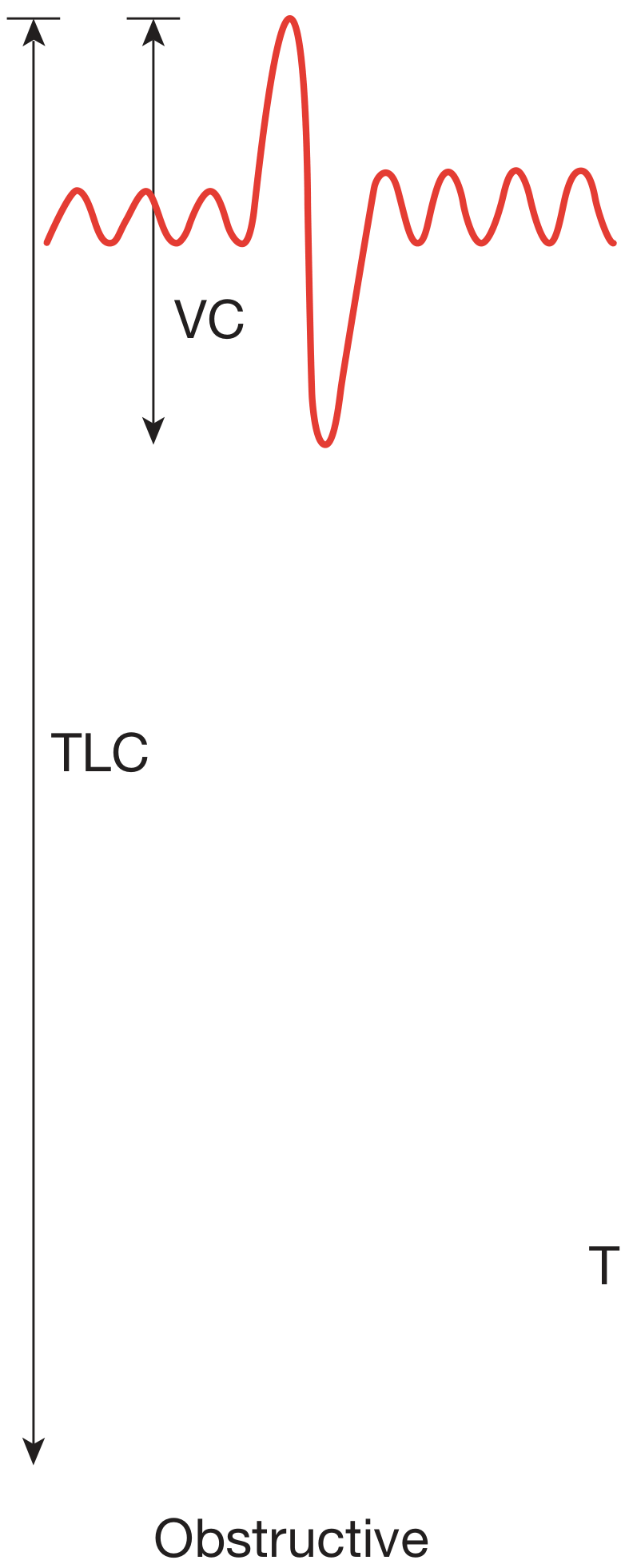
Obstructive pattern:
The forced expiratory maneuver is prolonged and concave - the FEV1 is markedly reduced while the FVC may be near normal, creating a "scooped-out" appearance.
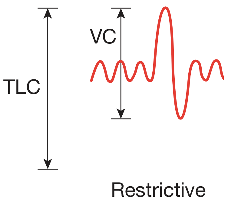
Restrictive pattern:
Both FEV1 and FVC are reduced. The curve reaches its endpoint quickly and is steep but small - TLC and VC are both brought down.
Flow-Volume Loop
Obstructive (Panel A - COPD): The expiratory limb is concave upward ("scooped"), indicating reduced flow particularly at low lung volumes. The loop is widened horizontally due to air trapping.
Restrictive (Panel B - ILD): The loop is narrow and vertically oriented (convex upward), with high flows relative to the reduced volume. The entire loop is shifted inward.
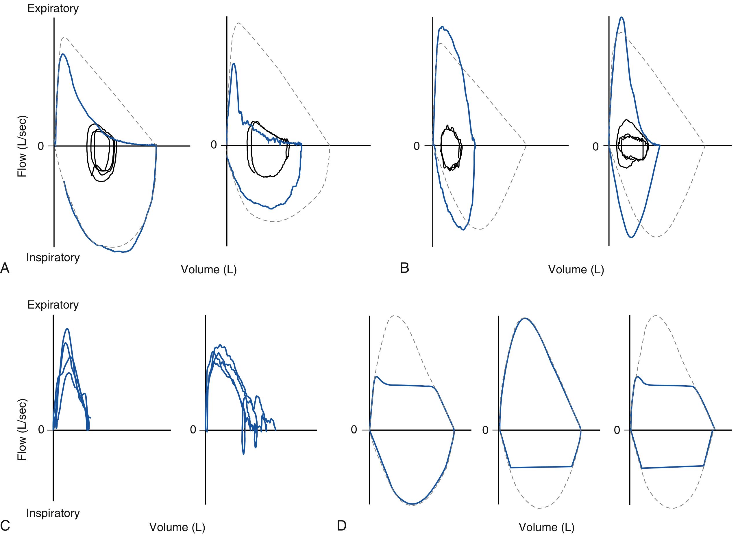
- Murray & Nadel's (p. 763): Panel A description - "maximal expiratory flow is concave upward...reflects a greater reduction in flow at low compared to high lung volumes." Panel B - "maximal expiratory flow appears steep and vertically oriented (convex upward) with increased flow at high lung volumes. Vital capacity is reduced."
VI. Grading Severity
According to Murray & Nadel's Textbook of Respiratory Medicine (Table 32.3):
| Severity | FEV1 (% predicted) |
|---|---|
| Mild | 70-LLN |
| Moderate | 60-69% |
| Moderately severe | 50-59% |
| Severe | 35-49% |
| Very severe | <35% |
This grading applies to both obstructive FEV1 and restrictive FVC/TLC impairment.
VII. Clinical Examples Relevant to CVTS Physiotherapy
| Condition | Type | Characteristic PFT |
|---|---|---|
| COPD | Obstructive | FEV1/FVC <0.70, increased TLC, RV |
| Asthma | Obstructive (reversible) | FEV1/FVC <0.70, +ve bronchodilator response |
| Pulmonary Fibrosis (IPF) | Restrictive | Low TLC, FEV1/FVC normal/elevated, low DLCO |
| Kyphoscoliosis | Restrictive (extra-parenchymal) | Low TLC, FEV1/FVC normal, DLCO normal |
| Post-lobectomy / thoracic surgery | Restrictive | Low FVC, low TLC proportionally |
| Bronchiectasis | Obstructive | Low FEV1/FVC, variable bronchodilator response |
VIII. Significance in Physiotherapy Practice
- Treatment planning: Obstructive conditions require airway clearance techniques (ACBT, PEP therapy, postural drainage), whereas restrictive conditions benefit from chest expansion exercises, breathing retraining, and maintaining mobility.
- Monitoring progress: Serial spirometry tracks improvement with bronchial hygiene or exercise training.
- Pre-surgical assessment: In CVTS patients, PFTs predict post-operative pulmonary risk. FEV1 > 1.5L and FVC > 1.5L are generally required for lung resection.
- Exercise prescription: PEFR and FEV1 guide safe exercise intensity for patients with COPD or ILD.
- Bronchodilator reversibility: A positive response (improvement in FEV1 by >200 mL and >12%) distinguishes asthma from fixed obstructive disease (COPD) - influencing which therapies are selected.
Summary (Exam Quick Recall)
Obstructive: FEV1/FVC LOW | FEV1 LOW | TLC/RV HIGH | "Can't get air OUT"Restrictive: FEV1/FVC NORMAL or HIGH | FVC LOW | TLC LOW | "Can't get air IN"
Sources:
- Murray & Nadel's Textbook of Respiratory Medicine, pp. 763-770 (General approach to interpretation, obstructive and restrictive ventilatory defects, flow-volume loop patterns)
- Bailey & Love's Short Practice of Surgery, 28th Edition, p. 998 (Spirometry and FVC interpretation in obstructive and restrictive defects)
- Sabiston Textbook of Surgery (Physiologic Evaluation): "In obstructive lung disease, FEV1/FVC is low (FEV1 is low, FVC is high); in restrictive disease, the ratio is normal or high"
This is a shared conversation. Sign in to Orris to start your own chat.