
Oculomotor nerve
oculomotor nerve CN III anatomy diagram
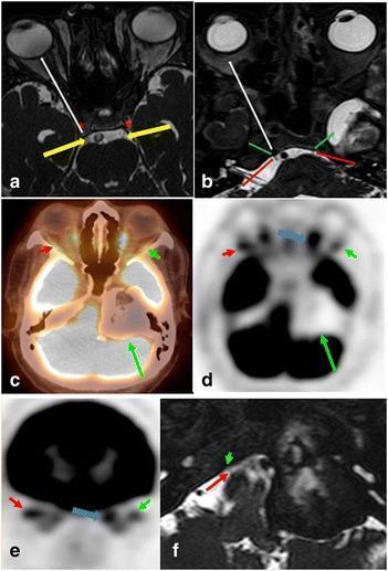
This composite of diagnostic images illustrates the neuroanatomy of the oculomotor (CN III) and abducens (CN VI) nerves and their clinical involvement in pathology. Panels (a) and (b) utilize axial FIESTA MRI to demonstrate normal anatomy: yellow arrows track CN III from its cisternal segment into the cavernous sinus (asterisk), while red arrows show CN VI entering Dorello’s canal (green arrow). The white arrows indicate their anterior trajectory toward the superior orbital fissure. Panels (c) through (e) present axial and coronal fused FDG PET/CT and PET-only images of a 73-year-old male with diplopia. A large, peripherally ossified, hypometabolic mass is visible in the left petrous apex and cerebellopontine angle (long green arrow). Functional denervation is demonstrated by reduced FDG uptake in the left lateral rectus muscle (short green arrow) compared to the normal right side (red arrow), with compensatory hypermetabolism in the left medial rectus muscle. High-resolution MRI (f) confirms the mass involves the expected anatomical course of the left abducens nerve, contrasting with the visible normal right nerve.
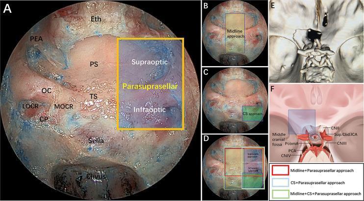
This composite educational graphic illustrates the surgical anatomy and modular corridors of the endoscopic endonasal parasuprasellar approach. Panel A shows an intraoperative endoscopic endonasal view with key landmarks labeled: posterior ethmoidal artery (PEA), planum sphenoidale (PS), optic canal (OC), tuberculum sellae (TS), medial and lateral optocarotid recesses (MOCR, LOCR), carotid protuberance (CP), and the sella. A yellow quadrangle delineates the parasuprasellar area, further divided by the optic nerve into supraoptic and infraoptic regions. Panels B–D provide comparative overlays of different surgical modules: midline (transsellar/transtuberculum), cavernous sinus (CS), and parasuprasellar corridors, coded by color (red, blue, green) to represent various combined surgical approaches. Panel E presents a 3D reconstruction of a postoperative CT scan demonstrating the extent of bone removal. Panel F is a schematic diagram illustrating the neurovascular relationships in the parasuprasellar area, including the optic nerve (CN II), oculomotor nerve (CN III), and supraclinoidal internal carotid artery (ICA). This material is intended for neurosurgical training in skull base surgery and endoscopic corridor selection.
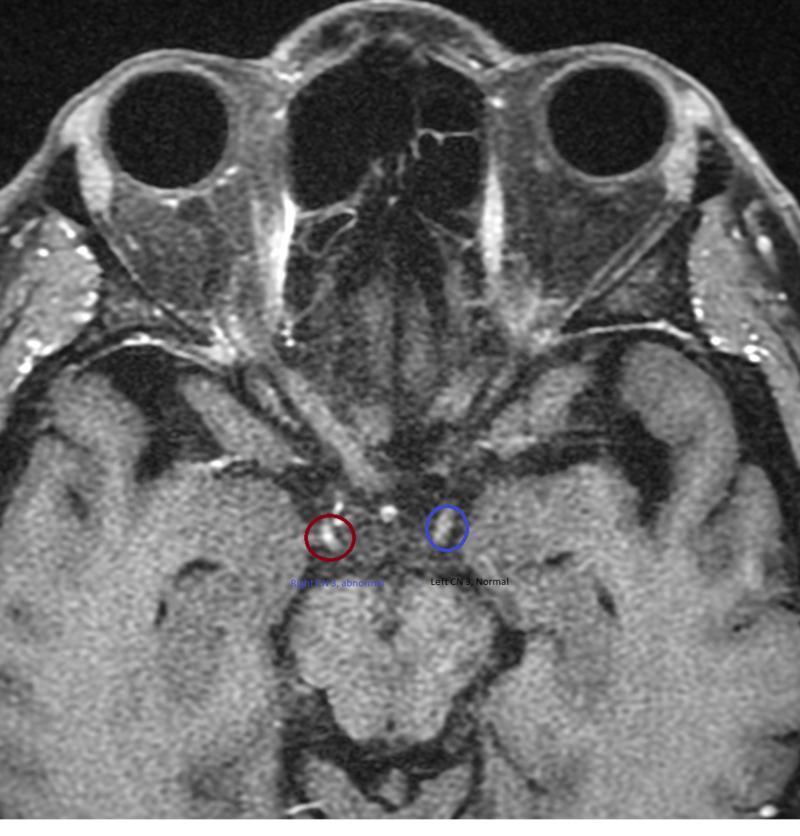
This diagnostic image is an axial T1-weighted post-contrast magnetic resonance imaging (MRI) of the head at the level of the midbrain and interpeduncular cistern. The primary focus is the comparison of the bilateral third cranial nerves (oculomotor nerve, CN III). The right CN III, highlighted by a red circle, exhibits focal abnormal enhancement, appearing as a hyperintense (bright) signal compared to the surrounding anatomy. In contrast, the left CN III, highlighted by a blue circle, shows normal signal intensity without evidence of enhancement. Other visible anatomical structures include the orbits and globes, the optic nerves and chiasm, and the brain parenchyma of the temporal lobes. The clinical significance of this finding is indicative of inflammation, demyelination, or compression of the oculomotor nerve, often associated with conditions such as Miller Fisher syndrome or ophthalmoplegic migraine. This comparison serves as a diagnostic educational tool for identifying abnormal nerve enhancement in neuroimaging.
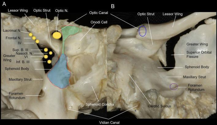
This medical anatomical diagram features two clinical photographs of a dry bone specimen, showing the osseous anatomy of the human orbital apex of the sphenoid bone. Image (A) provides an anterior view, highlighting the optic canal superomedially and the superior orbital fissure (SOF) inferolaterally. Key landmarks include the optic strut, which separates the optic nerve and ophthalmic artery from the SOF contents, and the maxillary strut, which separates the SOF from the foramen rotundum. Overlays illustrate the surgical decompression areas: the light green area denotes the medial wall of the optic canal, and the light blue area indicates the medial wall of the SOF. Image (B) presents a posterior view, showing the spatial relationship between the optic canal, SOF, foramen rotundum, and the carotid sulcus. The diagram identifies neurovascular structures passing through these apertures, including the optic nerve (CN II), oculomotor nerve (CN III), trochlear nerve (CN IV), and divisions of the trigeminal nerve (V1, V2). This resource is designed for advanced surgical planning in endoscopic endonasal decompression procedures.
oculomotor nerve palsy ptosis pupil dilation
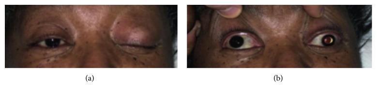
Clinical photograph in two panels (a and b) illustrating a complete left-sided third cranial nerve (oculomotor nerve) palsy. Panel (a) shows the patient attempting to open both eyes, revealing severe unilateral ptosis of the left upper eyelid, while the right eyelid remains normal. Panel (b) depicts the same patient with both upper eyelids passively retracted by an examiner. This manual elevation reveals significant anisocoria, with the left pupil demonstrating fixed mydriasis (dilation) compared to the reactive right pupil. Additionally, the left eye displays a characteristic 'down and out' gaze deviation (exotropia and hypotropia), indicating the loss of function in the superior, inferior, and medial recti, as well as the inferior oblique muscles. These visual findings are pathognomonic for a complete oculomotor nerve lesion, often associated with clinical emergencies such as pituitary apoplexy or an enlarging posterior communicating artery aneurysm.
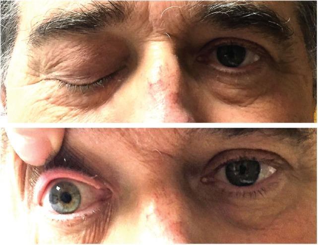
This clinical photograph consists of a two-panel vertical comparison demonstrating a cranial nerve III (oculomotor nerve) palsy. The top panel shows a front-facing view of a patient with complete right-sided ptosis, where the right upper eyelid is fully closed in contrast to the normal eyelid position and appearance of the left eye. The bottom panel shows the clinician manually elevating the right upper eyelid, revealing an ipsilateral fixed and dilated pupil (mydriasis) and a slightly bloodshot sclera. The left pupil appears smaller and reactive. These visual findings—ptosis and a large, non-reactive pupil—are characteristic signs of oculomotor nerve dysfunction, often associated with compressive lesions or infiltrative processes such as cavernous sinus involvement or skull base tumors. This material is used in medical education to teach the clinical presentation of multiple cranial neuropathies and the physical examination techniques for neuro-ophthalmological assessment.
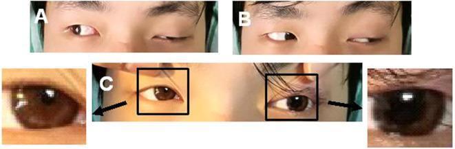
This composite clinical photograph illustrates the hallmark signs of a left oculomotor nerve (third cranial nerve) palsy in a 24-year-old male. Panel A demonstrates significant left-sided ptosis (drooping upper eyelid) and impaired adduction of the left eye when the patient attempts a rightward gaze, resulting in a divergent ocular alignment. Panel B shows preserved abduction of the left eye, indicating intact sixth cranial nerve function. Panel C provides a close-up comparison of the pupils, revealing anisocoria with a dilated, non-reactive left pupil (mydriasis) compared to the right. The combination of ptosis, 'down and out' resting position or inability to adduct, and pupillary involvement is characteristic of complete third nerve paralysis, often associated with compression or intrinsic lesions of the oculomotor nerve. This visual record is educational for identifying neuro-ophthalmic manifestations of cranial nerve pathology, specifically differentiating between pupil-sparing and pupil-involved palsies.
Oculomotor Nerve (CN III)
Overview
- General somatic efferent (GSE) - voluntary motor to most extraocular muscles
- General visceral efferent (GVE) - preganglionic parasympathetic output
Nucleus
| Subnucleus | Innervation |
|---|---|
| Superior rectus subnucleus | Contralateral superior rectus (crossed) |
| Inferior rectus subnucleus | Ipsilateral inferior rectus |
| Medial rectus subnucleus | Ipsilateral medial rectus |
| Inferior oblique subnucleus | Ipsilateral inferior oblique |
| Caudal central subnucleus (midline, unpaired) | Both levator palpebrae superioris muscles (bilateral) |
| Edinger-Westphal nucleus | Preganglionic parasympathetics to iris sphincter + ciliary muscle |
Course
- Fascicles arise from the nucleus and traverse the midbrain, passing through or near the red nucleus and close to the cerebral peduncles
- Exit the brainstem as rootlets from the lateral interpeduncular fossa (anterior midbrain surface, between midbrain and pons)
- Pass through the subarachnoid (interpeduncular) space, running between the superior cerebellar artery (below) and the posterior cerebral artery (above)
- Travel parallel to the posterior communicating artery (PCOM) - at risk from PCOM aneurysms here
- Enter the anterior edge of the tentorium cerebelli
- Pass in the lateral wall of the cavernous sinus (uppermost cranial nerve in the sinus)
- Enter the orbit via the superior orbital fissure (within the common tendinous ring)
- Divide into superior and inferior divisions just before entry
Terminal Distribution in the Orbit
- Levator palpebrae superioris
- Superior rectus
- Medial rectus (passes below the optic nerve)
- Inferior rectus
- Inferior oblique - this branch also gives off the branch to the ciliary ganglion (parasympathetic root)
- Sphincter pupillae (pupil constriction/miosis)
- Ciliary muscle (accommodation for near vision)
Clinical Findings: Complete CN III Palsy
| Feature | Explanation |
|---|---|
| Ptosis | Loss of levator palpebrae superioris |
| "Down and out" eye position | Unopposed lateral rectus (CN VI) + superior oblique (CN IV) |
| Inability to elevate, depress, or adduct the eye | Loss of SR, IR, MR, IO |
| Fixed dilated pupil (mydriasis) | Loss of parasympathetic sphincter pupillae |
| Cycloplegia | Loss of ciliary muscle (no accommodation) |
| Diplopia | Image from affected eye projected upward and medially |
Partial vs. Complete Palsy
- A partial palsy may present with any combination of ptosis, pupil dilation, or individual muscle weakness
- Frequent serial examinations are essential during the rapidly evolving phase
Pupil-Involved vs. Pupil-Sparing: Critical Distinction
| Type | Likely Etiology |
|---|---|
| Pupil-involved CN III palsy | Compressive lesion (aneurysm, tumor) - parasympathetic fibers on the surface are compressed first |
| Pupil-sparing CN III palsy | Microvascular ischemia (diabetes, hypertension) - ischemia affects the nerve core, sparing superficial parasympathetics |
Brainstem Fascicular Syndromes
| Syndrome | Fascicle + Structure Involved | Features |
|---|---|---|
| Weber's | CN III + cerebral peduncle | Ipsilateral CN III palsy + contralateral hemiparesis |
| Benedikt's | CN III + red nucleus | Ipsilateral CN III palsy + contralateral tremor/chorea |
| Nothnagel's | CN III + superior cerebellar peduncle | Ipsilateral CN III palsy + ipsilateral cerebellar ataxia |
| Claude's | CN III + red nucleus + superior cerebellar peduncle | CN III palsy + contralateral hemiataxia |
Nuclear Lesion: Key Distinguishing Features
- Bilateral ptosis - because the single caudal central subnucleus serves both levators
- Contralateral superior rectus weakness - because that subnucleus decussates to innervate the contralateral SR
- Isolated nuclear palsy is rare; usually other brainstem signs are present (infarction, hemorrhage, tumor, demyelination)
Causes by Anatomical Location
| Location | Causes |
|---|---|
| Nucleus/fascicle (midbrain) | Infarction, hemorrhage, demyelination, tumor |
| Subarachnoid space / interpeduncular fossa | PCOM aneurysm, meningitis, tumor, herniation (uncus compressing nerve against tentorium) |
| Cavernous sinus | Carotid aneurysm, carotid-cavernous fistula, cavernous sinus thrombosis, pituitary adenoma, meningioma, metastasis, herpes zoster, Tolosa-Hunt syndrome |
| Orbit / superior orbital fissure | Trauma, tumor |
| Systemic (microvascular) | Diabetes, hypertension (pupil-sparing) |
Aberrant Regeneration
- Elevation of the eyelid on downgaze or adduction (Pseudo-von Graefe sign)
- Pupil constriction on attempted adduction, elevation, or depression