
Sickle cell anemia Pathophysiology Anaesthesia considerations Anaesthesia management Management of Sickle cell crisis
sickle cell anemia pathophysiology diagram sickling
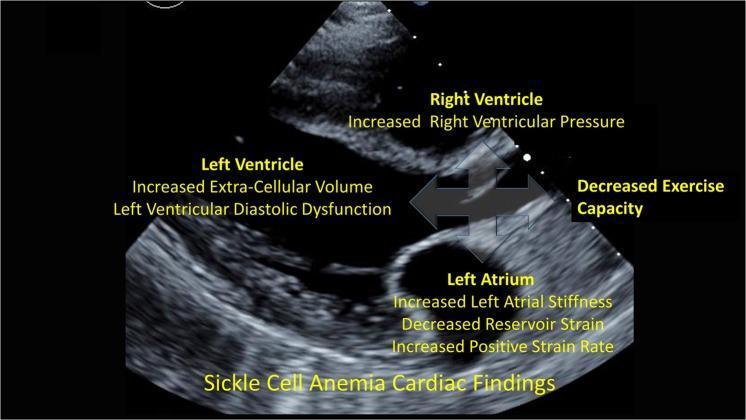
This diagnostic image is an annotated echocardiogram in the parasternal long-axis view, functioning as a pathophysiology diagram to illustrate cardiac involvement in Sickle Cell Anemia (SCA). The image labels three primary cardiac chambers with their associated clinical and functional abnormalities. The Right Ventricle is annotated with increased right ventricular pressure. The Left Ventricle is associated with increased extra-cellular volume and left ventricular diastolic dysfunction, suggesting interstitial myocardial fibrosis or edema. The Left Atrium highlights increased left atrial stiffness, decreased reservoir strain, and increased positive strain rate. Centrally, a four-way arrow signifies the interrelated nature of these dysfunctions, culminating in decreased exercise capacity. This educational visual demonstrates how diffuse myocardial changes in SCA lead to impaired chamber compliance and global functional impairment. It is intended for intermediate to advanced medical learners focusing on cardiology, hematology, or the systemic manifestations of hemoglobinopathies.
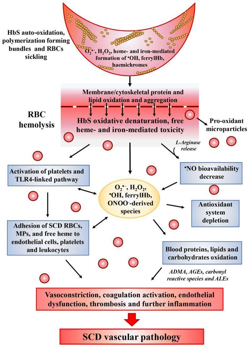
A medical pathophysiology diagram illustrating the oxidative stress cascade in Sickle Cell Disease (SCD) and its progression to vascular pathology. The diagram follows a vertical flow starting with a sickled red blood cell (RBC) undergoing HbS auto-oxidation and polymerization, leading to the generation of reactive oxygen species (ROS) such as H2O2, ferrylHb, and hemichromes. This triggers a central event of RBC hemolysis, characterized by membrane lipid oxidation, protein aggregation, and the release of pro-oxidant microparticles and L-arginase. Downstream pathways depict the systemic consequences, including the activation of platelets and the TLR4-linked inflammatory pathway, adhesion of SCD RBCs to endothelial cells and leukocytes, and a significant decrease in nitric oxide (NO) bioavailability. The lower section highlights the depletion of the antioxidant system and the oxidative modification of blood components into ADMA, AGEs, and ALEs. The cascade culminates in clinical manifestations such as vasoconstriction, endothelial dysfunction, and thrombosis, collectively summarized as SCD vascular pathology. This illustration is designed for advanced medical education regarding the molecular mechanisms of hematologic disorders.
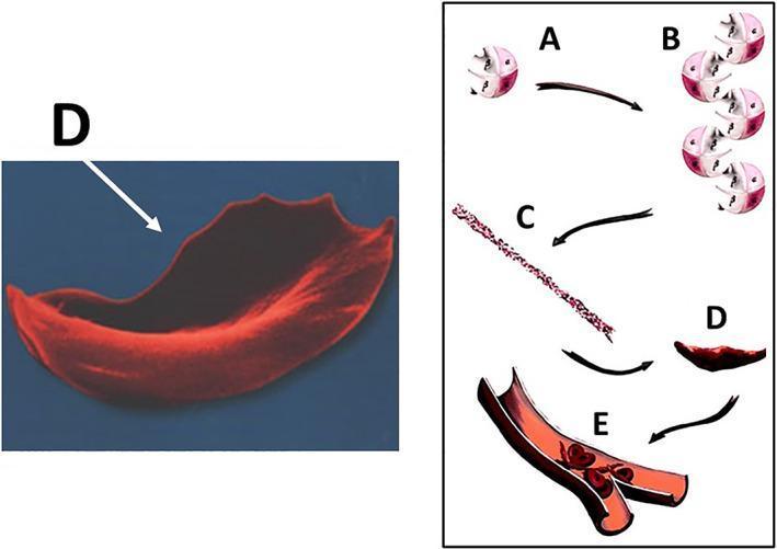
This educational graphic illustrates the molecular and cellular pathophysiology of sickle cell disease (SCD) leading to vaso-occlusion. The diagram is divided into a microscopic anatomical illustration and a high-magnification scanning electron micrograph. Steps A through D depict the hemoglobin S (HbS) polymerization process: (A) individual deoxygenated hemoglobin tetramers, (B) aggregation of hemoglobin molecules, (C) formation of long, rigid hemoglobin polymers or strands, and (D) the resulting transformation of a flexible biconcave red blood cell into a rigid, crescent or sickle-shaped erythrocyte. A corresponding scanning electron micrograph (labeled D) highlights the characteristic elongated, spicular morphology of a sickled cell. Section E shows a cross-section of a small bifurcating blood vessel where these rigid, abnormally shaped cells are aggregating, causing mechanical obstruction (vaso-occlusion) of the lumen. This process demonstrates the mechanism behind tissue ischemia and subsequent complications such as osteonecrosis in SCD patients. The content is suitable for medical students and clinicians studying hematology and musculoskeletal pathology.
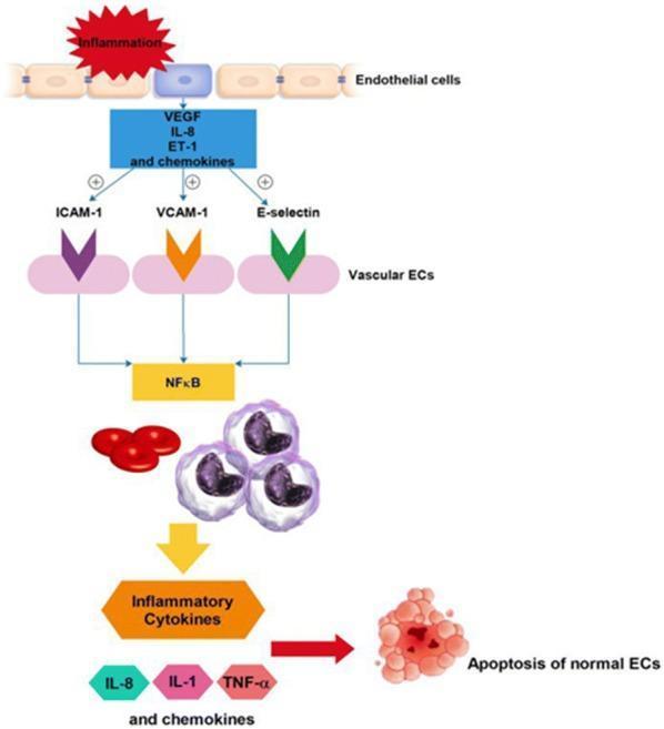
This pathophysiology diagram illustrates the signaling pathway of endothelial cell (EC) activation and apoptosis, particularly in the context of sickle cell disease (SCD) interactions with the vascular wall. The process begins with 'Inflammation' triggering the release of mediators including VEGF, IL-8, ET-1, and chemokines. These factors upregulate adhesion molecules on vascular ECs, specifically ICAM-1, VCAM-1, and E-selectin (represented by colored chevron icons). Activation of these molecules converges on the NF-̄Κβ signaling hub. This pathway facilitates the recruitment and interaction of homozygotic SS red blood cells (RBCs) and leukocytes (white blood cells). The subsequent cascade leads to the secretion of inflammatory cytokines, notably IL-8, IL-1, and TNF-α. The final stage of this inflammatory loop is the induction of apoptosis in normal endothelial cells, shown as a cluster of fragmented cells. The diagram serves as a model for understanding vaso-occlusive mechanisms and potential therapeutic targets like NF-̄Κβ inhibition in hematological disorders.
sickle cell disease anesthesia perioperative management
Sickle Cell Anemia: Complete Clinical Review
1. PATHOPHYSIOLOGY
Genetic Basis
- HbSS (sickle cell anemia): homozygous - most severe; >85% HbS, Hct 18-28%
- HbSC disease: compound heterozygous HbS + HbC; moderate severity (~50% HbS)
- HbS β-thalassemia: variable severity
- Sickle cell trait (HbAS): carrier; 35-40% HbS; benign clinically (sickle only at SpO₂ ~40%)
Molecular Mechanism of Sickling
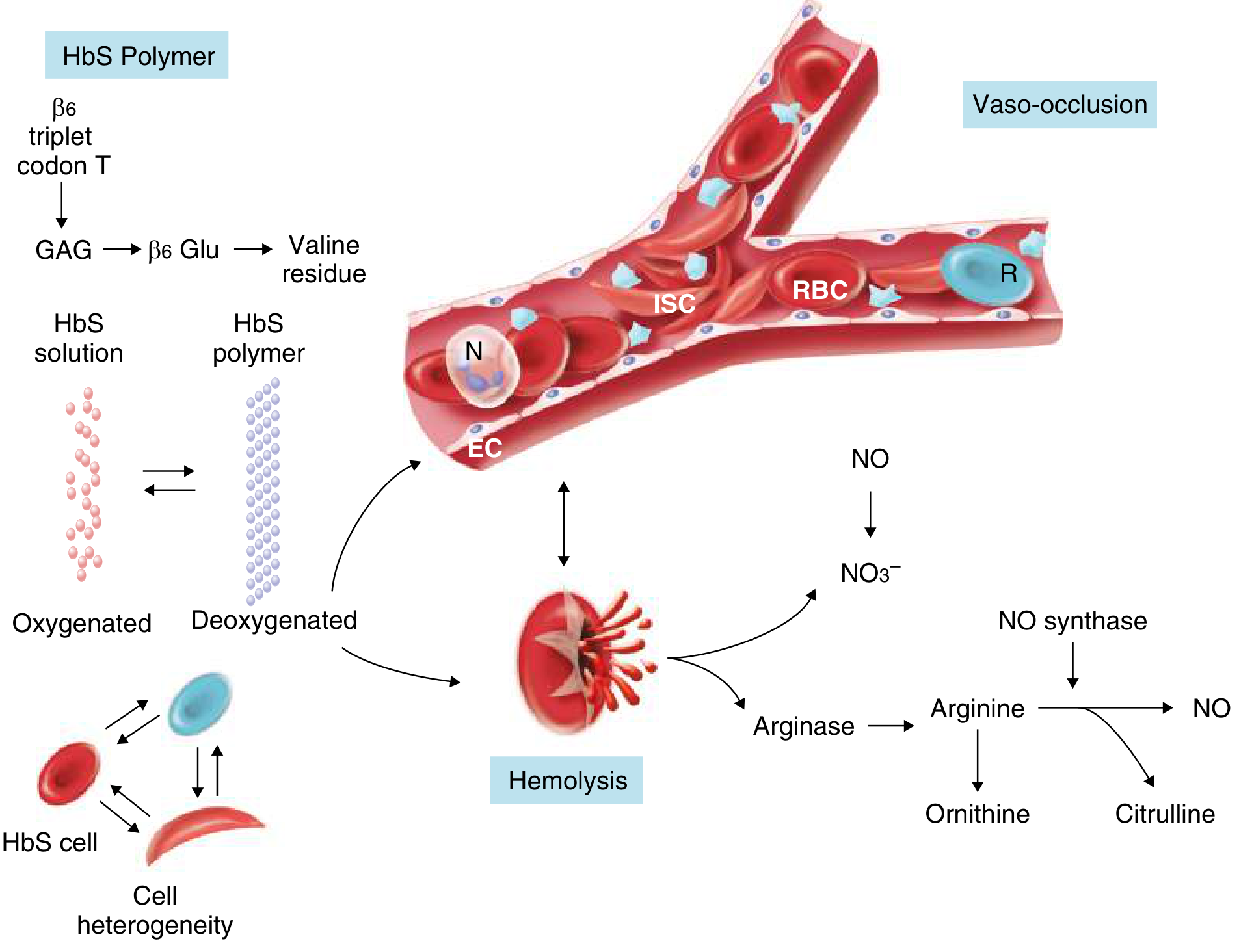
- Deoxygenation in capillaries → HbS polymerization → sickle shape
- Return to lungs → reoxygenation → polymer deaggregation (reversal)
- If transit is delayed → persistent sickling → membrane damage → vaso-occlusion
- Low pO₂ / low pH (acidosis)
- High temperature
- Dehydration (elevated intracellular HbS concentration)
- Low HbF levels
- Presence of HbF (fetal hemoglobin) - critical protective effect
- HbA (in heterozygotes)
- Hydroxyurea (increases HbF production)
Downstream Pathological Mechanisms
| Mechanism | Consequence |
|---|---|
| Microvascular obstruction | Tissue ischemia and infarction |
| RBC + platelet adhesion to endothelium | Vascular inflammation, thrombosis |
| Hemolysis with free Hb release | Nitric oxide (NO) scavenging → vasoconstriction |
| Arginase release from lysed RBCs | Depletes L-arginine (NO substrate) → further NO deficiency |
| Reactive oxygen species (ROS) generation | Oxidative stress, endothelial injury |
| Leukocyte activation | Cytokine release, inflammatory cascade |
| Reperfusion injury | Activates endothelium after ischemia |
| Reticulocyte adhesion ligands | Augment RBC-endothelial interactions |
Consequences of Chronic Hemolysis
- RBC lifespan reduced from 120 days → 12-17 days
- Chronic hemolytic anemia (Hb typically 6-9 g/dL in HbSS)
- Compensatory bone marrow hyperplasia
- High reticulocyte count; elevated LDH, bilirubin; low haptoglobin
- Functional asplenia (auto-infarction of spleen in childhood) → immunocompromised state
2. MULTISYSTEM COMPLICATIONS
| System | Complications |
|---|---|
| Hematologic | Hemolytic anemia, aplastic anemia, leukocytosis, alloimmunization |
| Pulmonary | Acute chest syndrome (ACS), pulmonary hypertension (10%), asthma (50%), fibrosis, pulmonary infarction, sleep apnea |
| Cardiac | Cardiomegaly, LVH, RV dilation, diastolic dysfunction, cor pulmonale, cardiomyopathy |
| Neurologic | Stroke (cerebral infarct - adolescents; hemorrhagic - adults), hemorrhage, aneurysm, meningitis |
| Renal | Papillary necrosis, inability to concentrate urine, glomerular sclerosis, renal failure, hematuria |
| Musculoskeletal | Painful vaso-occlusive crises, avascular necrosis, osteomyelitis, bone infarction, dactylitis |
| Spleen | Auto-infarction, hyposplenism, acute sequestration crisis |
| Hepatobiliary | Jaundice, cholelithiasis, hepatitis, cirrhosis, cholestasis |
| Ocular | Retinopathy, vitreous hemorrhage, vision loss |
| GU | Priapism, renal failure, infections |
| Psychosocial | Depression, anxiety, chronic pain disorder, opioid dependence |
3. ANAESTHESIA CONSIDERATIONS
Why SCD Patients Are High-Risk
Preoperative Assessment
- Frequency, severity, and pattern of vaso-occlusive crises
- Previous acute chest syndrome episodes
- Prior stroke or neurological events
- History of OSA, pulmonary hypertension
- Opioid tolerance and pain management history
- Response to prior anaesthetics
- Current medications (especially hydroxyurea)
- Vaccination status
- Full blood count (CBC) + reticulocyte count
- Serum creatinine (renal function)
- ECG
- Chest X-ray
- Echocardiogram - indicated for all but minor procedures; screen every 1-3 years (NT-proBNP >159 pg/mL or 6MWT <333 m suggests pulmonary hypertension)
- Type and screen (even for low-risk procedures given risk of hemolytic crisis)
- Liver iron content by MRI every 1-2 years in patients receiving repeated transfusions (not ferritin alone)
- Arterial blood gases if indicated
- Pulmonary: Pulmonary hypertension independently predicts mortality. ACS is the leading cause of death.
- Cardiac: LVH, diastolic dysfunction common. Symptoms overlap between SCD and pulmonary hypertension.
- Renal: Loss of concentrating ability → prone to dehydration. CKD affects drug dosing.
- Neurologic: Prior stroke may affect regional anaesthesia decision and positioning.
- Immune: Functional asplenia → susceptible to encapsulated organisms; ensure vaccinations up to date.
Preoperative Optimization
- Low-risk surgery (<30-60 min; superficial): no transfusion needed, or observe; if on hydroxyurea with Hb >9 g/dL, no transfusion required
- Intermediate-risk surgery (most intra-abdominal, head/neck, orthopaedic): simple transfusion to target Hb >10 g/dL (>100 g/L); as effective as exchange transfusion for reducing HbS to <30% in this setting
- High-risk surgery (cardiac, neurosurgery, major vascular, transplant): exchange transfusion to reduce HbS concentration to <30%; also required if baseline Hb already >10 g/dL (simple top-up would cause hyperviscosity)
- Red cell exchange (RCE) regardless of Hb level recommended for HbSS patients undergoing high-risk surgery
4. ANAESTHESIA MANAGEMENT
Intraoperative Principles
- Hypoxia - maintain SpO₂ >95%; use supplemental oxygen
- Hypothermia - active warming; warm IV fluids; monitor temperature
- Hypovolaemia/dehydration - maintain normovolaemia with IV fluids
- Hypotension - rapid treatment; hypoperfusion is a sickling trigger
- Acidosis - avoid hyperventilation-induced alkalosis paradoxically; target normal pH; avoid metabolic acidosis
- Stasis/venous stasis - careful positioning, avoid vena caval compression
- Infection
- Hypothermia
Choice of Anaesthetic Technique
- All anaesthetic techniques are acceptable provided the above principles are respected
- Regional/neuraxial anaesthesia is preferred where feasible:
- Superior perioperative pain control (important in opioid-tolerant patients)
- Resulting vasodilation may reduce sickling tendency
- Spinal anaesthesia for caesarean section associated with less blood loss and reduced postoperative opioid requirement
- Note: Treat hypotension from spinal quickly to avoid hypoperfusion
- Avoid epinephrine in local anaesthetic solutions (vasoconstriction)
- General anaesthesia: acceptable; all standard agents are suitable; maintain normothermia, normoxia, normocarbia
Positioning
- Avoid high-risk positions that promote venous stasis: prone position, lithotomy for extended duration
- Avoid vena caval compression (especially in pregnant patients)
- Careful extremity positioning to prevent circulatory compromise
Tourniquets
- Avoid tourniquets if possible - cause distal metabolic acidosis and regional hypoxemia → promote sickling
- If absolutely required for surgical success, may be used with extreme caution; ensure adequate exsanguination before inflation
Contrast Media
- Use iso-osmolar or hypo-osmolar contrast agents; hyperosmolar contrast can precipitate sickling
Monitoring
- Standard ASA monitoring + temperature probe
- Pulse oximetry is essential (target SpO₂ >95%)
- Consider arterial line for high-risk procedures
- Urine output monitoring
Fluid Management
- Maintain normovolaemia - both hypovolaemia (sickling) and hypervolaemia (pulmonary oedema in impaired cardiac function) are harmful
- Use balanced crystalloids; avoid hypotonic solutions
Postoperative Care
- Continue supplemental oxygen until fully awake and SpO₂ stable
- Optimal multimodal analgesia (opioids + NSAIDs + regional) - note: avoid NSAIDs in renal dysfunction
- Prevent dehydration, hypothermia, hypoxia, acidosis
- Incentive spirometry - reduces risk of ACS
- Venous thromboprophylaxis - indicated
- Monitor Hb and renal function
- Acute chest syndrome can develop 2-3 days postoperatively - requires supportive treatment
- Careful sedation monitoring if opioids used
5. MANAGEMENT OF SICKLE CELL CRISIS
Types of Sickle Cell Crisis
| Type | Key Feature | Reticulocytes | Common Cause |
|---|---|---|---|
| Vaso-occlusive (VOC) | Pain; ischemic infarction | Normal/elevated | Dehydration, infection, hypoxia, cold, stress |
| Hemolytic | Acute drop in Hb + Hct | Elevated (reticulocytosis) | Accelerated hemolysis |
| Aplastic | Acute drop in Hb | Low/absent | Parvovirus B19 (>90% of cases) |
| Sequestration | Acutely enlarged spleen + Hb ≥2 g/dL below baseline | Variable | Rapid splenic trapping of RBCs |
| Acute Chest Syndrome (ACS) | New pulmonary infiltrate + fever/respiratory symptoms | - | Infection (Chlamydia, Mycoplasma), fat embolism, infarction |
A. Vaso-Occlusive Crisis (VOC) / Painful Crisis
- Mild-moderate pain: oral NSAIDs (if renal function normal) + oral opioids (oxycodone, morphine, hydrocodone)
- Moderate-severe / hospitalized:
- IV opioids: morphine or hydromorphone as first-line
- Use fentanyl if hepatic or renal dysfunction
- Patient-controlled analgesia (PCA) with as-needed breakthrough doses; schedule if not using PCA
- Ketamine (low-dose) if poor opioid response (sub-anaesthetic, anti-hyperalgesic)
- NSAIDs (with adequate renal function) for opioid-sparing effect
- Epidural analgesia for severe VOC
- IV fluids for dehydration; avoid fluid overload
- Goal: maintain normovolaemia (specific fluid type/volume evidence is limited)
- Supplemental O₂ for SpO₂ <95%; goal SpO₂ >95%
- Incentive spirometry to prevent ACS
- Treat underlying infection with appropriate antibiotics
- Rewarm if cold exposure
- Not routinely indicated for simple VOC alone
- Consider if associated severe anemia, ACS, or organ dysfunction
B. Acute Chest Syndrome (ACS)
- Admission (all cases)
- Oxygen to maintain SpO₂ >95%
- IV antibiotics: IV cephalosporin (cefuroxime or cefotaxime) + oral macrolide (covers atypical organisms)
- Analgesia + IV fluids (see VOC management above)
- Incentive spirometry
- Transfusion:
- Simple transfusion for moderate illness
- Exchange transfusion for severe ACS or unresponsive cases
- Avoid transfusion if no other indication (risk of volume overload)
- Diuretics if fluid overloaded
- Respiratory support (non-invasive or mechanical ventilation) if severe
- High-dose dexamethasone is controversial
C. Aplastic Crisis
- Admission; isolate (parvovirus is contagious - risk to immunocompromised contacts)
- IV fluids
- Simple transfusion if severe anemia
- Parvovirus serology + PCR for confirmation
- Self-limiting (erythropoiesis recovers within 7-10 days)
D. Splenic Sequestration Crisis
- Admit; serial abdominal examinations
- IV fluid resuscitation (aggressive)
- Transfusion - cautious (release of sequestered RBCs on recovery can cause hyperviscosity)
- May require splenectomy for recurrent episodes or hemodynamic instability
E. Admission Criteria (Harriet Lane)
- Fever ≥40°C, WBC >30,000 or <5,000, Hb <5 g/dL, platelets <100,000, abnormal SpO₂, infiltrate on CXR
- Signs of ACS, stroke, or organ complications
- Pain not manageable in outpatient setting
- Aplastic crisis
- Splenic sequestration
Long-Term Disease-Modifying Therapy
| Drug | Mechanism | Use |
|---|---|---|
| Hydroxyurea | Increases HbF production | Reduces VOC frequency/severity, ACS, transfusion requirements |
| Voxelotor | Increases HbS-oxygen affinity | Reduces sickling and hemolysis |
| Crizanlizumab | Anti-P-selectin antibody | Reduces VOC |
| L-glutamine | Reduces oxidative stress | Reduces ACS |
| Bone marrow transplant | Curative | Severe SCD in eligible patients |
| Gene therapy | Emerging curative | Under investigation |