
Causes of dysphagia detailly
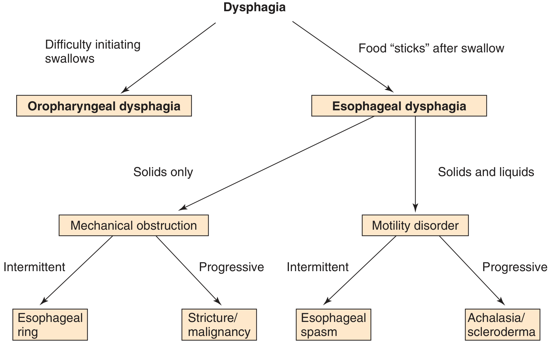
causes of dysphagia classification diagram
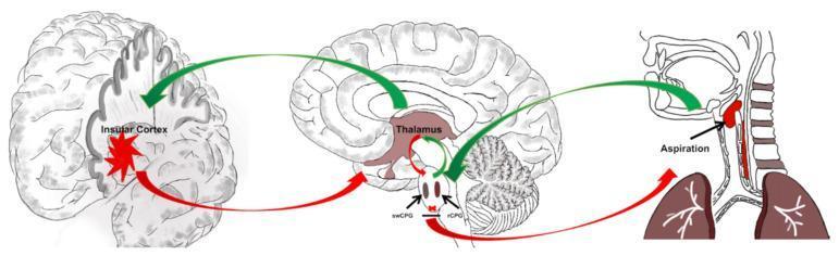
This pathophysiology diagram illustrates a hypothesized neurological mechanism for post-stroke dysphagia (PSD) and aspiration following a lesion in the insular cortex. The visual is divided into three sections: a cortical view, a sagittal brain section, and a peripheral anatomical view. The process begins with sensory inputs from peripheral organs (tongue, bronchial, and esophagus) directed via green arrows (promotion) to the brainstem. In the brainstem, the swallowing center pattern generator (swCPG) and respiratory center pattern generator (rCPG) are depicted as adjacent ovals with a red horizontal line indicating reciprocal inhibition. Information flows from the brainstem to the thalamus, which serves as a central relay station, and subsequently to the insular cortex. A red starburst in the insular cortex represents a stroke or disturbance. Red arrows indicate that this cortical damage disrupts the thalamic and brainstem pathways, specifically interfering with the swCPG/rCPG reciprocal inhibition. The final outcome is shown in a cross-sectional view of the throat and lungs, where a red bolus is seen entering the trachea, labeled 'Aspiration,' signifying a failure of airway protection mechanisms.
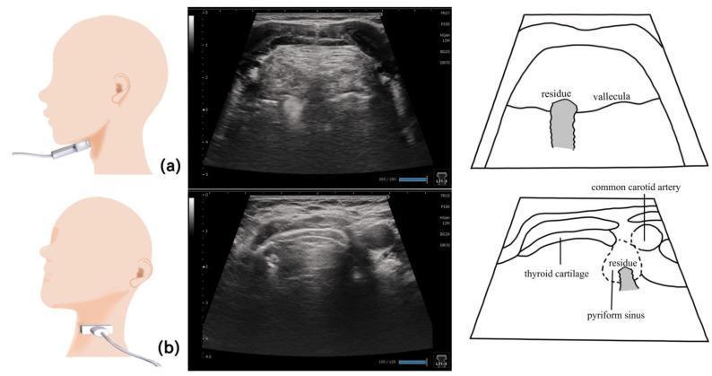
This composite educational image illustrates the use of ultrasonography for evaluating pharyngeal residue in patients with dysphagia. It is divided into two sections, (a) and (b), each featuring a probe placement diagram, a transverse ultrasound scan, and a corresponding labeled schematic. Row (a) demonstrates the visualization of the epiglottic vallecula. The probe is positioned submentally at the level of the hyoid bone. The ultrasound reveals a distinct hyperechoic area within the vallecular space, indicating food or liquid residue. Row (b) illustrates the assessment of the pyriform sinus. The probe is placed lower on the lateral neck at the level of the laryngeal prominence. Key anatomical landmarks include the thyroid cartilage and the common carotid artery, which serve as orientation points for the pyriform sinus. In this view, a smaller hyperechoic residue is visible within the sinus. This diagnostic imaging modality provides a non-invasive method for detecting post-swallow residue, aiding in the clinical management of swallowing disorders.
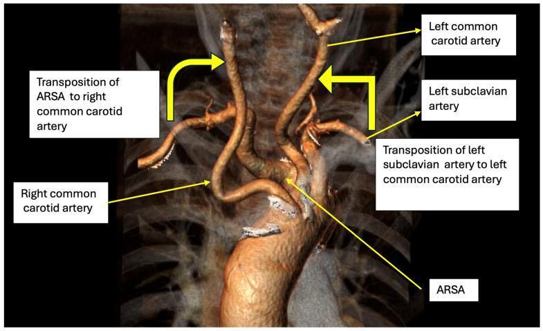
This diagnostic image is a 3D CT angiography reconstruction of the aortic arch and supra-aortic vessels, illustrating a treatment plan for Dysphagia Lusoria. The primary pathology shown is an Aberrant Right Subclavian Artery (ARSA), which originates as the last branch of the aortic arch and takes a retroesophageal course. The diagram annotates a surgical strategy for bilateral carotid-to-subclavian transposition. Key anatomical labels include the Right Common Carotid Artery and Left Common Carotid Artery, with yellow arrows indicating the proposed transposition of both the ARSA and the Left Subclavian Artery to their respective ipsilateral common carotid arteries. This vascular reconstruction highlights the relationship between the aberrant vessel and the esophageal compression site. The image serves as a clinical algorithm for hybrid surgical management, focusing on the debranching and revascularization of the upper extremities to relieve symptomatic compression while maintaining distal perfusion. Target audience includes vascular surgeons and radiologists studying congenital aortic arch anomalies.
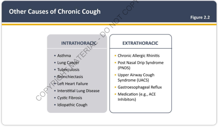
Summary : This figure presents a categorized list of other causes of chronic cough, divided into intrathoracic and extrathoracic origins, as shown in Figure 2.2. list diagram: # Categories : ## Intrathoracic : • Asthma • Lung Cancer • Tuberculosis • Bronchiectasis • Left Heart Failure • Interstitial Lung Disease • Cystic Fibrosis • Idiopathic Cough ## Extrathoracic : • Chronic Allergic Rhinitis • Post Nasal Drip Syndrome (PNDS) • Upper Airway Cough Syndrome (UACS) • Gastroesophageal Reflux • Medication (e.g., ACE Inhibitors) # Layout : • Two adjacent boxes, each with a heading and bulleted list. • Left box: "INTRATHORACIC" causes. • Right box: "EXTRATHORACIC" causes. # Figure Label : • Figure 2.2 in upper right corner. # Analysis : • The figure clearly distinguishes between causes of chronic cough originating within the thorax (primarily pulmonary and cardiac conditions) and those outside the thorax (mainly upper airway, gastrointestinal, and medication-related causes). • Intrathoracic causes are more disease-specific, while extrathoracic causes include syndromes and external factors. • This classification aids in systematic differential diagnosis for chronic cough.
dysphagia flowchart oropharyngeal esophageal causes
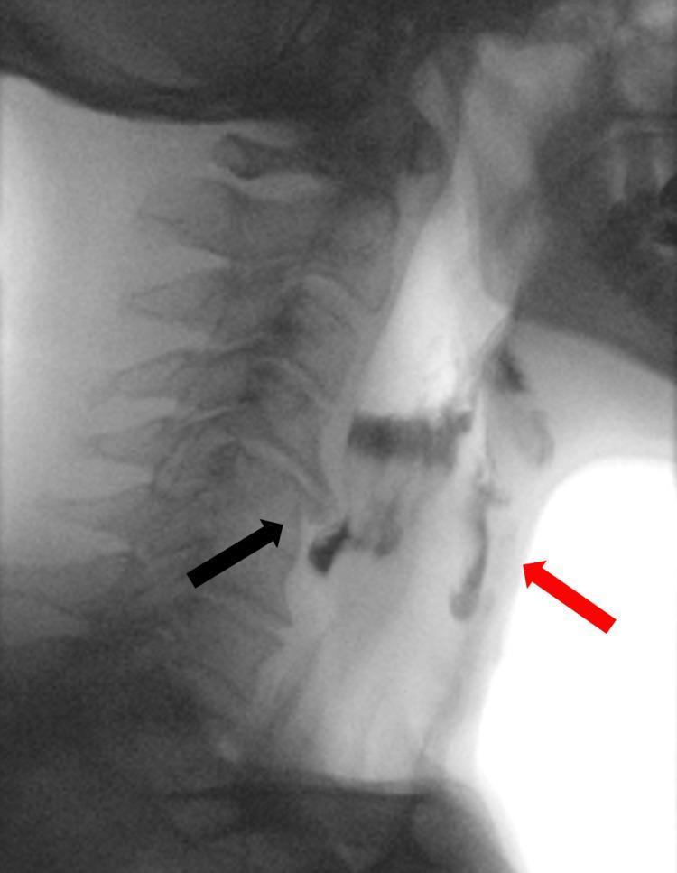
A lateral view videofluoroscopic swallowing study (VFSS) image demonstrating oropharyngeal dysphagia. The image captures the cervical spine and the pharyngeal/esophageal soft tissue structures during the transit of a radiopaque contrast bolus. A prominent anterior osteophyte at the C5 vertebral body level (indicated by a black arrow) causes focal extrinsic compression and narrowing of the proximal cervical esophagus. This mechanical obstruction leads to the stasis and pooling of the contrast material above the upper esophageal sphincter. Significantly, contrast is seen misdirected into the laryngeal vestibule and trachea (indicated by a red arrow), confirming laryngeal penetration and tracheal aspiration. This diagnostic image illustrates the clinical relationship between cervical spondylosis (DISH or severe osteophytosis) and mechanical dysphagia, highlighting the risk of respiratory complications due to impaired bolus clearance and subsequent airway entry.
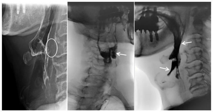
This diagnostic image consists of three lateral-view cervical esophagrams (fluoroscopic barium swallow study) demonstrating oropharyngeal dysphagia and mechanical obstruction. The first panel highlights prevertebral soft tissue swelling at the C3–C4 level (white circle), which causes extrinsic compression and anterior displacement of the esophagus. The subsequent panels illustrate functional swallowing impairment: radiopaque contrast material is seen abnormally pooling within the laryngeal vestibule and epiglottic space. White arrows indicate laryngeal penetration and aspiration, where contrast moves anteriorly into the airway rather than proceeding through the upper esophageal sphincter. These findings are clinically significant for diagnosing mechanical dysphagia, in this case secondary to an anteriorly extruded cervical disc. Key educational concepts include the radiographic identification of aspiration, the assessment of prevertebral soft tissue thickness, and the recognition of extrinsic esophageal compression in the context of cervical spine pathology.
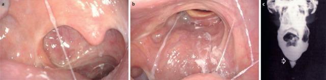
This composite educational clinical figure illustrates oropharyngeal and esophageal pathologies contributing to dysphagia. Image (a) is a transoral endoscopic view of an 87-year-old patient showcasing post-surgical changes following an extended partial resection of the right oropharynx and neck dissection. Visible features include a disrupted pharyngeal wall structure on the right and distorted mucosa consistent with radiotherapy and chemotherapy effects. Image (b) presents a transnasal endoscopic view of the velum and posterior oropharyngeal wall. It demonstrates significant scarring and asymmetrical contraction of the right-sided velum, with visible scar tissue extending across the posterior pharyngeal wall. Prominent white, viscous saliva is seen spanning the lumen, indicating impaired clearance and a transportation disorder. Image (c) is a contrast radiography (barium swallow) from a different patient, identifying tissue formations (indicated by an arrow) in the pharynx or esophagus that create mechanical obstacles. Together, these images teach how oncological treatments, scarring, and structural abnormalities disrupt the swallowing mechanism.
Causes of Dysphagia
I. OROPHARYNGEAL (TRANSFER) DYSPHAGIA
A. Neurogenic (Motility) Causes
| Condition | Mechanism |
|---|---|
| Cerebrovascular accident (stroke) | Most common cause; disrupts cortical and brainstem swallowing centers |
| Parkinson disease | Impaired oral and pharyngeal muscle coordination; delayed swallow reflex |
| Amyotrophic lateral sclerosis (ALS) | Progressive LMN + UMN degeneration affecting bulbar muscles |
| Multiple sclerosis | Demyelination of corticobulbar tracts |
| Brainstem tumors | Direct compression of swallowing centers in medulla/pons |
| Peripheral neuropathies | Cranial nerve IX, X, XII involvement |
| Cerebral palsy | Oral muscular incoordination; failure to trigger pharyngeal phase |
| Myasthenia gravis | Fatigable weakness of pharyngeal muscles (neuromuscular junction disorder) |
B. Myogenic (Muscle) Causes
| Condition | Mechanism |
|---|---|
| Polymyositis / Dermatomyositis | Inflammatory myopathy affecting striated pharyngeal muscles; dysphagia in up to 1/3 of cases |
| Myotonic dystrophy | Pharyngeal and esophageal smooth muscle dysfunction |
| Oculopharyngeal dystrophy | Progressive ptosis + pharyngeal muscle weakness (late-onset autosomal dominant) |
| Muscular dystrophies | Variable pharyngeal involvement |
C. Structural / Mechanical Causes
| Cause | Notes |
|---|---|
| Zenker diverticulum | Pulsion diverticulum at Killian's dehiscence; presents with regurgitation of undigested food + gurgling sound |
| Cricopharyngeal bar / achalasia of UES | Failure of UES to relax, causing obstruction |
| Carcinoma of pharynx / hypopharynx | Progressive solid food dysphagia; often with voice change |
| Cervical osteophytes (DISH) | Anterior osteophytes cause extrinsic compression; visible on videofluoroscopy |
| Retropharyngeal abscess | Infection causing posterior pharyngeal wall swelling |
| Thyromegaly / goiter | Extrinsic compression of hypopharynx or upper esophagus |
| Proximal esophageal web | Plummer-Vinson/Paterson-Brown-Kelly syndrome (iron deficiency + postcricoid web in middle-aged women) |
| Foreign body | Especially in children and elderly |
D. Congenital Causes
| Condition | Notes |
|---|---|
| Cleft lip and palate | Inadequate oral seal; nasal regurgitation during feeding |
| Laryngeal clefts | Communication between larynx and hypopharynx |
| Tracheoesophageal fistula | Direct communication; presents at birth |
| Esophageal atresia | Total obstruction; presents immediately after birth |
| Vascular rings | Aberrant subclavian artery, double aortic arch, anomalous left pulmonary artery compressing esophagus |
| Congenital vocal cord palsy | Aspiration during feeding in neonates |
E. Miscellaneous Oropharyngeal Causes
- Decreased saliva - from Sjogren syndrome, radiation therapy, anticholinergic medications (dry mouth impairs bolus formation)
- Poor dentition / ill-fitting dentures - disrupts mastication, oversized poorly chewed bolus
- Infective - Ludwig's angina, peritonsillar abscess, epiglottitis, deep neck space infections
II. ESOPHAGEAL DYSPHAGIA
A. Mechanical Obstruction
Intrinsic Lesions
| Cause | Clinical Clue |
|---|---|
| Esophageal carcinoma (adenocarcinoma / SCC) | Progressive dysphagia for solids → liquids; weight loss; risk factors: GERD, Barrett's (adenocarcinoma), alcohol/tobacco (SCC) |
| Peptic stricture (GERD-induced) | Long history of heartburn; distal esophagus; progressive |
| Eosinophilic esophagitis (EoE) | Now the most common cause of dysphagia in young adults/adolescents; recurrent food impaction; linear furrows + corrugated rings on endoscopy |
| Schatzki ring (lower esophageal ring) | Episodic dysphagia for solids, typically at the beginning of a meal; usually at GEJ |
| Esophageal webs | Upper/mid esophagus; associated with Plummer-Vinson syndrome |
| Medication-induced stricture | NSAIDs, doxycycline, bisphosphonates, potassium chloride |
| Diverticula (mid-esophageal, epiphrenic) | Traction or pulsion; food pouching |
| Benign tumors (leiomyoma) | Smooth, intramural; progressive solid dysphagia |
| Foreign body impaction | Acute sudden-onset dysphagia |
| Radiation-induced stricture | History of mediastinal/neck irradiation |
Extrinsic Compression
| Cause | Notes |
|---|---|
| Mediastinal mass / lymphadenopathy | Lymphoma, metastatic nodes, sarcoidosis |
| Vascular compression - Dysphagia lusoria | Aberrant right subclavian artery (most common), aortic aneurysm; compression of posterior esophagus |
| Spinal osteophytes | Anterior cervical spondylosis causing mechanical compression |
| Lung cancer / mediastinal tumors | Extrinsic encasement of esophagus |
B. Motility (Neuromuscular) Disorders
| Disorder | Mechanism / Features |
|---|---|
| Achalasia | Failure of LES relaxation + absent peristalsis due to loss of myenteric neurons. Dysphagia for both solids + liquids, nocturnal regurgitation of undigested food, weight loss. Barium shows "bird-beak" narrowing |
| Distal esophageal spasm (DES) | Simultaneous (non-peristaltic) contractions of the distal esophagus; intermittent dysphagia + chest pain; "corkscrew esophagus" on barium |
| Jackhammer (hypercontractile) esophagus | Very high amplitude peristaltic contractions (>8000 mmHg on HRM); chest pain + dysphagia |
| Nutcracker (high-pressure) esophagus | High-amplitude peristalsis; chest pain is predominant |
| Hypertensive LES | Increased LES pressure with intact peristalsis |
| Scleroderma (systemic sclerosis) | Smooth muscle replaced by collagen in lower 2/3 of esophagus; aperistalsis of lower esophagus + incompetent LES → severe GERD → peptic stricture + Barrett's; Raynaud's phenomenon is a clue |
| Chagas disease | Trypanosoma cruzi destroys myenteric ganglia; produces secondary achalasia (endemic in Latin America) |
| CREST syndrome | Calcinosis, Raynaud's, Esophageal dysmotility, Sclerodactyly, Telangiectasia |
| Severe ineffective esophageal motility (IEM) | Low-amplitude peristalsis; commonly associated with GERD and elderly patients |
C. Inflammatory Causes
| Cause | Notes |
|---|---|
| GERD-related esophagitis | Erosions, ulcerations, stricture in lower esophagus |
| Eosinophilic esophagitis | Immune/food allergen-driven; eosinophilic infiltration of mucosa |
| Infectious esophagitis | Candida albicans (most common; immunocompromised), HSV, CMV, HIV-associated; odynophagia is more prominent than dysphagia |
| Pill esophagitis | Doxycycline, NSAIDs, bisphosphonates, slow-release KCl |
| Radiation esophagitis | Acute inflammation or chronic stricture formation |
D. Systemic/Autoimmune Causes Affecting Esophagus
| Condition | Mechanism |
|---|---|
| Scleroderma | Fibrosis of smooth muscle; lower esophageal aperistalsis |
| Systemic lupus erythematosus (SLE) | Mild dysphagia; oral mucosal ulcers |
| Dermatomyositis | Striated muscle inflammation; associated with underlying malignancy |
| Mixed connective tissue disease | Combined features of SLE + scleroderma + polymyositis |
| Sjögren syndrome | Xerostomia (dry mouth) → impaired bolus formation |
III. FUNCTIONAL DYSPHAGIA
- Visceral hyperalgesia
- Abnormal sensory perception (even normal bolus transit triggers dysphagia sensation)
- Central sensitization
- Psychiatric/anxiety disorders amplifying symptoms
Summary: Key Diagnostic Clues
| Clue | Likely Cause |
|---|---|
| Difficulty initiating swallow, coughing/choking | Oropharyngeal (neurological) |
| Solids only, progressive | Mechanical obstruction (stricture, carcinoma) |
| Solids only, intermittent | Schatzki ring, EoE |
| Solids + liquids, intermittent | Motility disorder (DES, jackhammer) |
| Solids + liquids, progressive + weight loss | Achalasia or malignancy |
| Heartburn history + dysphagia | Peptic stricture or scleroderma |
| Young adult + food impaction | Eosinophilic esophagitis |
| Gurgling after eating | Zenker diverticulum |
| Raynaud's + dysphagia | Scleroderma / CREST |
| Iron-deficiency anaemia + postcricoid dysphagia in middle-aged woman | Plummer-Vinson / Paterson-Brown-Kelly |
- Sleisenger & Fordtran's GI and Liver Disease, Chapter 13 & 30 - Oropharyngeal and Esophageal Dysphagia
- Yamada's Textbook of Gastroenterology, 7th ed., Box 30.1 and 30.2
- Harrison's Principles of Internal Medicine, 22nd ed., Chapter 333
- Cummings Otolaryngology Head & Neck Surgery, Chapter 68
- Scott-Brown's Otorhinolaryngology Head & Neck Surgery, Chapter on Dysphagia and Aspiration
What si extramural and intramural causes
esophageal leiomyoma intramural tumor endoscopy
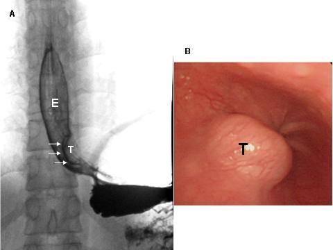
This composite medical image displays a case of esophageal leiomyoma through two modalities: diagnostic radiology and endoscopy. Panel A is a contrast esophagogram (barium swallow) showing an intramural tumor (T) in the distal esophagus (E) near the gastroesophageal junction. The lesion presents as a smooth, well-defined filling defect with sharp angles at its superior and inferior borders, characteristic of a benign intramural mass. Panel B is an intraluminal endoscopic photograph of the same tumor (T). It reveals a submucosal, rounded, protruding mass with an intact, smooth overlying mucosal surface, which is typical for a leiomyoma. The image illustrates key diagnostic features used to differentiate benign mesenchymal esophageal tumors from malignant mucosal lesions. These findings are clinically significant for planning surgical enucleation, often performed via video-assisted thoracoscopic surgery (VATS).
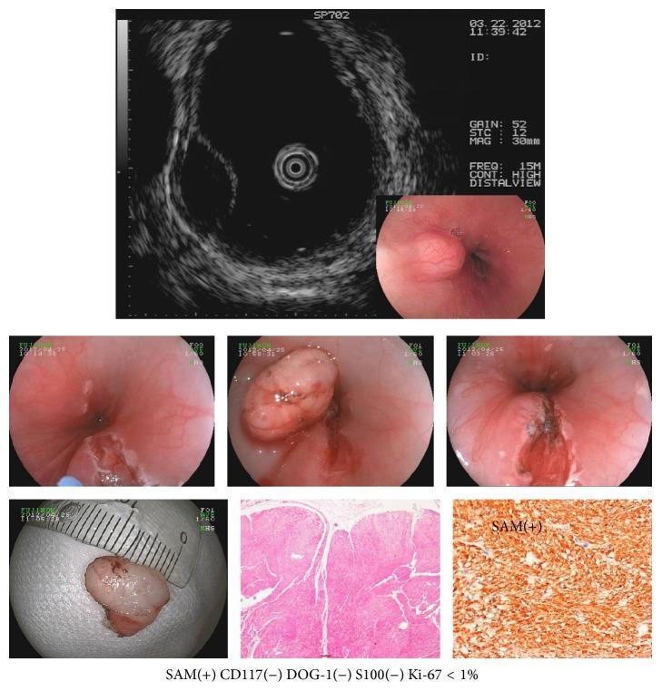
Educational composite image documenting an esophageal leiomyoma through multiple modalities. The top row features an Endoscopic Ultrasonography (EUS) scan showing a hypoechoic, well-circumscribed mass originating from the muscularis propria, alongside an initial endoscopic view of a subepithelial bulge. The middle row presents white-light endoscopy images of a raised, smooth-surfaced, hemispherical subepithelial tumor (SET) protruding into the esophageal lumen, showing intact overlying mucosa and normal vascularity. The bottom row displays a gross specimen measured against a centimeter scale, followed by histopathological analysis. The Hematoxylin and Eosin (H&E) stain shows fascicles of spindle cells with eosinophilic cytoplasm and minimal atypia. A corresponding immunohistochemistry (IHC) slide labeled 'SAM(+)' shows strong diffuse positive staining for smooth muscle actin (SMA), while the accompanying text notes negative results for CD117, DOG-1, and S100, and a low Ki-67 proliferation index (<1%), confirming the diagnosis of a benign mesenchymal tumor of myogenic origin.
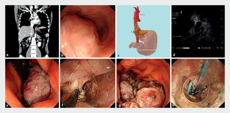
This composite figure illustrates the multi-modal diagnostic and surgical management of an esophageal leiomyoma at the esophagogastric junction. (a) Coronal CT scan shows a low-density mass (red arrow) near the mediastinal-abdominal transition. (b) White-light endoscopy reveals a large submucosal bulge with smooth, intact overlying mucosa. (c) A 3D anatomical reconstruction maps the 4.3 cm x 6.2 cm tumor's relationship to the aorta and stomach. (d) Endoscopic ultrasound (EUS) characterizes the lesion as a hypoechoic mass originating from the muscularis propria. (e-f) Intraoperative endoscopic images demonstrate the resection process: the tumor is detached from the muscular layers using a HookKnife and snare-assisted traction. (g) The excised specimen is shown segmented within the gastric lumen prior to extraction. (h) The final panel shows the closure of the mucosal defect using the kissing suture technique with endoscopic clips and sutures. This series highlights the progression from diagnostic imaging and EUS localization to complete endoscopic resection of a gastrointestinal stromal-type tumor.
Extramural vs. Intramural Causes of Dysphagia
INTRAMURAL CAUSES
Pathology within the esophageal wall itself - involving the mucosa, submucosa, or muscularis propria. The lesion narrows the lumen from inside.
1. Benign Intramural Tumors
| Tumor | Details |
|---|---|
| Leiomyoma | Most common benign esophageal tumor (~60-65% of benign tumors). Arises from smooth muscle of the muscularis propria (80% intramural). Peaks at age 30-59; men > women. Located in middle and lower esophagus. Slow-growing; over 50% are asymptomatic. Barium shows smooth filling defect with acute angle at wall junction. Endoscopy: smooth normal mucosa overlying a mobile mass. Biopsy often fails due to subepithelial location |
| GIST (Gastrointestinal Stromal Tumor) | Arises from interstitial cells of Cajal; submucosal |
| Granular cell tumor | Submucosal, pale yellow nodule; usually mid-esophagus |
| Fibrovascular polyp | Large pedunculated intraluminal polyp; upper esophagus; can cause regurgitation of the mass |
| Lipoma | Submucosal; compressible on endoscopy |
| Hemangioma / lymphangioma | Rare vascular malformations within the wall |
| Neurofibroma / schwannoma | Rare; neural sheath tumors of the wall |
| Duplication cyst | Congenital; submucosal cyst in the esophageal wall |
| Varices | Submucosal dilated veins (portal hypertension); can cause dysphagia when large |
2. Malignant Intramural Tumors
| Tumor | Details |
|---|---|
| Esophageal carcinoma (squamous cell or adenocarcinoma) | Mucosa is abnormal (ulcerated, irregular, friable) - distinguishes from benign intramural; progressive dysphagia solids → liquids; weight loss |
| Metastatic tumor to esophageal wall | Rare; hematogenous spread from lung, breast, melanoma |
| Leiomyosarcoma | Malignant smooth muscle tumor; rapid onset of dysphagia; mucosal ulceration is a warning sign |
3. Inflammatory/Fibrotic Intramural Causes
| Cause | Mechanism |
|---|---|
| Peptic stricture | GERD → chronic mucosal injury → submucosal fibrosis → lumen narrowing in distal esophagus |
| Eosinophilic esophagitis | Eosinophilic infiltration of mucosa and submucosa → stiffness, rings, furrows, stricture |
| Schatzki ring | Thin fibrotic mucosal ring at the gastroesophageal junction |
| Radiation-induced stricture | Transmural fibrosis of the esophageal wall after radiotherapy |
| Pill-induced stricture | NSAIDs, doxycycline, bisphosphonates → mucosal ulceration → stricture formation |
| Caustic/corrosive injury | Acid or alkali ingestion → transmural necrosis → circumferential stricture |
| Anastomotic stricture | Post-esophagectomy; fibrosis at surgical anastomosis |
| Esophageal webs | Thin mucosal shelf (not full thickness); upper esophagus; Plummer-Vinson syndrome |
| Diverticula | Zenker (pharyngeal), mid-esophageal traction, epiphrenic pulsion; food accumulates in pouch |
EXTRAMURAL CAUSES
Pathology outside the esophageal wall compressing it from the outside. The esophageal mucosa and wall are intrinsically normal but get narrowed by external pressure.
1. Vascular Causes
| Cause | Details |
|---|---|
| Dysphagia lusoria | Aberrant right subclavian artery (most common) courses behind the esophagus, compressing its posterior wall; presents with dysphagia for solids; diagnosed on CT angiography |
| Double aortic arch | Congenital vascular ring encircling both esophagus and trachea; presents in infancy |
| Thoracic aortic aneurysm | Enlarged aorta compresses mid/lower esophagus; pulsatile compression on barium |
| Anomalous left pulmonary artery (pulmonary sling) | Compresses esophagus from in front (anterior compression - rare) |
| Enlarged left atrium | In severe mitral stenosis, the enlarged left atrium can compress the mid-esophagus |
2. Mediastinal Masses / Lymphadenopathy
| Cause | Details |
|---|---|
| Lymphoma | Mediastinal lymphadenopathy from Hodgkin's or NHL; encases and compresses the esophagus |
| Metastatic lymphadenopathy | Lung cancer, breast cancer, esophageal carcinoma spread to mediastinal nodes |
| Sarcoidosis | Hilar/mediastinal adenopathy compressing esophagus |
| Lung carcinoma | Direct invasion or extrinsic compression from a mediastinal lung mass |
| Thymoma / mediastinal germ cell tumor | Anterior mediastinal masses compressing esophagus |
| Mediastinal fibrosis | Fibrosing mediastinitis (e.g., histoplasmosis) encasing the esophagus |
3. Spinal / Bony Causes
| Cause | Details |
|---|---|
| Cervical osteophytes (DISH / spondylosis) | Anterior osteophytes at C3-C6 compress the posterior pharynx and upper esophagus; visible on videofluoroscopy; more common in elderly |
| Spinal tumors | Vertebral metastases extending anteriorly |
4. Thyroid and Neck Causes
| Cause | Details |
|---|---|
| Goiter / thyromegaly | Enlarged thyroid gland compresses the cervical esophagus from the front or sides |
| Retrosternal goiter | Extends into superior mediastinum; compresses both trachea and esophagus |
| Thyroid malignancy | Direct invasion or compression |
| Parathyroid/cervical masses | Rare causes of extrinsic cervical esophageal compression |
5. Subdiaphragmatic / Adjacent Organ Causes
| Cause | Details |
|---|---|
| Hiatal hernia | Large paraesophageal hernia can cause mechanical distortion |
| Hepatomegaly / splenomegaly | Rare; can distort the gastroesophageal junction |
| Pancreatic pseudocyst / tumor | Compresses the lower esophagus from below |
| Inflammatory masses | Perigastric or periesophageal abscess, pancreatitis |
Summary Comparison
| Feature | Intramural | Extramural |
|---|---|---|
| Origin | Within esophageal wall (mucosa/submucosa/muscularis) | Outside the esophageal wall |
| Mucosa on endoscopy | May be normal (benign) or ulcerated/friable (malignant) | Normal - smooth bulge from outside |
| Best diagnostic tool | Upper endoscopy + EUS | Barium esophagram / CT scan |
| Biopsy possible? | Sometimes (mucosa intact in benign tumors = usually fails) | No - mucosa is normal |
| Amenable to dilation? | Yes (strictures, rings) | Usually not - source is external |
| Examples | Leiomyoma, peptic stricture, carcinoma, EoE | Vascular rings, lymphoma, goiter, osteophytes |
List out most common causes only
Most Common Causes of Dysphagia
Oropharyngeal Dysphagia
| # | Cause |
|---|---|
| 1 | Stroke (CVA) - single most common cause of oropharyngeal dysphagia |
| 2 | Parkinson disease |
| 3 | Zenker diverticulum |
| 4 | Pharyngeal/laryngeal carcinoma |
| 5 | Cricopharyngeal bar / UES dysfunction |
| 6 | Myasthenia gravis |
| 7 | Cervical osteophytes (elderly) |
Neuromuscular disease accounts for ~80% of oropharyngeal dysphagia cases. - Rosen's Emergency Medicine
Esophageal Dysphagia - Intramural
| # | Cause | Key Clue |
|---|---|---|
| 1 | Eosinophilic esophagitis (EoE) | Most common in young adults/adolescents; recurrent food impaction |
| 2 | Esophageal carcinoma (adeno or SCC) | Progressive solids → liquids; weight loss |
| 3 | Peptic stricture (GERD-induced) | Long heartburn history; distal esophagus |
| 4 | Schatzki ring | Episodic, solids only, start of meal |
| 5 | Leiomyoma | Smooth submucosal mass; normal overlying mucosa |
| 6 | Achalasia | Solids + liquids; nocturnal regurgitation |
| 7 | Distal esophageal spasm | Intermittent; chest pain + dysphagia |
| 8 | Scleroderma | Raynaud's + heartburn + aperistalsis |
Esophageal Dysphagia - Extramural
| # | Cause | Key Clue |
|---|---|---|
| 1 | Mediastinal lymphadenopathy (lymphoma, lung Ca mets) | Most common extramural cause |
| 2 | Thyroid goiter / retrosternal goiter | Neck swelling; compresses cervical esophagus |
| 3 | Dysphagia lusoria (aberrant right subclavian artery) | Most common vascular cause |
| 4 | Thoracic aortic aneurysm | Pulsatile compression on barium swallow |
| 5 | Lung carcinoma | Mediastinal invasion / nodal compression |
Quick Memory Aid
Oropharyngeal → Think NEUROLOGICAL (stroke, Parkinson's, MG)
Intramural     → Think EoE (young) / Ca (old) / Achalasia (motility)
Extramural     → Think LYMPH NODES / GOITER / VESSELS