Explain me classification of digestive system
Reading File
Finding Sources
Finding Sources
Reading File
Finding Sources
Reading File
Reading File
Reading File
Finding Sources
Reading File
Reading File
Loading Image
Loading Image
Now I have rich information from multiple authoritative textbooks. Here is a comprehensive classification of the digestive system:
Classification of the Digestive System
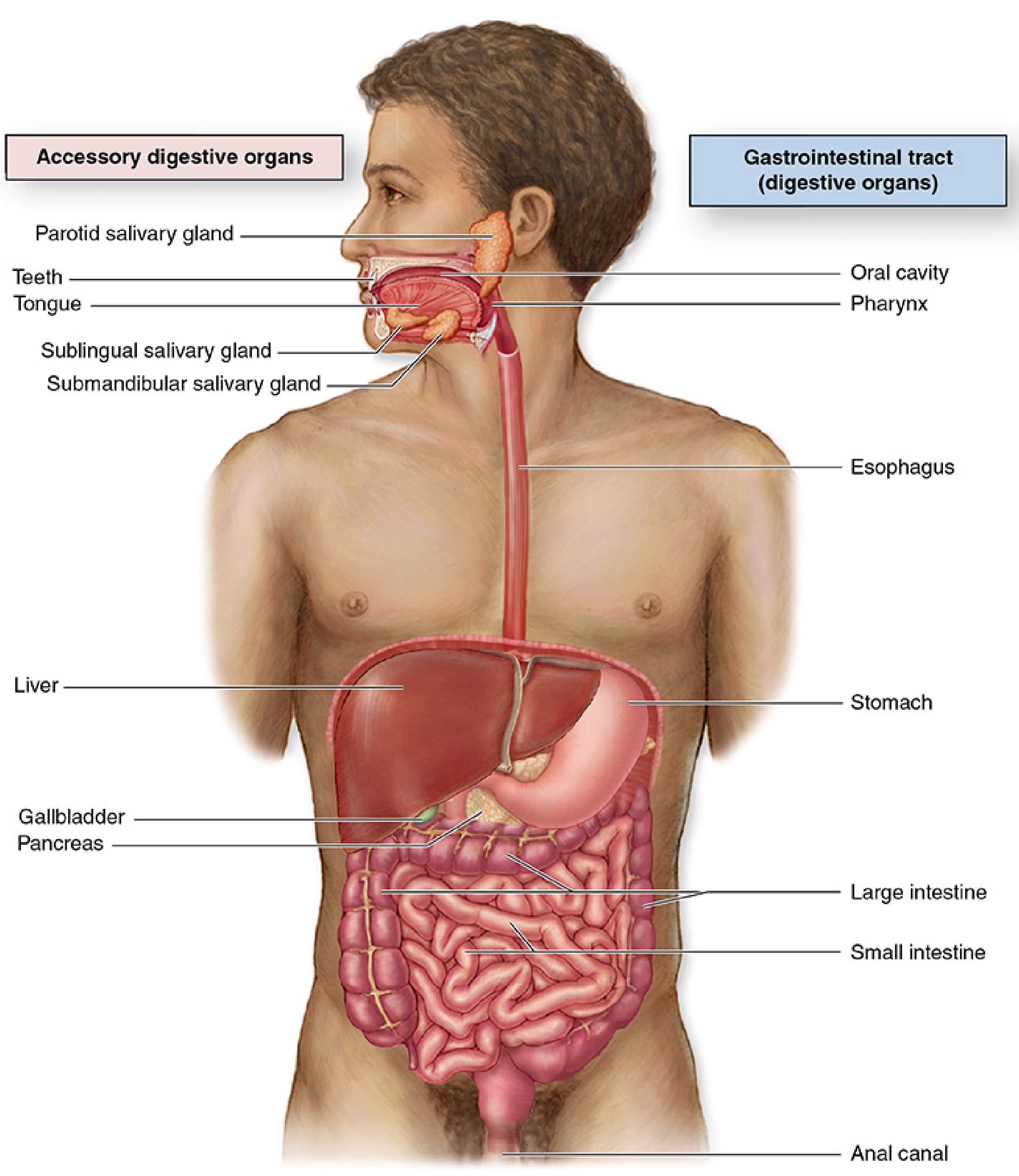
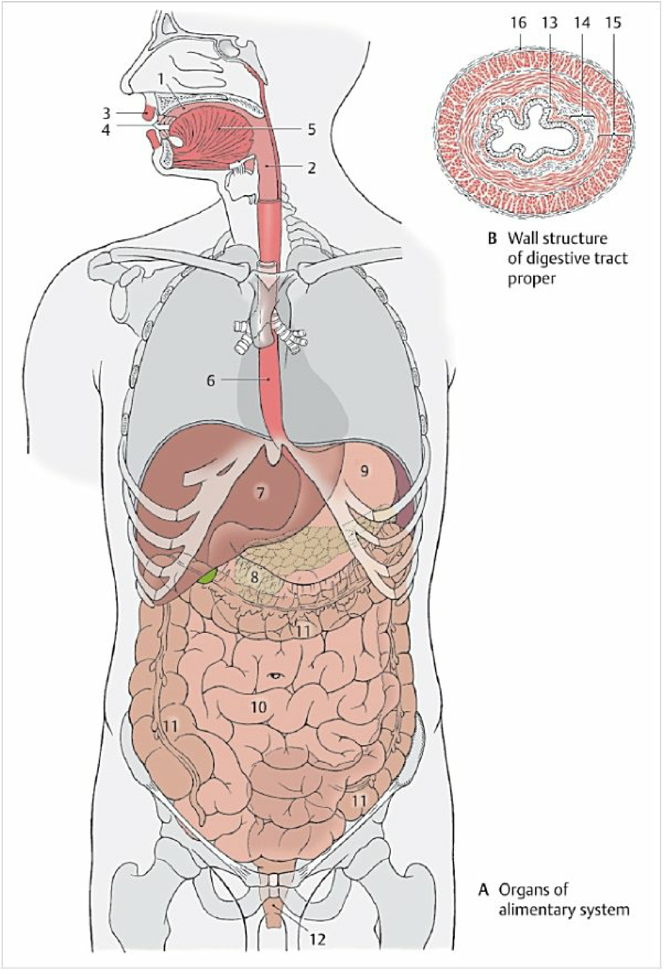
The digestive system consists of the alimentary canal (GI tract) and its accessory organs. Its major functions include transport of ingested food and water, secretion of digestive fluids, absorption of nutrients, and excretion of indigestible material.
Part 1: Gastrointestinal (GI) Tract / Alimentary Canal
A continuous muscular tube running from mouth to anus (~9 meters long), divided into segments each morphologically specialized for specific digestive functions.
1.1 Upper GI Tract
| Organ | Key Function |
|---|---|
| Oral cavity (mouth) | Ingestion, mastication, bolus formation; lined by stratified squamous epithelium |
| Pharynx | Rapid passage of food bolus from oral cavity to esophagus |
| Esophagus | Peristaltic transport of bolus to stomach |
| Stomach | Enzymatic and acid digestion; begins protein breakdown via pepsin + HCl |
1.2 Small Intestine (~6–7 m)
| Segment | Key Feature |
|---|---|
| Duodenum | Receives bile and pancreatic juice; major site of chemical digestion |
| Jejunum | Principal site of nutrient absorption (large surface area via villi/microvilli) |
| Ileum | Absorbs vitamin B₁₂, bile salts; contains Peyer's patches (immune function) |
1.3 Large Intestine (~1.5 m)
| Segment | Key Feature |
|---|---|
| Cecum + appendix | Receives ileal contents via ileocecal valve |
| Ascending colon | Absorbs water and electrolytes |
| Transverse colon | Continues absorption; fermentation by gut bacteria |
| Descending colon | Moves feces distally |
| Sigmoid colon | Stores feces before defecation |
| Rectum | Fecal storage |
| Anal canal | Final exit; internal (smooth) and external (skeletal) sphincters |
Part 2: Accessory Digestive Organs
These organs are not part of the canal itself but secrete substances essential for digestion. They empty into the GI tract via ducts.
2.1 Salivary Glands
- Parotid (largest) — serous secretion; parotid (Stensen) duct opens opposite upper 2nd molar
- Submandibular — mixed serous/mucous; Wharton's duct opens at sublingual caruncle
- Sublingual — mainly mucous; multiple short ducts
- Minor salivary glands — scattered in oral submucosa (buccal, labial, lingual, palatine)
2.2 Pancreas
- Exocrine function: secretes digestive enzymes (amylase, lipase, proteases) and bicarbonate via pancreatic duct into the duodenum
- Endocrine function: islets of Langerhans → insulin, glucagon (not digestive, but co-located)
2.3 Liver
- The largest internal organ and largest glandular mass in the body
- Acts as an exocrine organ: produces bile (stored in gallbladder), which emulsifies fats for lipid digestion
- Additional roles: metabolism of nutrients, plasma protein synthesis, detoxification, iron and vitamin storage
2.4 Gallbladder
- Stores and concentrates bile between meals
- Releases bile into duodenum via the bile duct in response to fat ingestion (cholecystokinin stimulus)
Part 3: Embryological Classification (Foregut / Midgut / Hindgut)
From a developmental perspective, the gut tube is divided into three regions based on the embryonic primitive gut:
| Region | Adult Derivatives | Blood Supply |
|---|---|---|
| Foregut | Pharynx, esophagus, stomach, proximal duodenum, liver, gallbladder, pancreas | Celiac artery |
| Midgut | Distal duodenum → right 2/3 of transverse colon | Superior mesenteric artery |
| Hindgut | Left 1/3 of transverse colon → upper anal canal | Inferior mesenteric artery |
Part 4: Wall Structure of the GI Tract
All segments of the digestive tract (esophagus to anus) share a common four-layer wall organization:
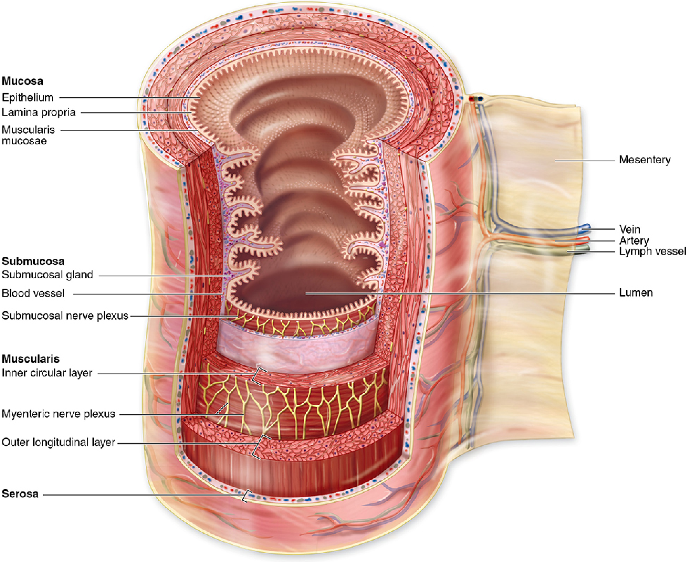
| Layer | Components |
|---|---|
| Mucosa | Epithelium + lamina propria + muscularis mucosae |
| Submucosa | Dense connective tissue; Meissner (submucosal) plexus |
| Muscularis | Inner circular + outer longitudinal smooth muscle; Auerbach (myenteric) plexus between them |
| Serosa / Adventitia | Peritoneal covering (serosa) where intraperitoneal; fibrous adventitia elsewhere |
Summary Classification at a Glance
DIGESTIVE SYSTEM
├── Alimentary Canal (GI Tract)
│   ├── Upper GI: Oral cavity → Pharynx → Esophagus → Stomach
│   ├── Small intestine: Duodenum → Jejunum → Ileum
│   └── Large intestine: Cecum → Colon → Rectum → Anal canal
└── Accessory Organs
    ├── Salivary glands (parotid, submandibular, sublingual)
    ├── Pancreas
    ├── Liver
    └── Gallbladder
Sources: Histology: A Text and Atlas, 8e (Pawlina/Ross); Color Atlas of Human Anatomy, Vol. 2 — Internal Organs; Junqueira's Basic Histology, 17e; Langman's Medical Embryology, 14e.
This is a shared conversation. Sign in to Orris to start your own chat.