Pulmonary function test. Prepare me for MD anesthesia final practicals. Pulmonary function test viva with pulmonary function test real graphs and interpretations and possible viva questions.

Reading File
Finding Sources
Searching Images

pulmonary function test spirometry flow volume loop normal obstructive restrictive

Searching the Web

pulmonary function test interpretation spirometry PFT viva MD anesthesia practical

Reading File
Finding Sources
Searching Images

spirometry flow volume curve obstructive COPD restrictive fibrosis

Reading File
Reading File
Reading File
Reading File
Reading File
Finding Sources
Reading File
Loading Image
Finding Sources
Loading Image
Reading File
Finding Sources
Loading Image
Loading Image
Finding Sources

Pulmonary Function Tests - MD Anesthesia Final Practical Viva Guide

Sources: Miller's Anesthesia 10e, Barash Clinical Anesthesia 9e, Fishman's Pulmonary Diseases, Murray & Nadel's Respiratory Medicine

SECTION 1 - LUNG VOLUMES AND CAPACITIES (The Foundation)

Start here - examiners almost always ask you to label this first.
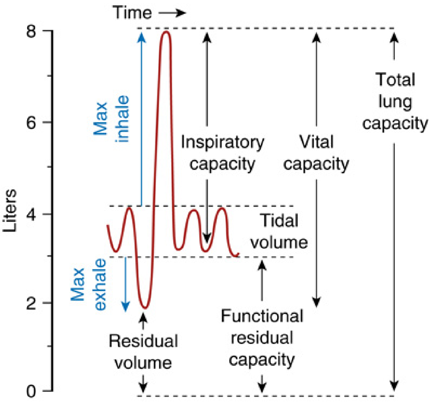
Lung Volumes Diagram - Miller's Anesthesia 10e

Key Definitions (rattle these off quickly)

Volume/CapacityNormal ValueDefinition
TV (Tidal Volume)~500 mLVolume per normal breath
IRV~3000 mLExtra volume above tidal inspiration
ERV~1200 mLExtra volume below tidal expiration
RV (Residual Volume)~1200 mLVolume remaining after maximal exhalation
IC (Inspiratory Capacity)TV + IRV = ~3500 mL
FRC (Functional Residual Capacity)ERV + RV = ~2400 mLVolume at end of normal expiration
VC (Vital Capacity)IRV + TV + ERV = ~4700 mL
TLC (Total Lung Capacity)~6000 mLAll volumes combined
Critical viva point: RV, FRC, and TLC cannot be measured by spirometry alone - you need nitrogen washout, helium dilution, or body plethysmography (Boyle's law).

SECTION 2 - SPIROMETRY GRAPHS

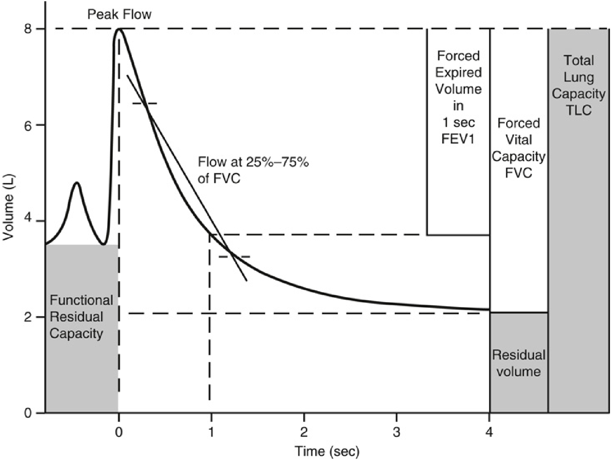
Graph 1: Volume-Time Curve (Spirogram)

Spirometry Volume-Time Graph - Miller's Anesthesia 10e
What you're reading: The patient inhales to TLC, then exhales forcefully. The graph plots volume exhaled (y-axis) against time (x-axis).
Key measurements from this curve:
  • FVC = total volume forcefully exhaled
  • FEV1 = volume exhaled in first 1 second
  • FEV1/FVC ratio = the single most important ratio in PFT interpretation
  • FEF25-75% = forced expiratory flow between 25-75% of FVC (sensitive marker for small-airway disease)
  • Peak Flow = highest flow rate achieved during forced expiration

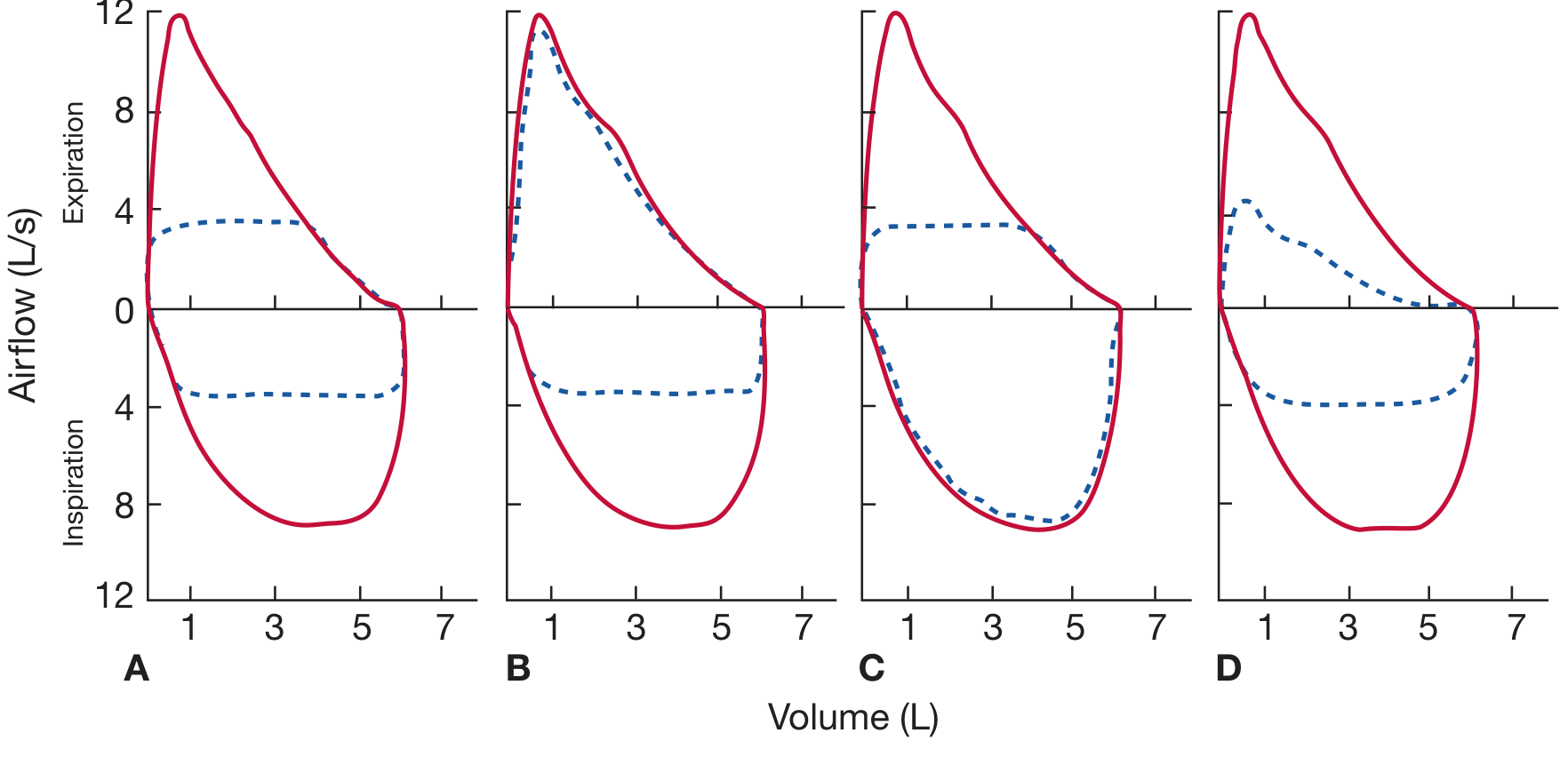
Graph 2: Flow-Volume Loops - Normal vs Pathological (Fishman's)

Flow-Volume Loops - Normal, Fixed, Variable Extrathoracic, Variable Intrathoracic, COPD - Fishman's Pulmonary Diseases
How to read a flow-volume loop:
  • Upper half (above zero) = expiration
  • Lower half (below zero) = inspiration
  • X-axis = lung volume (right = TLC, left = RV)
  • Red solid = normal; Blue dashed = pathological
PanelPatternInterpretation
ABoth limbs truncated/flattenedFixed upper airway obstruction (tracheal stenosis)
BInspiratory limb flattened, expiratory preservedVariable extrathoracic obstruction (vocal cord paralysis, goitre)
CExpiratory limb flattened, inspiratory preservedVariable intrathoracic obstruction (tracheomalacia, intrathoracic tracheal tumour)
DReduced loop size, scooped-out expiratory limbCOPD / small-airway obstruction

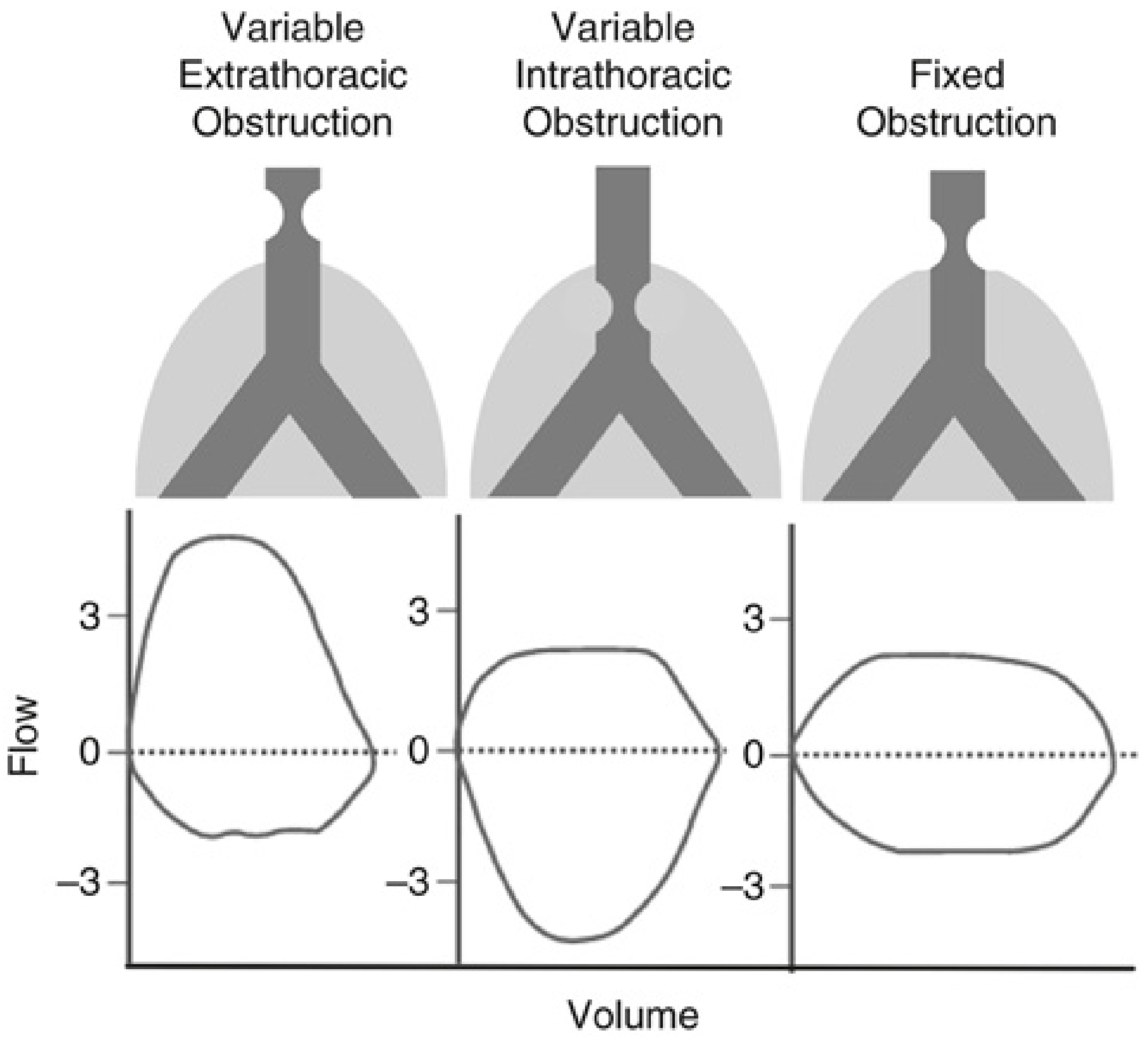
Graph 3: Upper Airway Obstruction - Fixed vs Variable (Miller's Anesthesia 10e)

Flow-Volume Loops - Variable Extrathoracic, Variable Intrathoracic, Fixed Obstruction - Miller's Anesthesia
The physiology behind the shapes:
  • Variable extrathoracic obstruction (e.g., vocal cord paresis): During forced inspiration, intratracheal pressure drops below atmospheric, causing the extrathoracic trachea to collapse inward - so the inspiratory limb is flattened. During expiration, tracheal pressure exceeds atmospheric, so obstruction lessens.
  • Variable intrathoracic obstruction (e.g., tracheomalacia): During forced expiration, pleural pressure exceeds intraluminal pressure, compressing the intrathoracic segment - so the expiratory limb is flattened. During inspiration, transmural pressure holds the airway open.
  • Fixed obstruction (e.g., tracheal scar): The geometry doesn't change during the respiratory cycle - both limbs are truncated equally (the classic "box-shaped" loop).

SECTION 3 - INTERPRETATION FRAMEWORK

Step-by-Step Approach (Say this in the viva)

Step 1: Is the FEV1/FVC ratio reduced? (< 0.70 or < LLN)
  • YES → Obstructive pattern
  • NO → go to Step 2
Step 2: Is FVC reduced?
  • YES, with normal FEV1/FVC → Restrictive pattern (confirm with TLC)
  • Both FVC and FEV1/FVC low → Mixed pattern
Step 3: Grade severity (using FEV1 % predicted for obstruction; TLC % predicted for restriction)

The Master Comparison Table (Barash Clinical Anesthesia 9e)

ParameterObstructive DiseaseRestrictive Disease
DefinitionSmall airway obstruction to expiratory flowProportional decrease in all lung volumes
FVCNormal or slightly ↑↓↓↓
FEV1Normal or slightly ↓↓↓↓
FEV1/FVC↓↓↓ (< 0.70)Normal
FEF25-75%↓↓↓Normal
FRCNormal or ↑ (gas trapping)↓↓↓
TLCNormal or ↑ (air trapping)↓↓↓
RV
- Barash Clinical Anesthesia 9e, Table 15-2

COPD GOLD Staging (must know for viva)

Post-bronchodilator FEV1/FVC < 0.70, then:
GOLD StageFEV1 % PredictedSeverity
I≥ 80%Mild
II50-79%Moderate
III30-49%Severe
IV< 30%Very severe

SECTION 4 - SPECIFIC PFT PATTERNS AND THEIR CAUSES

Obstructive Pattern Diseases

DiseaseFEV1FVCFEV1/FVCBronchodilatorDLCOLung Volumes
Chronic bronchitisNo responseNormal
EmphysemaNo response↓↓↑↑
Asthma↑↑ (reversible)Normal↑ (in attack)
Small-airway diseaseNormal/↓NormalNormal/↓VariableNormalNormal
- Fishman's Pulmonary Diseases, Table 31-19

Restrictive Pattern Diseases

Intrinsic (parenchymal): Pulmonary fibrosis, sarcoidosis, pneumoconiosis
  • FVC ↓, FEV1 ↓, FEV1/FVC normal or increased, TLC ↓, DLCO ↓
Extrinsic (extraparenchymal):
  • Chest wall: kyphoscoliosis, obesity, ankylosing spondylitis
  • Pleural: effusion, fibrosis
  • Neuromuscular: myasthenia gravis, Guillain-Barré, diaphragm palsy
  • In neuromuscular disease: FVC ↓, but DLCO is preserved (key distinguishing feature)

DLCO Patterns (a favourite viva topic)

ConditionDLCO
Emphysema↓↓ (loss of alveolar surface area)
Pulmonary fibrosis↓ (thickened membrane)
Pulmonary hypertension↓ (reduced capillary bed)
Pulmonary embolism
AsthmaNormal or ↑
ObesityNormal
Polycythemia / left-to-right shunt (increased Hb, more binding sites)
Anemia↓ (less Hb to bind CO)
Supine position
Normal DLCO: ~25 mL CO/min/mmHg (single-breath method) - Miller's Anesthesia 10e

SECTION 5 - ANESTHESIA-SPECIFIC APPLICATIONS

Pre-operative Risk Stratification (Most Important for Anesthesia Viva)

PFT ParameterThresholdClinical Significance
FEV1< 60% predictedIncreased risk of serious postoperative pulmonary complications
FEV1 for pneumonectomy> 2 L (or > 60%)Minimum to tolerate pneumonectomy
FEV1 for lobectomy> 1.5 LMinimum to tolerate lobectomy
DLCO< 60% predictedIncreased risk of postoperative pulmonary complications → needs CPET
VO2 max (CPET)< 15 mL/kg/minHigh risk for postoperative pulmonary complications
Anaerobic threshold< 10 mL/kg/minHigh risk for postoperative complications
VE/VCO2 ratio> 35More accurate predictor of postoperative risk
- Miller's Anesthesia 10e, pp. 1302-1307

Effect of Anesthesia on PFT Parameters

  • FRC decreases by 400-500 mL on induction (supine position + loss of respiratory muscle tone)
  • This FRC reduction causes airway closure and V/Q mismatch → hypoxemia
  • FRC reduction is greater in: obesity, elderly, pregnancy, pulmonary disease
  • SpO2 < 81% occurs in ~20% of patients for up to 5 minutes after induction
  • 50% of anesthesia-related deaths relate to hypoxemia

Preoperative Optimization Based on PFT

FindingAnesthetic Implication
FEV1/FVC < 0.70 (obstructive)Optimize bronchodilators, consider regional anesthesia
Reversible obstruction (asthma)Pre-treat with bronchodilator/steroids before surgery
Fixed obstruction (COPD)No benefit from bronchodilators in reversibility; manage air trapping
Restrictive patternAnticipate reduced FRC; careful fluid management; postop ventilation planning
FEV1 < 60%High risk; regional > general; minimize respiratory depression

SECTION 6 - POSSIBLE VIVA QUESTIONS & ANSWERS

Q1. What is FRC and why is it important for the anesthesiologist?

FRC is the volume of gas in the lungs at the end of normal passive expiration (ERV + RV, approximately 2400 mL). It is the oxygen reserve during apnea - a larger FRC means a longer safe apnea time. Anesthesia reduces FRC by ~400-500 mL due to loss of intercostal muscle tone and diaphragm cephalad shift. In obese patients this reduction is even greater, causing rapid desaturation on induction.

Q2. A patient with known COPD comes for elective laparotomy. How do you interpret his PFT and what anesthetic implications does it have?

  • FEV1/FVC < 0.70 post-bronchodilator confirms COPD
  • Severity graded by FEV1 % predicted (GOLD I-IV)
  • FEV1 < 60% = high risk for postoperative pulmonary complications
  • Air trapping: TLC ↑, RV ↑, FRC ↑
  • DLCO reduced in emphysema (not in pure chronic bronchitis)
  • Anesthetic implications: regional preferred, avoid prolonged positive pressure ventilation, risk of pneumothorax from high airway pressures, anticipate delayed extubation, postoperative respiratory failure risk

Q3. What is the difference between obstructive and restrictive PFT patterns?

Obstructive: FEV1/FVC < 0.70. FVC may be normal or slightly reduced. FEV1 is reduced. TLC and FRC are increased due to gas trapping. Causes: COPD, asthma, bronchiectasis.
Restrictive: FEV1/FVC is normal (often > 0.80). Both FEV1 and FVC are proportionally reduced. TLC is reduced (< 80% predicted). Causes: pulmonary fibrosis, obesity, kyphoscoliosis, neuromuscular disease.

Q4. How do you distinguish asthma from COPD on PFT?

Reversibility test: administer a short-acting bronchodilator (salbutamol 400 mcg inhaled). After 15 minutes, repeat spirometry.
  • Positive reversibility = FEV1 increase ≥ 200 mL AND ≥ 12% from baseline → suggests asthma
  • No significant reversibility → suggests COPD (fixed obstruction)
Additional points: DLCO is reduced in emphysema but normal or elevated in asthma. TLC is increased in both (gas trapping), but more severely in emphysema.

Q5. What does a flow-volume loop showing flattening of the inspiratory limb suggest?

Variable extrathoracic obstruction - e.g., bilateral vocal cord paralysis, subglottic stenosis, or extrinsic compression by a goitre. During forced inspiration, the segment above the thoracic inlet collapses because intraluminal pressure falls below atmospheric. This is an important pre-anesthetic finding - it predicts difficult intubation and potential need for awake fibreoptic intubation.

Q6. What is DLCO and what does it measure?

Diffusing capacity for carbon monoxide measures the functional alveolar-capillary surface area available for gas exchange. CO is used because its partial pressure in blood is near zero (so transfer is limited only by membrane, not flow) and it has 200x the affinity for Hb compared to O2.
DLCO is measured using the single-breath technique. Normal value: ~25 mL CO/min/mmHg.
Clinically: DLCO < 60% predicted indicates significant gas exchange impairment and warrants CPET before high-risk surgery.

Q7. What are the criteria for positive bronchodilator reversibility?

FEV1 increases by ≥ 200 mL AND ≥ 12% from the pre-bronchodilator value. Some newer evidence (ATS/ERS 2022) suggests using only absolute change or a different threshold, but the classic criterion remains the one expected in an exam setting.

Q8. What is FEF25-75% and what is its clinical significance?

Forced expiratory flow between 25% and 75% of FVC. It reflects flow through the middle and smaller airways. It is more sensitive than FEV1 for detecting early or mild small-airway disease. However, it has high variability and is effort-dependent, so it is not used alone for diagnosis. A marked reduction in FEF25-75% with normal FEV1/FVC suggests early obstructive disease.

Q9. How do you assess a patient for lung resection surgery?

Three-step assessment:
  1. Spirometry: FEV1 > 2L (or > 60%) for pneumonectomy; > 1.5L for lobectomy
  2. DLCO: Should be > 60% predicted
  3. CPET (if FEV1 or DLCO are borderline): VO2 max > 15 mL/kg/min = acceptable risk; < 10 mL/kg/min = prohibitive risk
Also calculate predicted postoperative (ppo) FEV1 = preoperative FEV1 × (1 - fraction of functional lung segments removed). A ppoFEV1 > 40% is generally acceptable.

Q10. What body plethysmography measures and why is it preferred over nitrogen washout?

Body plethysmography applies Boyle's law: the patient sits in an airtight chamber and breathes against a closed shutter. As the diaphragm contracts, thoracic volume increases and box pressure increases proportionately, allowing calculation of FRC. It is preferred because it measures all gas in the lungs including trapped gas (behind obstructed airways), whereas nitrogen washout and helium dilution only measure communicating lung volumes - so it gives a higher (more accurate) FRC/TLC in COPD.

Q11. What is the significance of a "scooped-out" expiratory limb on the flow-volume loop?

It is the characteristic shape in obstructive airways disease - the expiratory limb shows a rapid early peak flow followed by a concave scooped curve, reflecting dynamic airway collapse. The greater the scooping, the more severe the obstruction. In pure restriction, the loop is smaller but maintains a normal convex shape.

Q12. What are acceptability criteria for a valid spirometry result?

Per ATS standards:
  1. Rapid, abrupt start with high peak expiratory flow
  2. Duration ≥ 6 seconds in adults (or until plateau)
  3. No cough, extra breaths, or artifacts
  4. Maximum 8 attempts; best of at least 3 acceptable maneuvers; top 2 FVC values should be within 150 mL of each other

SECTION 7 - QUICK-FIRE MNEMONICS

For obstructive pattern: "ODEN" - Obstruction, Decreased FEV1/FVC, Emphysema/Asthma causes, Normal or elevated TLC
For what spirometry CANNOT measure: "FRAT" - FRC, RV, Absolute TLC need plethysmography
DLCO reduced in: emphysema, fibrosis, pulmonary HTN, pulmonary embolism, anemia DLCO normal/increased in: asthma, obesity, polycythemia, exercise, supine position
For lung resection: "2-1.5-60" - pneumonectomy needs FEV1 > 2L, lobectomy > 1.5L, DLCO > 60%

SECTION 8 - RAPID REFERENCE NORMAL VALUES

ParameterNormal Value
FVC> 80% predicted
FEV1> 80% predicted
FEV1/FVC> 0.70 (or > LLN)
TLC80-120% predicted
DLCO> 70% predicted (> 60% for surgical risk)
RV/TLC ratio< 0.35 (elevated in air trapping)
FEF25-75%> 60% predicted

References: Miller's Anesthesia 10e (pp. 1302-1307), Barash Clinical Anesthesia 9e (Table 15-2, pp. 1138-1140), Fishman's Pulmonary Diseases and Disorders (Chapter 31), Murray & Nadel's Textbook of Respiratory Medicine (Chapter 32)

Good luck with your finals! Focus especially on: (1) interpreting the flow-volume loop patterns for upper airway obstruction - this is a highly visual, examiner-favourite topic in anesthesia finals; (2) the bronchodilator reversibility test distinguishing asthma from COPD; and (3) preoperative PFT thresholds for lung resection - these come up in almost every practical.

Pulmonary function test, classified pulmonary function test, what are the bedside pulmonary function .

Finding Sources
Finding Sources
Searching the Web

classification of pulmonary function tests bedside PFT anesthesia types spirometry lung volumes diffusion

Finding Sources
Finding Sources
Finding Sources
Finding Sources
Finding Sources
Reading Web Page

https://pmc.ncbi.nlm.nih.gov/articles/PMC7807994

Finding Sources
Finding Sources
Reading File
Reading Web Page

https://www.slideshare.net/slideshow/bedside-pulmonary-funct…

I now have all the content needed. Let me compile the comprehensive answer.

Classification of Pulmonary Function Tests + Bedside PFTs

Sources: Fishman's Pulmonary Diseases, Miller's Anesthesia 10e, Barash Clinical Anesthesia 9e, BJA Education (PMC7807994)

CLASSIFICATION OF PULMONARY FUNCTION TESTS

PFTs are broadly classified into four major categories:

CLASS 1 - TESTS OF VENTILATORY MECHANICS (Spirometry)

These are the most commonly used PFTs. They measure volumes and flow rates during breathing maneuvers.

A. Static Tests (no airflow - measures volumes at rest)

Measures volumes while airflow is arrested - useful for defining elastic properties of the lung.
TestWhat It Measures
Tidal Volume (TV)Volume per normal breath (~500 mL)
Inspiratory Reserve Volume (IRV)Extra volume above TV on max inspiration
Expiratory Reserve Volume (ERV)Extra volume below TV on max expiration
Vital Capacity (VC)IRV + TV + ERV
Inspiratory Capacity (IC)TV + IRV
Slow VC (relaxed)VC measured without force
Note: RV, FRC, and TLC cannot be measured by spirometry - they need plethysmography, nitrogen washout, or helium dilution.

B. Dynamic Tests (during airflow - measures flow and timed volumes)

Introduced in 1933 with MVV; dynamic tests reflect flow-resistive (obstructive) properties.
Per Fishman's, the four dynamic test categories are:
CategoryParameters
Forced Vital Capacity (FVC) maneuverFVC, FEV1, FEV1/FVC, FEF25-75%, FET
Flow-Volume CurvesPeak expiratory flow, loop shapes for upper/lower airway
Maximal Voluntary Ventilation (MVV)Maximum L/min in 12-15 sec, reflects overall ventilatory capacity
Airway Resistance (Raw)Measured by plethysmography; specific conductance (SGaw)
Additional dynamic tests:
  • Airway reactivity (bronchial provocation - methacholine, histamine)
  • Small airway function (closing volume, closing capacity, nitrogen slope)

CLASS 2 - TESTS OF LUNG VOLUMES (Static Absolute Volumes)

These measure volumes that cannot be obtained by spirometry - specifically all volumes containing RV.
MethodPrincipleWhat It MeasuresAdvantage
Body PlethysmographyBoyle's law - pressure-volume in sealed boxFRC (= TLC if combined with spirometry)Measures ALL gas including trapped gas - preferred in COPD
Nitrogen WashoutConservation of nitrogen mass; patient breathes 100% O2 until N2 disappearsFRCEasier, less equipment needed
Helium DilutionClosed-circuit with known helium concentrationFRCMeasures only communicating lung volumes
Key exam point: In COPD, plethysmography gives a higher FRC/TLC than nitrogen washout or helium dilution because trapped gas is measured. The difference between the two estimates is a measure of gas trapping.

CLASS 3 - TESTS OF GAS TRANSFER / DIFFUSING CAPACITY

TestPrincipleNormal ValueClinical Use
DLCO (Diffusing Capacity for CO)Single-breath: inhale dilute CO, hold 10 sec, measure exhaled CO~25 mL CO/min/mmHgAlveolar-capillary membrane integrity
DLCO/VA (KCO)DLCO corrected for alveolar volumeAdjusts for lung sizeDifferentiates loss of lung volume vs membrane disease
Oxygen diffusing capacity (DLO2)Estimated: DLO2 = DLCO × 1.23-Rarely used clinically
DLCO reduced in: emphysema, pulmonary fibrosis, pulmonary hypertension, pulmonary embolism, anemia DLCO elevated in: polycythemia, left-to-right shunt, exercise, supine position, mild asthma DLCO normal: pure asthma, obesity, chest wall restriction, neuromuscular disease

CLASS 4 - TESTS OF GAS EXCHANGE AND CONTROL OF BREATHING

TestWhat It Measures
Arterial Blood Gas (ABG)PaO2, PaCO2, pH, SaO2 - overall gas exchange efficiency
Alveolar-arterial (A-a) gradientDetects V/Q mismatch, shunt, diffusion defect
Pulse oximetryBedside SpO2 - peripheral oxygen saturation
Ventilatory response curvesCO2 and O2 response curves - assesses central drive
Closing Volume / Closing CapacityVolume at which small airways begin to close

CLASS 5 - EXERCISE TESTING (Cardiopulmonary Exercise Testing - CPET)

The most integrative PFT - evaluates respiratory, cardiovascular, and musculoskeletal systems together.
ParameterNormalHigh Risk
VO2 max> 20 mL/kg/min< 15 mL/kg/min
Anaerobic threshold (AT)> 14 mL/kg/min< 10 mL/kg/min
VE/VCO2 slope< 30> 35
- Miller's Anesthesia 10e, pp. 1306-1307

SUMMARY: COMPLETE CLASSIFICATION AT A GLANCE

PULMONARY FUNCTION TESTS
│
├── 1. SPIROMETRY (Ventilatory Mechanics)
│   ├── Static: VC, TV, IRV, ERV, IC
│   └── Dynamic: FVC, FEV1, FEV1/FVC, FEF25-75%, MVV, Flow-Volume Loops
│
├── 2. LUNG VOLUMES (Absolute)
│   ├── Body Plethysmography (Boyle's law)
│   ├── Nitrogen Washout
│   └── Helium Dilution
│       → Gives: FRC, RV, TLC
│
├── 3. GAS TRANSFER
│   └── DLCO (Single-breath CO method)
│       → DLCO/VA = KCO
│
├── 4. GAS EXCHANGE & VENTILATORY CONTROL
│   ├── ABG / A-a gradient
│   ├── Pulse oximetry
│   ├── Closing volume
│   └── Ventilatory response curves (CO2/O2 drives)
│
└── 5. EXERCISE TESTING (CPET)
    → VO2 max, Anaerobic threshold, VE/VCO2

BEDSIDE PULMONARY FUNCTION TESTS

These are simple, no-equipment or minimal-equipment tests performed at the patient's bedside. Per the BJA Education review on PFTs in anaesthetic practice, bedside PFTs include: "ventilatory frequency, cough strength and its effectiveness, pulse oximetry, arterial blood gas measurement, and point-of-care measurements of lung volumes."

Complete List of Bedside PFTs (with interpretations)


1. Snider's Match Blowing Test

How to do it:
  • Patient mouth wide open, chin supported, no head tilt, no air movement in room
  • Match held 6 inches (15 cm) from the mouth at the same level
  • Patient blows the match out forcefully
ResultInterpretation
Cannot blow out matchMBC < 60 L/min; FEV1 < 1.6 L - poor respiratory reserve
Can blow out matchMBC > 60 L/min; FEV1 > 1.6 L - adequate reserve
Modified Snider's test:
DistanceMBC Correlation
3 inchesMBC > 40 L/min
6 inchesMBC > 60 L/min
9 inchesMBC > 150 L/min
Historical note: One of the earliest preoperative pulmonary tests described - BJA Education notes this "provides a simple measure of FEV1 or maximum breathing capacity." Some clinicians held the match at arm's length to correct for the subject's height.

2. Forced Expiratory Time (FET)

How to do it:
  • Patient takes a deep breath → exhales maximally and forcefully
  • Stethoscope placed over the trachea and listen to the duration of expiratory breath sounds
FETInterpretation
3-5 secondsNormal
< 3 secondsRestrictive lung disease (small FVC, finished quickly)
> 6 secondsObstructive lung disease (air trapping, prolonged emptying)
The FET correlates with FEV1/FVC ratio - a prolonged FET reliably predicts obstruction.

3. Seberese's (Sabrasez's) Single Breath Count Test

How to do it:
  • Patient takes a deep breath (maximal inspiration)
  • Counts aloud: 1, 2, 3 ... as fast as possible in a single breath until breath runs out
CountInterpretation
30-40Normal vital capacity
< 15Significantly reduced VC
  • Indicates vital capacity indirectly
  • Useful for monitoring trends pre- and post-operatively
  • Simple, repeatable, no equipment needed

4. Seberese's (Sabrasez's) Breath Holding Test

How to do it:
  • Patient takes a normal tidal inspiration and holds it
  • Time (in seconds) is recorded
DurationVC CorrelationClinical Significance
≥ 40 secondsNormal (VC ~4800 mL)Normal reserve
25-30 sec~3500 mL VCAcceptable
20-25 sec~3000 mL VCBorderline
15-20 sec~2500 mL VCReduced reserve
10-15 sec~2000 mL VCHigh risk
5-10 sec~1500 mL VCVery high risk
< 15 seconds< 2000 mL VCContraindication to elective surgery
Normal VC = 3100-4800 mL or 60-70 mL/kg

5. Cough Test

How to do it:
  • Patient takes a deep breath, then coughs forcefully
  • Assess: ability to cough, strength, effectiveness
Inadequate cough if:
  • FVC < 20 mL/kg
  • FEV1 < 15 mL/kg
Clinical importance for anesthesiologist:
  • Inadequate cough = inability to clear secretions post-extubation
  • Risk of aspiration, atelectasis, and postoperative pneumonia
  • Guides decision on extubation readiness and postoperative physiotherapy need

6. Debono's Whistle Test

How to do it:
  • Patient blows into a whistle-like device or pursed-lip breathing test
  • Assesses expiratory reserve and expiratory flow rate
  • Normal: able to produce a sustained whistle
  • Reduced: indicates poor expiratory reserve

7. Wright's Peak Flow Meter

How to do it:
  • Patient takes maximal inspiration → exhales forcefully and rapidly into the peak flow meter
  • Reads Peak Expiratory Flow Rate (PEFR) in L/min
Normal PEFRSeverity
Men: 550-650 L/min
Women: 350-500 L/min
< 200 L/minSevere obstruction
< 150 L/minRisk of respiratory failure
  • Simple monitoring tool for asthma (diurnal variation > 20% = asthma)
  • Pre- and post-bronchodilator comparison at bedside

8. Spirometry with a Pocket-Sized Spirometer

  • Handheld devices (e.g., MicroSpiro, Vitalograph) can be used at bedside
  • Gives FVC, FEV1, FEV1/FVC ratio
  • Acceptable for screening; less accurate than laboratory spirometry

Additional Bedside Assessments (from BJA Education / Miller's)

TestWhat It Assesses
Respiratory RateNormal 12-20/min; tachypnea (>25) predicts respiratory failure
Pulse Oximetry (SpO2)Peripheral oxygen saturation; quick V/Q mismatch screen
Arterial Blood Gas (ABG)PaO2, PaCO2, pH - definitive gas exchange assessment
FRC by ventilatorModified nitrogen washout on modern ICU ventilators
6-Minute Walk TestFunctional cardiorespiratory reserve
Stair climbing testSimple surrogate for VO2 max: 2 flights ≈ VO2 max ~12 mL/kg/min

QUICK COMPARISON TABLE: BEDSIDE PFTs

TestNo. EquipmentWhat It MeasuresNormalAbnormal =
Snider's MatchMatchMBC / FEV1Blows out at 15 cmCan't blow = FEV1 < 1.6L
Forced Expiratory TimeStethoscopeFEV1/FVC pattern3-5 sec>6 sec = obstruction
Single Breath CountNothingVital capacity30-40 counts<15 = low VC
Breath Holding TestWatchVC / respiratory reserve≥ 40 sec<15 sec = C/I to surgery
Cough TestNothingExpiratory strengthStrong, effectiveFVC <20 mL/kg = inadequate
Debono's WhistleWhistleExpiratory flowSustained whistleInability = poor reserve
Wright's Peak FlowPeak flow meterPEFR400-650 L/min<200 = severe obstruction
Pocket SpirometerSpirometerFVC, FEV1Normal predictedObstructive/restrictive

VIVA PEARLS ON CLASSIFICATION AND BEDSIDE PFTs

Q: How do you classify PFTs? Into 5 groups: (1) Spirometry - static and dynamic (2) Absolute lung volumes - plethysmography/washout (3) Diffusing capacity - DLCO (4) Gas exchange and ventilatory control - ABG, oximetry (5) Exercise testing - CPET.
Q: Why can't you measure FRC by spirometry? Because spirometry can only measure volumes from TLC down to RV - it cannot measure the volume that remains after full expiration (RV). FRC = ERV + RV, and since RV is unknown, FRC requires body plethysmography or a dilution method.
Q: Which bedside test is most reliable for preoperative screening? The breath holding test gives a reasonable estimate of VC and respiratory reserve. Breath hold < 15 seconds is a contraindication to elective surgery. The match test gives a quick rough estimate of FEV1 and MBC. Forced expiratory time quickly screens for obstruction vs restriction.
Q: What is the difference between static and dynamic PFTs? Static tests measure volumes at zero airflow - they characterize elastic (compliance) properties of the lung. Dynamic tests measure volumes and flow during active breathing - they characterize resistive (obstructive) properties. Most clinical PFTs combine both: spirometry starts with a static VC then transitions to the dynamic FVC maneuver.
Q: In a busy ward setting, which three bedside tests would you use preoperatively? (1) Breath holding test - estimates VC and reserve; < 15 sec = high risk; (2) Forced expiratory time - distinguishes obstructive (>6 sec) from restrictive (<3 sec); (3) Cough test - assesses ability to clear secretions post-extubation. Together these screen ventilatory reserve, airway mechanics, and airway protection.

Prepare me for md finals practicals,. Iv fluids RL , NS , D5, sterofundin iso ,DNS,NS , 3 percent NS, colloids , PCM infusion with possible viva question and how to present in viva

Reading File
Finding Sources
Finding Sources
Searching the Web

IV fluids composition RL NS D5 sterofundin colloids DNS 3% NS anesthesia viva comparison table

Finding Sources
Finding Sources
Reading File
Finding Sources
Finding Sources
Reading File
Finding Sources
Finding Sources
Reading File
Finding Sources
Reading File
Reading Web Page

https://www.medscape.org/viewarticle/503138

Reading File
Finding Sources
I now have comprehensive textbook content. Let me compile the full answer:

IV Fluids - MD Anesthesia Final Practical Viva Guide

Sources: Miller's Anesthesia 10e (Chapter 43), Morgan & Mikhail's Clinical Anesthesiology 7e, Medscape IV Fluids Review

HOW TO PRESENT AN IV FLUID IN VIVA

When the examiner puts a bag of IV fluid in front of you, follow this structured approach - every time, without fail:
"Sir/Ma'am, this is a bag of [Name]. It is a [crystalloid/colloid], [isotonic/hypotonic/hypertonic] solution. Its composition is... Its osmolality is... Its pH is... It is used for... Key clinical considerations are..."
This structure shows you know the pharmacology, not just the name. The examiner will stop you and ask questions along the way - that's expected.

PART 1 - THE MASTER COMPOSITION TABLE

(Memorize this - it is the core of every IV fluid viva)
FluidNa⁺ (mEq/L)Cl⁻ (mEq/L)K⁺ (mEq/L)Ca²⁺ (mEq/L)BufferGlucoseOsmolality (mOsm/L)pHType
Plasma138-14297-1074-54.5-5HCO₃⁻ 24-285-2957.4-
0.9% NS15415400None03085.0Isotonic
Ringer's Lactate (RL)13010943Lactate 280273-2756.5Isotonic
D5W0000None50 g/L252 (effective 0)4.0Hypotonic
DNS (D5 + 0.9% NS)15415400None50 g/L5604.5Hypertonic
3% NaCl51351300None010265.0Hypertonic
0.45% NaCl (Half NS)777700None01545.0Hypotonic
Sterofundin ISO14012742.5Acetate 24 + Malate 50~3095.1-5.9Isotonic
5% Albumin130-160130-160<10None03096.4-7.4Isotonic colloid
6% HES (Hydroxyethyl Starch)15415400None03105.5Isotonic colloid
Gelofusine (4%)15412500None03087.4Isotonic colloid
Dextran 40 (10%)15415400None03114.0-6.5Hypertonic colloid

PART 2 - INDIVIDUAL FLUID PRESENTATIONS


1. Normal Saline (0.9% NaCl)

How to present:
"This is 0.9% Normal Saline. It is an isotonic, unbalanced crystalloid. It contains Na⁺ 154 mEq/L and Cl⁻ 154 mEq/L. Osmolality is 308 mOsm/L, pH is 5.0. It distributes to the extracellular compartment - approximately 25% remains intravascular, 75% moves to the interstitium."
Key facts:
  • Called "normal" but is not physiologically normal - plasma Cl⁻ is 97-107 mEq/L; NS has 154 mEq/L (supraphysiologic)
  • The excess chloride reduces the Strong Ion Difference (SID) → causes hyperchloremic, non-anion gap metabolic acidosis with large volumes
  • 1 L NS gives ~308 mOsm of solute - stays in ECF
Indications:
  • Hypovolemia, hemorrhage resuscitation
  • Hypochloremic, hypokalemic metabolic alkalosis (vomiting, NGT suction)
  • Blood transfusion diluent (compatible; RL causes Ca²⁺-mediated clotting)
  • Drug dilution (many drugs incompatible with RL)
  • Correction of hyponatremia (relative to hypotonic NS)
Contraindications / cautions:
  • Avoid large volumes in: renal failure, hyperchloremia, hypernatremia
  • Avoid if at risk of hyperchloremic acidosis (trauma, massive resuscitation)
  • Not ideal for maintenance - causes fluid retention (no buffer mechanism)
Viva trap question: "Why is NS pH 5.0 if it contains only NaCl?" - Because dissolved CO₂ from the atmosphere forms carbonic acid during sterilization/storage.

2. Ringer's Lactate (RL) / Hartmann's Solution

How to present:
"This is Ringer's Lactate, also called Hartmann's solution. It is an isotonic, balanced crystalloid. Composition: Na⁺ 130, Cl⁻ 109, K⁺ 4, Ca²⁺ 3 mEq/L, with lactate 28 mEq/L as buffer. Osmolality 273-275 mOsm/L, pH 6.5. It most closely mimics plasma composition among commonly available fluids."
Why it is "balanced": The reduced Na⁺ and Cl⁻ (relative to NS) are compensated by lactate anion. When metabolized by the liver, lactate → HCO₃⁻ (1:1), preventing acidosis.
Key facts (Miller's Anesthesia 10e):
  • Lactate metabolized primarily by hepatic oxidation or gluconeogenesis at up to 200 mmol/hour
  • Contains racemic D- and L-lactate (D-lactate in trace amounts - concerns about encephalopathy not confirmed at clinical doses)
  • Avoid in severe liver failure - lactate cannot be metabolized
  • Slightly hypotonic (275 vs 285-295 mOsm/L of plasma) - minimal free water effect
  • Does not cause hyperchloremic acidosis
  • Contains Ca²⁺ - incompatible with blood transfusion (Ca²⁺ chelates citrate anticoagulant, can cause clotting in giving set)
  • Contains K⁺ 4 mEq/L - use with caution in hyperkalemia (renal failure, burns)
Indications:
  • Fluid resuscitation (surgical losses, burns, trauma, diarrhea)
  • Preferred intraoperative maintenance fluid
  • Burns (Parkland formula: 4 mL/kg/% BSA burned in first 24h, half in first 8h)
  • GI losses (closest match to intestinal fluid composition)
  • Hypovolemia in most routine surgical cases
Contraindications:
  • Severe liver failure (cannot metabolize lactate)
  • Hyperkalemia (contains K⁺ 4 mEq/L - though clinically minor)
  • Blood transfusion via same IV line
  • Head injury (slightly hypotonic - theoretical concern of cerebral edema; use NS instead)

3. D5W (5% Dextrose in Water)

How to present:
"This is 5% Dextrose in Water - D5W. It contains 50 grams of glucose per litre in sterile water. Initial osmolality is 252 mOsm/L, but once glucose is metabolized, it becomes effectively free water. pH is 4.0. It is functionally hypotonic."
Key facts:
  • 1 L D5W provides 200 kcal (not meaningful caloric support - maintenance glucose only)
  • Once glucose is taken up by cells: distributes as free water → 1/12 remains intravascular, 8/12 goes intracellular, 3/12 to interstitium
  • Glucose 5 g/dL = 278 mmol/L → same as glucose in plasma but as a bolus this rapidly equilibrates
  • Do not use for resuscitation - minimal intravascular volume expansion
Indications:
  • Hypoglycemia
  • Hypernatremia (provides free water to dilute Na⁺)
  • DKA (after initial NS resuscitation, when glucose < 250 mg/dL - to prevent hypoglycemia while continuing insulin)
  • Maintenance fluid to provide calories and prevent ketosis (often used as D5 in 0.45% NS or D5 in 0.225% NS for maintenance)
  • Vehicle for IV medications (e.g., amiodarone infusion)
Contraindications:
  • Head injury (hypotonic → worsens cerebral edema)
  • Acute stroke
  • Trauma (free water worsens edema)
  • Burns (hypovolemia worsens)
  • Liver failure (impaired glucose metabolism)
  • Never use for resuscitation - no oncotic/effective osmotic pressure

4. DNS (Dextrose Normal Saline = D5 + 0.9% NS)

How to present:
"This is DNS - Dextrose Normal Saline, containing 5% dextrose in 0.9% normal saline. It is a hypertonic solution with osmolality approximately 560 mOsm/L. Na⁺ 154, Cl⁻ 154, glucose 50 g/L. pH ~4.5."
Key facts:
  • The dextrose component adds 252 mOsm/L on top of NS's 308 mOsm/L → hypertonic initially
  • Once glucose is metabolized → effectively becomes 0.9% NS
  • Provides both sodium and calories - used when both fluid and limited energy are needed
Indications:
  • Maintenance fluid in paediatrics (classic: 4-2-1 rule maintenance often uses D5/0.45% NS)
  • Post-operative maintenance when caloric support needed along with electrolyte replacement
  • Hypoglycemia with mild hyponatremia
  • Patients who are NPO for extended periods
Cautions:
  • Same hyperchloremic acidosis risk as NS in large volumes
  • Hyperglycemia risk (monitor glucose, especially diabetics and post-cardiac surgery)

5. Sterofundin ISO (Balanced Crystalloid)

How to present:
"Sterofundin ISO is a balanced, isotonic crystalloid. Its composition closely mimics plasma: Na⁺ 140, Cl⁻ 127, K⁺ 4, Ca²⁺ 2.5 mEq/L, with acetate 24 and malate 5 mEq/L as buffers. Osmolality ~309 mOsm/L, pH 5.1-5.9. It is similar to PlasmaLyte."
Why it is superior to NS (per Miller's Anesthesia):
  • Chloride level (127) is much closer to plasma (97-107) - less risk of hyperchloremic acidosis
  • Uses acetate and malate instead of lactate - metabolized by liver, muscle, heart
  • Acetate has faster metabolism than lactate (300 mmol/h vs 200 mmol/h)
  • Does not require hepatic metabolism predominantly - useful in liver failure
  • Does not contain Ca²⁺ → compatible with blood transfusion (unlike RL)
  • Near-physiologic SID → maintains acid-base balance
Comparison with RL:
FeatureRLSterofundin ISO
BufferLactateAcetate + Malate
Ca²⁺Yes (3 mEq/L)Yes (2.5 mEq/L)
Blood compatibleNoNo
Liver metabolism neededYes (lactate)Less (acetate/malate metabolized widely)
Useful in liver failureCautiousBetter
Osmolality275 mOsm/L309 mOsm/L
Indications:
  • General perioperative fluid replacement
  • Preferred when large volumes needed - less acidosis risk than NS
  • When hepatic lactate metabolism is impaired
  • Increasingly replacing NS in ICU and OR

6. 3% NaCl (Hypertonic Saline)

How to present:
"This is 3% Sodium Chloride, a hypertonic saline solution. It contains Na⁺ 513, Cl⁻ 513 mEq/L. Osmolality 1026 mOsm/L - approximately 3.5 times that of plasma. It is a highly concentrated hypertonic solution administered in small volumes via central venous access."
Mechanism of action:
  • Creates a strong osmotic gradient → draws free water from cells into the intravascular space
  • Reduces intracellular volume → reduces cerebral edema
  • Rapidly corrects hyponatremia
Indications:
  • Severe symptomatic hyponatremia (seizures, coma, Na⁺ < 120 mEq/L with symptoms)
  • Raised intracranial pressure / cerebral edema (alternative to mannitol; preferred in some units)
  • Neurosurgery (TBI, SAH)
  • After cardiac surgery with hyponatremia
Critical rules:
  • Must be given via central venous catheter (peripheral extravasation → tissue necrosis)
  • Rate of correction: max 8-10 mEq/L per 24 hours (risk of osmotic demyelination syndrome / central pontine myelinolysis if corrected too fast)
  • Monitor serum Na⁺ every 2-4 hours during infusion
  • Do not use in hypernatremia, heart failure, renal failure
Viva pearl: "What is the risk of rapidly correcting hyponatremia with 3% NaCl?" → Osmotic demyelination syndrome (ODS), previously called central pontine myelinolysis - myelin sheaths in pons rupture due to rapid osmotic shifts; causes quadriplegia, dysarthria, dysphagia.

PART 3 - COLLOIDS

Classification of Colloids

COLLOIDS
│
├── NATURAL
│   └── Albumin (4-5%, 20-25%)
│
└── SYNTHETIC
    ├── Gelatins (Gelofusine, Haemaccel)
    ├── Hydroxyethyl Starch (HES) - Voluven, Volulyte
    └── Dextrans (Dextran 40, Dextran 70)

Why Colloids Expand Plasma Volume Better Than Crystalloids

Colloids contain large molecules (molecular weight > 30,000 Da) that cannot cross the capillary membrane under normal conditions → they remain in the intravascular space → generate colloid osmotic (oncotic) pressure → retain fluid intravascularly.
1:3 rule (old teaching): 1 L crystalloid expands plasma by ~250-300 mL; 1 L colloid expands plasma by ~700-1000 mL. However, Miller's notes that in inflammation/sepsis, the glycocalyx is damaged, colloids leak out, and this advantage is lost.

7. Albumin (5% and 25%)

How to present:
"Albumin is a natural colloid derived from pooled human plasma. The 5% albumin solution is iso-oncotic, with osmolality ~309 mOsm/L. The 25% albumin is hyperoncotic and draws fluid from the interstitium into the intravascular space. Na⁺ 130-160 mEq/L."
Property5% Albumin25% Albumin
Oncotic pressure~20 mmHg (= plasma)~70 mmHg (hyperoncotic)
Volume effect1:1 (1L in = 1L expansion)1:4 to 1:5 (draws extra fluid)
UseVolume replacementHypoalbuminemia, HRS, SBP
Indications:
  • Spontaneous bacterial peritonitis (SBP) with cirrhosis
  • Hepatorenal syndrome (HRS)
  • Large volume paracentesis (>5L) - prevents paracentesis-induced circulatory dysfunction
  • Burns (after first 24h, replacing colloid loss)
  • Hypoalbuminemia when serum albumin < 2 g/dL with clinical manifestations
  • SAFE trial: No harm vs NS in ICU; may benefit patients with TBI (trend toward harm with albumin in TBI)
Disadvantages:
  • Expensive
  • Risk of viral transmission (though heat-treated)
  • Anaphylactoid reactions (rare)

8. Hydroxyethyl Starch (HES) - Voluven 6%, Volulyte

How to present:
"Voluven is 6% Hydroxyethyl Starch in 0.9% NaCl. HES is a synthetic polysaccharide derived from waxy maize starch. It has a molecular weight of ~130 kDa. It is an isotonic colloid with good plasma expansion properties."
Pharmacology:
  • Cleared by amylases (serum, tissue) → excreted renally
  • C2:C6 ratio and degree of substitution determine half-life and renal clearance
  • Voluven (130/0.4) has lower molecular weight and degree of substitution than older HES → less tissue accumulation
Indications:
  • Hypovolemia and plasma volume expansion
  • Perioperative volume replacement
Contraindications (critical for viva - MHRA/EMA restrictions 2023):
  • Banned/severely restricted in: sepsis, renal impairment, ICU patients, intracranial surgery
  • 6Cs/CHEST trial evidence: HES associated with increased AKI and need for renal replacement therapy, increased mortality in septic patients
  • Do not use in: critical illness, sepsis, renal failure, liver failure, intracranial bleeding
Viva answer on HES: "I would not use HES in this septic ICU patient because the 6S and CHEST trials showed increased AKI and mortality with HES compared to Ringer's acetate in sepsis."

9. Gelatin Solutions (Gelofusine, Haemaccel)

How to present:
"Gelofusine is a 4% succinylated gelatin solution in 0.9% NaCl. Molecular weight ~30,000 Da. Na⁺ 154, Cl⁻ 125 mEq/L. Osmolality 308 mOsm/L, pH 7.4. Haemaccel is a 3.5% polygeline in electrolyte solution."
PropertyGelofusineHaemaccel
Gelatin typeSuccinylatedUrea-linked polygeline
Ca²⁺06.25 mEq/L
Blood compatibleYesNo (Ca²⁺ present)
Anaphylaxis riskLowLow (higher than gelofusine)
Duration of effect3-4 hours3-4 hours
Key points:
  • Cheaper and more available than albumin
  • Smaller molecules → leak across capillaries faster → shorter duration of effect
  • Lower anaphylaxis risk than dextrans
  • No effect on coagulation (unlike dextrans and HES)
  • Not restricted like HES - can use in wider range of patients

10. Dextrans (Dextran 40, Dextran 70)

How to present:
"Dextran is a synthetic polysaccharide colloid. Dextran 40 is a 10% solution in NS (MW ~40,000 Da); Dextran 70 is a 6% solution (MW ~70,000 Da). Both have osmolality ~310 mOsm/L."
Unique properties:
  • Anti-thrombotic / anti-sludging effects: Dextran 40 reduces red cell aggregation, improves microcirculation - used in peripheral vascular surgery, free flap surgery
  • Coats platelets and vessel walls → inhibits coagulation - risk of bleeding
  • Excreted renally - can cause tubular obstruction (dextran nephropathy)
Side effects:
  • Anaphylaxis - highest risk among colloids (1:3000 to 1:50,000)
  • Coagulopathy (max dose 1.5 g/kg/day for Dextran 70; 20 mL/kg for Dextran 40)
  • Interfere with blood cross-matching (coat RBCs - blood bank must be aware)
  • Dextran nephropathy with high doses
  • Pulmonary edema

PART 4 - IV PARACETAMOL (PCM) INFUSION

How to Present:

"This is Perfalgan or IV Paracetamol - acetaminophen 10 mg/mL in 100 mL vial (i.e., 1 gram in 100 mL). It is an analgesic and antipyretic for IV use. It is given as a 15-minute infusion."

Composition:

  • Paracetamol 10 mg/mL (1 g/100 mL)
  • Mannitol as excipient (for solubility)
  • pH: 5.5 (adjusted with hydrochloric acid/NaOH)
  • Osmolality: ~290 mOsm/L

Dosing:

PatientDoseFrequencyMax Daily
Adult ≥ 50 kg1 g (100 mL)Every 4-6 hours4 g/day
Adult < 50 kg15 mg/kgEvery 4-6 hours60 mg/kg/day
Child 10-50 kg15 mg/kgEvery 4-6 hours60 mg/kg/day
Child < 10 kg7.5 mg/kgEvery 4-6 hours30 mg/kg/day
Administration: Infuse over 15 minutes (slow infusion reduces peak concentration-related side effects).

Mechanism of Action:

  • Inhibits prostaglandin synthesis centrally (COX-1 and COX-2 in CNS - weak peripheral effect)
  • Acts on endocannabinoid system (AM404 metabolite activates TRPV1 channels)
  • May act on serotonergic descending pain pathways
  • No peripheral anti-inflammatory action (unlike NSAIDs) - no COX inhibition in peripheral tissues at clinical doses

Advantages over oral PCM in perioperative setting:

  • Faster Tmax (end of infusion vs 30-60 min for oral)
  • Reliable absorption (no GI uncertainty in perioperative period)
  • Can be given when patient is NPO
  • Opioid-sparing effect: reduces morphine requirement by 20-30%
  • No GI side effects, no platelet effects (unlike NSAIDs)
  • Safe in renal impairment (unlike NSAIDs)

Indications (perioperative):

  • Part of multimodal analgesia protocol
  • Post-operative pain (mild-moderate)
  • Antipyresis in ICU
  • Opioid-sparing strategy (reduces PONV, respiratory depression risk)

Contraindications / Cautions:

  • Hepatic impairment / active liver disease - reduce dose or avoid
  • Alcoholism (increased NAPQI formation)
  • G6PD deficiency (relative)
  • Paracetamol poisoning: NAPQI accumulates → hepatic necrosis → treat with N-acetylcysteine (NAC)
  • Drug interactions: warfarin (prolonged INR with chronic use), isoniazid (increases NAPQI)

Hepatotoxicity Mechanism (must know):

Normal: Paracetamol → sulfation/glucuronidation (safe) + small % → NAPQI → immediately conjugated by glutathione Overdose: Glutathione depleted → NAPQI accumulates → centrilobular hepatic necrosis Treatment: NAC - replenishes glutathione stores

PART 5 - CRYSTALLOID vs COLLOID: THE KEY VIVA COMPARISON

FeatureCrystalloidColloid
Molecule sizeSmallLarge (>30,000 Da)
Capillary crossingYes - freelyNo (normally)
Intravascular stay25-30% (15-20 min)70-100% (hours)
Volume expansion3-4L needed per 1L plasma expansion~1L needed
Edema riskHigherLower
CostVery lowHigh
AnaphylaxisNonePossible
CoagulopathyNoneHES, Dextran
AcidosisNS → hyperchloremicMinimal
Effect on COPReducesMaintains/increases
In sepsis (inflamed glycocalyx)Redistributes rapidlyAlso leaks - benefit lost
Per Miller's Anesthesia 10e: "No clear consensus exists on which IV fluid is associated with best clinical outcomes in the perioperative setting."

PART 6 - POSSIBLE VIVA QUESTIONS & IDEAL ANSWERS

Q1: Why does large volume NS cause metabolic acidosis but RL does not? NS has Cl⁻ 154 mEq/L (supraphysiologic). Adding excess Cl⁻ reduces the Strong Ion Difference (SID = Na⁺ - Cl⁻). Per Stewart's model, a lower SID forces dissociation of water to H⁺ and OH⁻, producing acidosis - specifically a normal anion gap, hyperchloremic metabolic acidosis. RL has Cl⁻ only 109 mEq/L, with lactate as the compensating anion; lactate is metabolized to HCO₃⁻, maintaining or slightly raising pH.
Q2: Why is RL incompatible with blood? RL contains Ca²⁺ 3 mEq/L. Blood products are anticoagulated with citrate, which chelates Ca²⁺. When RL runs in the same IV line, the Ca²⁺ in RL competes with citrate, potentially overcoming the anticoagulation and causing clot formation in the IV tubing. Use NS or Sterofundin (Ca²⁺-free) with blood.
Q3: Why is D5W contraindicated in head injury? D5W distributes as free water once glucose is metabolized (it has no effective osmoles). Free water crosses the blood-brain barrier and enters brain cells down the osmotic gradient → worsens cerebral edema and raises ICP. In head injury, use isotonic fluids (NS or balanced crystalloids, kept isotonic or slightly hypertonic).
Q4: What is the 4-2-1 rule? Holliday-Segar formula for maintenance fluid rate:
  • First 10 kg: 4 mL/kg/hour
  • Next 10 kg (10-20 kg): 2 mL/kg/hour
  • Each kg above 20 kg: 1 mL/kg/hour Example: 25 kg child → (10×4) + (10×2) + (5×1) = 40 + 20 + 5 = 65 mL/hour
Q5: What is Sterofundin and why is it considered better than NS? Sterofundin ISO is a balanced crystalloid with composition closest to plasma - Na⁺ 140, Cl⁻ 127 (vs NS Cl⁻ 154), buffered with acetate and malate. It maintains acid-base balance, does not cause hyperchloremic acidosis, is metabolized widely (not just liver), and has near-physiologic osmolality (309 mOsm/L). It is preferred in large-volume resuscitation where acid-base preservation matters.
Q6: When would you use 25% albumin vs 5% albumin? 5% albumin is iso-oncotic - used for volume replacement when you want to maintain COP without drawing extra fluid from tissues (e.g., burns after 24h, post-paracentesis). 25% albumin is hyperoncotic - it actively draws fluid from the interstitium into the vascular space - used when you want to reduce tissue edema while expanding plasma volume - e.g., hepatorenal syndrome, hypoalbuminemia with ascites, severe malnutrition with anasarca.
Q7: Why is HES no longer recommended in critically ill patients? The 6S trial (NEJM 2012) in severe sepsis showed HES (130/0.42) vs Ringer's acetate → significantly higher 90-day mortality and higher need for renal replacement therapy with HES. The CHEST trial (NEJM 2012) in ICU patients showed HES (130/0.4) vs NS → significantly higher rates of acute kidney injury requiring renal replacement therapy. Result: EMA and MHRA restricted HES use in 2023 - banned in ICU/sepsis/renal failure patients.
Q8: What is the opioid-sparing effect of IV paracetamol? IV paracetamol reduces morphine consumption by approximately 20-30% in the postoperative period when used as part of multimodal analgesia. This translates to reduced opioid-related side effects: less PONV, less sedation, better respiratory function, earlier mobilization. It works synergistically with NSAIDs, regional blocks, and ketamine.
Q9: What is the antidote for paracetamol overdose and how does it work? N-acetylcysteine (NAC). Mechanism: NAC is a precursor to glutathione. It replenishes depleted glutathione stores, allowing NAPQI (the toxic intermediate metabolite of paracetamol formed via CYP2E1) to be safely conjugated and excreted. NAC is most effective within 8-10 hours of ingestion. Even given late (>24h), it has a hepatoprotective role.
Q10: What fluid would you choose for a 60 kg patient undergoing 4-hour abdominal surgery? I would choose Ringer's Lactate (or Sterofundin ISO) as the primary maintenance and replacement fluid. RL is balanced, isotonic, closest to plasma composition, does not cause hyperchloremic acidosis, and is appropriate for replacing third-space and insensible losses. I would supplement with IV paracetamol 1 g in 100 mL for multimodal analgesia, and use colloid (gelatin or albumin) only if hemodynamic instability persists after adequate crystalloid loading. I would guide resuscitation with goal-directed fluid therapy using stroke volume variation (SVV) if an arterial line is in place.

QUICK MEMORY AID

"BORN SAD" - Crystalloids to know:
  • Balanced = Sterofundin, PlasmaLyte
  • Osmolality comparison: NS 308 > Sterofundin 309 > RL 275 > D5W 252
  • RL = No blood transfusion (Ca²⁺ issue)
  • NS = Hyperchloremic acidosis in large volumes
  • Sodium 154 in NS; 130 in RL; 140 in Sterofundin; 513 in 3% NaCl
  • Acidosis from NS - hyperchloremic, normal anion gap
  • D5W = Free water once metabolized - avoid in head injury
Colloids order of anaphylaxis risk: Dextrans > HES > Gelatins > Albumin (least risk)
References: Miller's Anesthesia 10e, Chapter 43 (IV Fluid and Electrolyte Therapy); Morgan & Mikhail's Clinical Anesthesiology 7e; Medscape IV Fluids Review; SAFE Trial (NEJM 2004); 6S Trial (NEJM 2012); CHEST Trial (NEJM 2012)
This is a shared conversation. Sign in to Orris to start your own chat.