
Explain me brachial plexus block with anatomy as anesthesia pg
Reading File
Now let me search the medical library for brachial plexus block content across anesthesia and anatomy textbooks simultaneously.
Finding Sources
Finding Sources
Finding Sources
Excellent - I found content in Miller's Anesthesia 10e, Barash Clinical Anesthesia 9e, and Morgan & Mikhail. Let me read all key sections in parallel.
Reading File
Reading File
Reading File
Good content from Morgan & Mikhail and Miller's. Let me read more of the Miller's block to get supraclavicular, infraclavicular, and axillary details, plus fetch the key anatomy image.
Reading File
Loading Image
Loading Image
Excellent content and images. Let me now read the rest of the Miller's section covering supraclavicular, infraclavicular, and axillary approaches, plus complications.
Reading File
Reading File
I have rich content now. Let me fetch a couple more key images and read the Morgan & Mikhail section on infraclavicular and axillary approaches for completeness.
Reading File
Loading Image
Now let me get the infraclavicular and axillary block content from Morgan & Mikhail and Miller's.
Reading File
Finding Sources
I now have comprehensive content from Miller's Anesthesia, Morgan & Mikhail, and Barash. Let me also get the axillary block content and complications from Morgan & Mikhail.
Reading File
I now have comprehensive material from multiple anesthesia textbooks. Let me compile the complete, PG-level answer.
Brachial Plexus Block - Complete Guide for Anesthesia PG
1. Anatomy of the Brachial Plexus
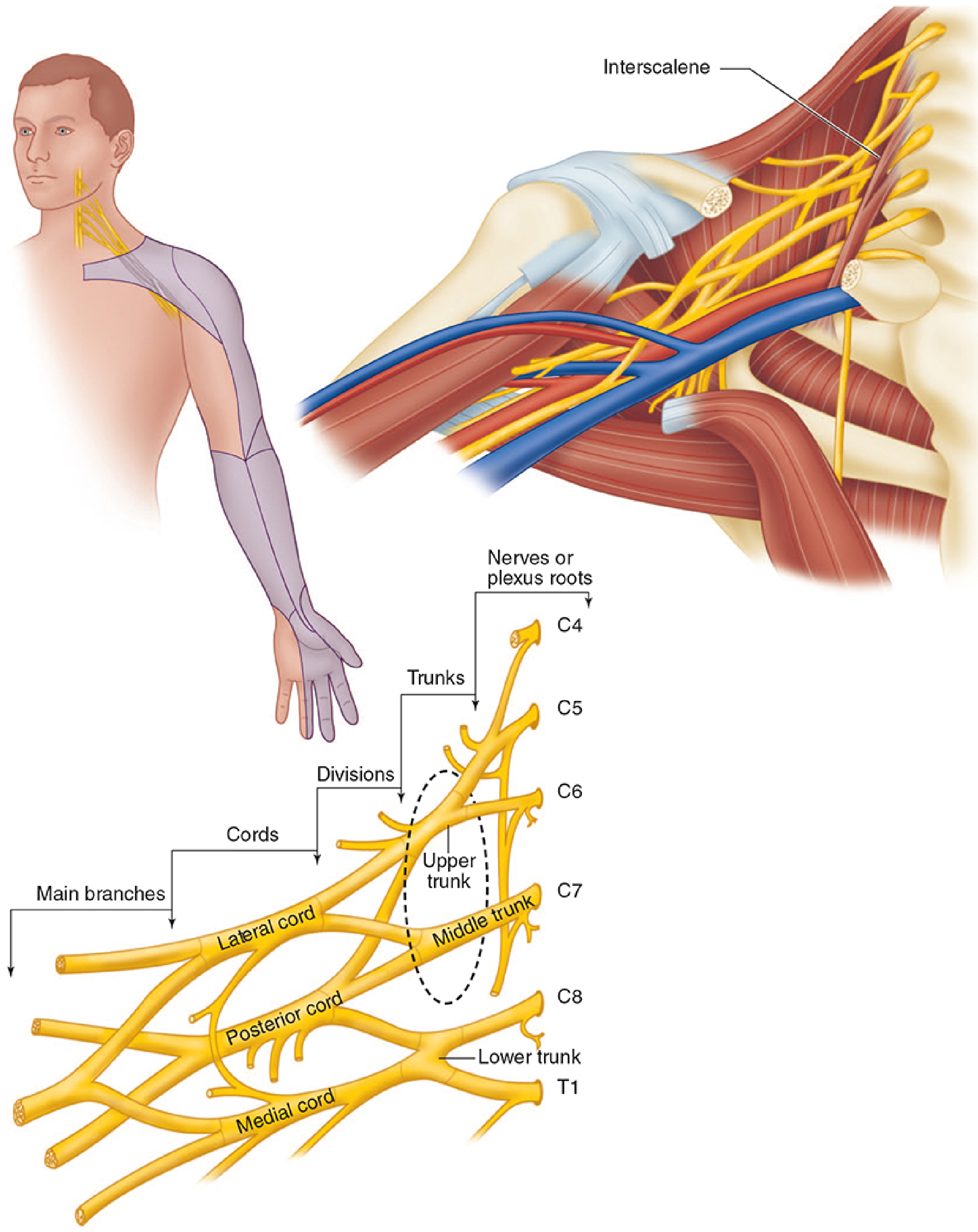
The brachial plexus is formed by the ventral rami (anterior primary divisions) of C5, C6, C7, C8, and T1. Minor contributions from C4 and T2 may occur. The organization follows the mnemonic RTDCB: Roots → Trunks → Divisions → Cords → Branches (terminal nerves).
Organization
| Level | Components | Location |
|---|---|---|
| Roots | C5, C6, C7, C8, T1 | Exit intervertebral foramina |
| Trunks | Superior (C5+C6), Middle (C7), Inferior (C8+T1) | Between anterior & middle scalene muscles |
| Divisions | Anterior & posterior from each trunk (6 total) | Over lateral border of 1st rib, under clavicle |
| Cords | Lateral, Medial, Posterior | Named relative to axillary artery; below clavicle |
| Terminal nerves | Median, Ulnar, Radial, Musculocutaneous, Axillary | Lateral border of pectoralis minor |
Cord-to-Terminal Nerve Formation
- Lateral cord → lateral head of median nerve + musculocutaneous nerve
- Medial cord → medial head of median nerve + ulnar nerve + medial brachial cutaneous + medial antebrachial cutaneous nerves
- Posterior cord → axillary nerve + radial nerve
Pre-terminal Branches (Important - often missed)
- Dorsal scapular nerve (C5) - rhomboids
- Long thoracic nerve (C5, C6, C7) - serratus anterior
- Suprascapular nerve (C5, C6) - supraspinatus, infraspinatus, glenohumeral joint - branches from upper trunk, must be blocked for complete shoulder analgesia
- Nerve to subclavius (C5, C6)
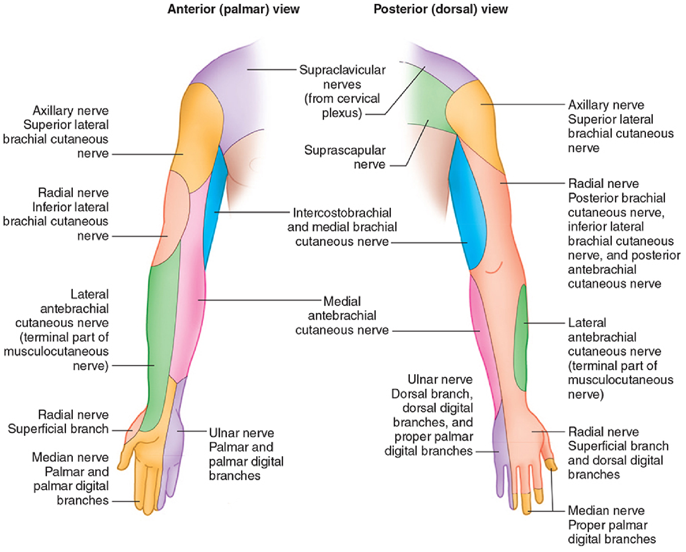
Nerve Sensory Distribution
2. Approaches to Brachial Plexus Block
The key principle is: where you inject determines what you block. Proximal blocks intercept roots/trunks (shoulder and above). Distal blocks intercept cords/terminal nerves (elbow and below).
| Approach | Anatomical Level Targeted | Surgical Indication |
|---|---|---|
| Interscalene | Roots / upper & middle trunks | Shoulder, proximal humerus |
| Supraclavicular | Distal trunks / proximal divisions | Elbow, forearm, hand ("spinal of the arm") |
| Infraclavicular | Cords | Elbow, forearm, hand; best for catheters |
| Axillary | Terminal nerves | Hand, wrist, forearm distal to elbow |
3. Interscalene Block
Anatomy
The brachial plexus trunks lie between the anterior scalene and middle scalene muscles in the neck, visualized at the C6 level. The phrenic nerve (C3-C5) lies directly on the anterior scalene muscle just medial to the interscalene groove.
Indication
Shoulder surgery, proximal humerus fractures, AC joint procedures. Not suitable for hand/wrist surgery because C8-T1 (inferior trunk, ulnar distribution) is frequently spared.
US-Guided Technique
- Patient supine, head turned 30-45° away from the side to be blocked
- High-frequency linear probe placed at the level of the cricoid cartilage (C6)
- Trace the plexus cephalad from a supraclavicular view until the trunks appear as hypoechoic structures between the scalene muscles - the "stoplight" sign (three trunks stacked vertically)
- In-plane or out-of-plane needle approach; out-of-plane avoids the long thoracic and dorsal scapular nerves inside the middle scalene
- Local anesthetic volume: 10-20 mL surrounds the trunks
Key Point for Shoulder Coverage
The supraclavicular nerve (C3-C4, from cervical plexus) covers the "cape" area of the shoulder. A separate cervical plexus block or subcutaneous infiltration along the clavicle is needed for complete shoulder anesthesia.
Complications
- Ipsilateral phrenic nerve block - near 100% incidence with conventional volumes; causes hemidiaphragmatic paralysis. Contraindicated in contralateral phrenic nerve palsy, severe COPD/restrictive disease (FEV1 or FVC <50%)
- Recurrent laryngeal nerve block (hoarseness)
- Horner syndrome (ptosis, miosis, anhidrosis)
- Cervical sympathetic chain block
- Vertebral artery injection (risk of CNS toxicity)
- Intrathecal/epidural injection
- Spinal cord injury with high-volume injection
4. Supraclavicular Block
Anatomy
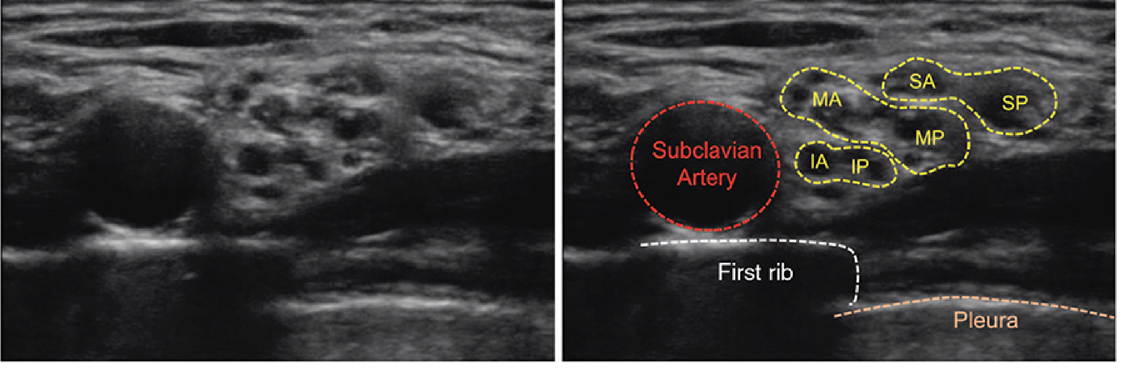
At the lateral border of the first rib, all six divisions of the brachial plexus are compact in a space <1 cm, making this the most efficient location for complete upper extremity anesthesia. The subclavian artery lies medial/deep, the first rib is deep (bony backstop), and the pleura is just beyond the rib.
Indication
Operations on elbow, forearm, and hand. Sometimes called the "spinal of the arm" due to rapid onset and dense block.
Not ideal for: shoulder surgery (suprascapular nerve, which is a proximal branch of upper trunk, is not reliably blocked).
US-Guided Technique
- Patient supine, head turned 30° away
- High-frequency linear probe placed in the supraclavicular fossa, angled slightly toward thorax
- Subclavian artery identified as pulsatile structure; brachial plexus appears as "cluster of grapes" - multiple hypoechoic nodules, just superficial and posterolateral to the artery
- First rib is a hyperechoic line deep to the artery; pleura lies just beyond and moves with respiration
- Needle inserted posterolateral in-plane toward subclavian artery; aim for the "corner pocket" between artery, plexus, and first rib - this ensures inferior trunk (ulnar nerve distribution) is adequately blocked
- Local anesthetic: 20-30 mL, injected in multiple locations to encircle plexus
Complications
- Pneumothorax (most feared; risk ~0.5-1%, reduced but not eliminated by US)
- Phrenic nerve palsy (~50% incidence - less than interscalene but still significant)
- Subclavian artery puncture
- Horner syndrome
- Ulnar sparing (if "corner pocket" injection is missed)
5. Infraclavicular Block
Anatomy
Below the clavicle, the brachial plexus is now organized as three cords arranged around the axillary artery:
- Lateral cord - lateral/cephalad to artery
- Medial cord - medial/caudad to artery
- Posterior cord - posterior to artery
All three cords lie deep to the pectoralis major and pectoralis minor muscles. The subclavian artery becomes the axillary artery at the lateral border of the first rib.
Indication
Surgery at or distal to the elbow. Preferred approach for perineural catheter placement (superior to supraclavicular and axillary catheters due to muscle and fascial layers securing it).
US-Guided Technique
- Patient supine; arm abducted 90° (dramatically improves visualization)
- High-frequency linear or small curvilinear probe placed parasagittal, 2 cm medial and 2 cm caudad to coracoid process
- Axillary artery and vein seen in cross-section; cords appear as hyperechoic bundles:
- Lateral cord - cephalad to artery
- Medial cord - caudad to artery
- Posterior cord - posterior (deep) to artery
- A long (10-cm) needle inserted 1-3 cm cephalad to probe tip, angled steeply
- Optimal injection: between artery and posterior cord ("U" of local anesthetic wrapping around artery); a single 30-mL injection at this location is equivalent to individual cord injections
- Catheter tip placed posterior to artery for continuous infusion
Complications
- Vascular puncture (axillary artery or vein)
- Pneumothorax (less common than supraclavicular)
- Hematoma
- Contraindicated: ipsilateral subclavian vascular catheter, transvenous pacemaker
6. Axillary Block
Anatomy
At the lateral border of pectoralis minor, the cords have divided into terminal branches surrounding the axillary artery in fascial compartments:
- Median nerve - anterior/medial to artery
- Radial nerve - posterior to artery
- Ulnar nerve - medial to artery
- Musculocutaneous nerve - already departed and lies within the coracobrachialis muscle (must be separately targeted)
- Axillary nerve - already branched proximally (not blocked by axillary approach)
- Medial brachial & antebrachial cutaneous nerves - branch proximally (spared)
Because the nerves are separated by fascial compartments at this level, multiple injections are usually required.
Indication
Surgery on hand, wrist, and forearm distal to elbow. Most superficial and safe approach.
Not suitable for: arm tourniquet at the upper arm (intercostobrachial nerve, T2, is spared and must be blocked separately with subcutaneous infiltration along the axilla).
US-Guided Technique
- Patient supine, arm abducted 90°, elbow flexed, forearm externally rotated (supinated)
- High-frequency linear probe placed in axilla, perpendicular to arm
- Axillary artery pulsates in center; veins are compressible; nerves are hyperechoic
- Individual injections around each nerve (5-10 mL per nerve)
- Musculocutaneous nerve located as oval hyperechoic structure within coracobrachialis muscle - requires separate injection
- Total volume: 25-40 mL
Complications
- Axillary artery/vein puncture (most common; hematoma formation)
- Nerve injury
- Local anesthetic systemic toxicity (LAST) - highly vascular area
7. Phrenic Nerve-Sparing Approaches for Shoulder Surgery
Patients with significant pulmonary disease cannot tolerate interscalene block (phrenic nerve palsy). Alternatives:
- Superior trunk block - targets C5-C6 at the level where suprascapular nerve branches; 5-15 mL deep to omohyoid muscle; significantly less phrenic nerve involvement
- Anterior suprascapular nerve block - blocks the suprascapular nerve proximal to suprascapular notch; combined with infraclavicular block for complete shoulder coverage
8. Local Anesthetics Used
| Drug | Concentration | Duration | Onset |
|---|---|---|---|
| Ropivacaine | 0.375-0.5% (surgical), 0.2% (analgesia) | 8-12 h | Moderate |
| Bupivacaine | 0.25-0.5% | 8-16 h | Moderate |
| Lidocaine | 1-1.5% (±epi) | 2-4 h | Rapid |
| Mepivacaine | 1-1.5% | 4-6 h | Rapid |
Adding epinephrine (1:200,000) prolongs duration and serves as an intravascular injection marker. Dexamethasone 4-8 mg (IV or perineural) reliably extends block duration by 6-8 hours.
9. Contraindications (General)
- Patient refusal
- Local infection at injection site
- Severe coagulopathy (relative; varies by approach depth)
- True local anesthetic allergy
- Pre-existing severe neurological deficit (relative)
- Bilateral blocks (combined phrenic nerve palsy risk)
10. Complications Overview
| Complication | Most Common Approach |
|---|---|
| Phrenic nerve palsy | Interscalene (>95%), Supraclavicular (~50%) |
| Pneumothorax | Supraclavicular > Infraclavicular |
| Horner syndrome | Interscalene, Supraclavicular |
| Recurrent laryngeal nerve | Interscalene |
| Vascular puncture | Axillary, Infraclavicular |
| LAST | All (especially axillary - high vascularity) |
| Nerve injury | All approaches |
| Spinal/epidural injection | Interscalene |
Summary - "Which Block for Which Surgery?" (Exam Table)
| Surgery | Best Approach | Why |
|---|---|---|
| Shoulder arthroplasty / RCR | Interscalene | Targets C5-C6 roots + upper trunk |
| Clavicle ORIF | Interscalene + cervical plexus | Supraclavicular nerve (C3-C4) supplementation needed |
| Shoulder (high-risk lung) | Superior trunk / suprascapular + infraclavicular | Phrenic-sparing |
| Elbow / forearm | Supraclavicular or infraclavicular | Dense block at division/cord level |
| Wrist / hand | Infraclavicular or axillary | Cord / terminal nerve level |
| Catheter for prolonged analgesia | Infraclavicular | Best catheter stability |
| Pediatric or obese arm | Axillary | Superficial, safe |
Sources: Miller's Anesthesia 10e, Morgan & Mikhail's Clinical Anesthesiology 7e, Barash Clinical Anesthesia 9e
Okay
Got it! Let me know if you have any questions about brachial plexus blocks, want me to go deeper on any specific approach, or if there's another topic you'd like to explore.
Create a summary PDF on block selection by surgery
Reading File
Running Command
ls -la /home/daytona/workspace/ 2>/dev/null && mkdir -p /home/daytona/workspace/brachial-plexus-pdf
Writing File
~/brachial-plexus-pdf/generate_pdf.py
from reportlab.lib.pagesizes import A4
from reportlab.lib import colors
from reportlab.lib.units import cm, mm
from reportlab.lib.styles import getSampleStyleSheet, ParagraphStyle
from reportlab.lib.enums import TA_CENTER, TA_LEFT, TA_JUSTIFY
from reportlab.platypus import (
    SimpleDocTemplate, Paragraph, Spacer, Table, TableStyle,
    HRFlowable, KeepTogether
)
from reportlab.pdfbase import pdfmetrics
from reportlab.pdfbase.ttfonts import TTFont

OUTPUT = "/home/daytona/workspace/brachial-plexus-pdf/Brachial_Plexus_Block_Selection.pdf"

# --- Colours ---
DARK_BLUE   = colors.HexColor("#1A3557")
MED_BLUE    = colors.HexColor("#2E6DA4")
LIGHT_BLUE  = colors.HexColor("#D6E8F7")
ACCENT      = colors.HexColor("#E8511A")
PALE_GREY   = colors.HexColor("#F4F6F9")
MID_GREY    = colors.HexColor("#D0D8E4")
WHITE       = colors.white
GREEN_DARK  = colors.HexColor("#1A5741")
GREEN_LIGHT = colors.HexColor("#D4EDDA")
AMBER_DARK  = colors.HexColor("#7A4F00")
AMBER_LIGHT = colors.HexColor("#FFF3CD")
RED_DARK    = colors.HexColor("#7A1A1A")
RED_LIGHT   = colors.HexColor("#FADADD")

doc = SimpleDocTemplate(
    OUTPUT,
    pagesize=A4,
    leftMargin=1.8*cm, rightMargin=1.8*cm,
    topMargin=2*cm, bottomMargin=2*cm,
    title="Brachial Plexus Block Selection",
    author="Orris Medical"
)

W = A4[0] - 3.6*cm   # usable width

styles = getSampleStyleSheet()

def S(name, **kw):
    return ParagraphStyle(name, **kw)

TITLE_STYLE = S("Title",
    fontSize=20, textColor=WHITE, alignment=TA_CENTER,
    fontName="Helvetica-Bold", spaceAfter=4)

SUBTITLE_STYLE = S("Subtitle",
    fontSize=11, textColor=LIGHT_BLUE, alignment=TA_CENTER,
    fontName="Helvetica", spaceAfter=2)

SECTION_STYLE = S("Section",
    fontSize=13, textColor=WHITE, alignment=TA_LEFT,
    fontName="Helvetica-Bold", leftIndent=6, spaceAfter=2)

BODY = S("Body",
    fontSize=9, textColor=colors.HexColor("#1C1C1C"),
    fontName="Helvetica", leading=13, spaceAfter=3)

BOLD_BODY = S("BoldBody",
    fontSize=9, textColor=DARK_BLUE,
    fontName="Helvetica-Bold", leading=13, spaceAfter=3)

SMALL = S("Small",
    fontSize=7.5, textColor=colors.HexColor("#444444"),
    fontName="Helvetica", leading=11)

FOOTER = S("Footer",
    fontSize=7, textColor=colors.HexColor("#888888"),
    fontName="Helvetica-Oblique", alignment=TA_CENTER)

TH = S("TH",
    fontSize=8.5, textColor=WHITE, fontName="Helvetica-Bold",
    alignment=TA_CENTER, leading=11)

TD = S("TD",
    fontSize=8, textColor=colors.HexColor("#1C1C1C"),
    fontName="Helvetica", alignment=TA_LEFT, leading=11)

TD_C = S("TDC",
    fontSize=8, textColor=colors.HexColor("#1C1C1C"),
    fontName="Helvetica", alignment=TA_CENTER, leading=11)

TD_B = S("TDB",
    fontSize=8, textColor=DARK_BLUE, fontName="Helvetica-Bold",
    alignment=TA_LEFT, leading=11)

WARN = S("Warn",
    fontSize=7.5, textColor=RED_DARK, fontName="Helvetica-Bold",
    alignment=TA_CENTER, leading=10)

NOTE = S("Note",
    fontSize=7.5, textColor=GREEN_DARK, fontName="Helvetica-Bold",
    alignment=TA_LEFT, leading=10)

story = []

# ========================================================
# HEADER BANNER
# ========================================================
header_data = [[
    Paragraph("BRACHIAL PLEXUS BLOCK SELECTION", TITLE_STYLE),
]]
header_table = Table([[Paragraph("BRACHIAL PLEXUS BLOCK SELECTION", TITLE_STYLE)]], colWidths=[W])
header_table.setStyle(TableStyle([
    ("BACKGROUND", (0,0), (-1,-1), DARK_BLUE),
    ("TOPPADDING",    (0,0), (-1,-1), 14),
    ("BOTTOMPADDING", (0,0), (-1,-1), 4),
    ("LEFTPADDING",   (0,0), (-1,-1), 10),
    ("RIGHTPADDING",  (0,0), (-1,-1), 10),
    ("ROUNDEDCORNERS", [6]),
]))
story.append(header_table)

sub_table = Table([[Paragraph("Quick Reference Guide for Anesthesia | Based on Miller's 10e · Morgan & Mikhail 7e · Barash 9e", SUBTITLE_STYLE)]], colWidths=[W])
sub_table.setStyle(TableStyle([
    ("BACKGROUND", (0,0), (-1,-1), MED_BLUE),
    ("TOPPADDING",    (0,0), (-1,-1), 6),
    ("BOTTOMPADDING", (0,0), (-1,-1), 8),
    ("LEFTPADDING",   (0,0), (-1,-1), 10),
    ("RIGHTPADDING",  (0,0), (-1,-1), 10),
]))
story.append(sub_table)
story.append(Spacer(1, 10))

# ========================================================
# HELPER: section header
# ========================================================
def section_header(text):
    t = Table([[Paragraph(text, SECTION_STYLE)]], colWidths=[W])
    t.setStyle(TableStyle([
        ("BACKGROUND", (0,0), (-1,-1), MED_BLUE),
        ("TOPPADDING",    (0,0), (-1,-1), 5),
        ("BOTTOMPADDING", (0,0), (-1,-1), 5),
        ("LEFTPADDING",   (0,0), (-1,-1), 8),
        ("RIGHTPADDING",  (0,0), (-1,-1), 8),
    ]))
    return t

def mini_header(text, color=DARK_BLUE):
    style = S("MH", fontSize=9, textColor=WHITE, fontName="Helvetica-Bold",
              alignment=TA_LEFT, leading=11, leftIndent=4)
    t = Table([[Paragraph(text, style)]], colWidths=[W])
    t.setStyle(TableStyle([
        ("BACKGROUND", (0,0), (-1,-1), color),
        ("TOPPADDING",    (0,0), (-1,-1), 4),
        ("BOTTOMPADDING", (0,0), (-1,-1), 4),
        ("LEFTPADDING",   (0,0), (-1,-1), 8),
        ("RIGHTPADDING",  (0,0), (-1,-1), 8),
    ]))
    return t

def colored_cell(text, style, bg):
    t = Table([[Paragraph(text, style)]], colWidths=["100%"])
    t.setStyle(TableStyle([
        ("BACKGROUND", (0,0), (-1,-1), bg),
        ("TOPPADDING",    (0,0), (-1,-1), 3),
        ("BOTTOMPADDING", (0,0), (-1,-1), 3),
        ("LEFTPADDING",   (0,0), (-1,-1), 4),
        ("RIGHTPADDING",  (0,0), (-1,-1), 4),
    ]))
    return t

# ========================================================
# SECTION 1: ANATOMY OVERVIEW
# ========================================================
story.append(section_header("1. BRACHIAL PLEXUS ANATOMY OVERVIEW"))
story.append(Spacer(1, 6))

# Organisation table
anat_headers = ["Level", "Components", "Location", "Approach Targeting Here"]
anat_rows = [
    ["Roots", "C5, C6, C7, C8, T1", "Exit intervertebral foramina between C4-T1 vertebrae", "Interscalene"],
    ["Trunks", "Superior (C5+C6)\nMiddle (C7)\nInferior (C8+T1)", "Between anterior & middle scalene muscles", "Interscalene\n(upper/middle)"],
    ["Divisions", "Anterior & posterior\nfrom each trunk (6 total)", "Over lateral border of 1st rib,\nunder the clavicle", "Supraclavicular"],
    ["Cords", "Lateral, Medial, Posterior\n(named relative to axillary artery)", "Below clavicle,\nunder pectoralis minor", "Infraclavicular"],
    ["Terminal\nNerves", "Median, Ulnar, Radial\nMusculocutaneous, Axillary", "Lateral border of\npectoralis minor - axilla", "Axillary"],
]

anat_col = [2.4*cm, 4.2*cm, 5.8*cm, 3.8*cm]

anat_data = [[Paragraph(h, TH) for h in anat_headers]]
row_colors = [LIGHT_BLUE, PALE_GREY, LIGHT_BLUE, PALE_GREY, LIGHT_BLUE]
for i, row in enumerate(anat_rows):
    anat_data.append([Paragraph(str(c).replace("\n","<br/>"), TD_B if i==0 else TD) for c in row])

anat_table = Table(anat_data, colWidths=anat_col, repeatRows=1)
anat_table.setStyle(TableStyle([
    ("BACKGROUND", (0,0), (-1,0), DARK_BLUE),
    ("ROWBACKGROUNDS", (0,1), (-1,-1), [PALE_GREY, WHITE]),
    ("GRID", (0,0), (-1,-1), 0.4, MID_GREY),
    ("TOPPADDING",    (0,0), (-1,-1), 4),
    ("BOTTOMPADDING", (0,0), (-1,-1), 4),
    ("LEFTPADDING",   (0,0), (-1,-1), 5),
    ("RIGHTPADDING",  (0,0), (-1,-1), 5),
    ("VALIGN", (0,0), (-1,-1), "MIDDLE"),
]))
story.append(anat_table)
story.append(Spacer(1, 4))

# Cord → nerve mnemonic box
cord_style = S("CS", fontSize=8, textColor=DARK_BLUE, fontName="Helvetica",
               leading=12, alignment=TA_LEFT)
cord_text = (
    "<b>Cord → Terminal Nerve Formation:</b>  "
    "<b>Lateral cord</b> → Lat. head of median nerve + Musculocutaneous nerve  |  "
    "<b>Medial cord</b> → Med. head of median nerve + Ulnar nerve + Medial cutaneous nerves  |  "
    "<b>Posterior cord</b> → Axillary nerve + Radial nerve"
)
cord_box = Table([[Paragraph(cord_text, cord_style)]], colWidths=[W])
cord_box.setStyle(TableStyle([
    ("BACKGROUND", (0,0), (-1,-1), LIGHT_BLUE),
    ("TOPPADDING",    (0,0), (-1,-1), 6),
    ("BOTTOMPADDING", (0,0), (-1,-1), 6),
    ("LEFTPADDING",   (0,0), (-1,-1), 8),
    ("RIGHTPADDING",  (0,0), (-1,-1), 8),
    ("BOX", (0,0), (-1,-1), 0.8, MED_BLUE),
]))
story.append(cord_box)
story.append(Spacer(1, 10))

# ========================================================
# SECTION 2: MAIN BLOCK SELECTION TABLE
# ========================================================
story.append(section_header("2. BLOCK SELECTION BY SURGERY"))
story.append(Spacer(1, 6))

# colour key
key_style = S("KEY", fontSize=7.5, textColor=colors.HexColor("#1C1C1C"),
              fontName="Helvetica", alignment=TA_CENTER, leading=10)
key_data = [
    [colored_cell("■  SHOULDER", key_style, colors.HexColor("#BDD7EE")),
     colored_cell("■  UPPER ARM", key_style, colors.HexColor("#C6EFCE")),
     colored_cell("■  ELBOW / FOREARM", key_style, colors.HexColor("#FFEB9C")),
     colored_cell("■  WRIST / HAND", key_style, colors.HexColor("#FFC7CE")),
    ]
]
key_table = Table(key_data, colWidths=[W/4]*4)
key_table.setStyle(TableStyle([
    ("TOPPADDING",    (0,0), (-1,-1), 2),
    ("BOTTOMPADDING", (0,0), (-1,-1), 2),
    ("LEFTPADDING",   (0,0), (-1,-1), 2),
    ("RIGHTPADDING",  (0,0), (-1,-1), 2),
]))
story.append(key_table)
story.append(Spacer(1, 5))

# Surgery region headers + rows
# Format: region_color, surgery, block, level_targeted, LA_volume, notes
SHOULDER_C = colors.HexColor("#BDD7EE")
ARM_C      = colors.HexColor("#C6EFCE")
ELBOW_C    = colors.HexColor("#FFEB9C")
HAND_C     = colors.HexColor("#FFC7CE")

main_cols = [3.8*cm, 3.5*cm, 3.2*cm, 2.0*cm, 3.7*cm]
main_headers = ["Surgery / Procedure", "Preferred Block", "Level Targeted", "LA Vol (mL)", "Key Notes"]

main_rows = [
    # --- SHOULDER ---
    ("SHOULDER", None, None, None, None, SHOULDER_C),
    ("Shoulder arthroplasty (TSA/hemi)", "Interscalene", "Roots C5-C6 /\nUpper trunk", "15–25", "Gold standard; spares C8-T1 (ulnar)", SHOULDER_C),
    ("Rotator cuff repair (RCR)", "Interscalene", "Roots C5-C6 /\nUpper trunk", "15–25", "Excellent post-op analgesia 12-18 h", SHOULDER_C),
    ("Shoulder arthroscopy", "Interscalene", "Roots / Trunks", "15–20", "Add cervical plexus block if tourniquet at shoulder", SHOULDER_C),
    ("Clavicle ORIF", "Interscalene +\nCervical plexus", "C5-C6 +\nC3-C4 (supraclavicular n.)", "15–20 +\n5–10", "Supraclavicular nerve (C3-C4) covers medial clavicle", SHOULDER_C),
    ("AC joint procedures", "Interscalene +\nCervical plexus", "Upper trunk +\nC3-C4", "15–20 +\n5–10", "Often requires superficial cervical plexus supplement", SHOULDER_C),
    ("Shoulder (high respiratory risk)", "Superior trunk block OR\nAnterior suprascapular\n+ Infraclavicular", "C5-C6 / suprascapular n.\n+ Cords", "10–15 +\n25–30", "Phrenic-sparing; avoids hemidiaphragm paralysis", SHOULDER_C),
    # --- UPPER ARM ---
    ("UPPER ARM", None, None, None, None, ARM_C),
    ("Proximal humerus fracture ORIF", "Interscalene", "Roots / Upper trunk", "20–25", "C5-C7 block; tourniquet at shoulder needs cervical supplement", ARM_C),
    ("Mid-shaft humerus ORIF", "Interscalene OR\nSupraclavicular", "Roots/Trunks OR\nDivisions", "20–30", "Supraclavicular gives more reliable C8-T1 coverage distally", ARM_C),
    ("Arm tourniquet analgesia (any)", "Any + Intercostobrachial\nsupplementation", "T2 cutaneous coverage", "3–5\n(subcutaneous)", "T2 (intercostobrachial n.) always spared; ring infiltration in axilla", ARM_C),
    # --- ELBOW / FOREARM ---
    ("ELBOW / FOREARM", None, None, None, None, ELBOW_C),
    ("Elbow fracture / ORIF", "Supraclavicular OR\nInfraclavicular", "Divisions OR Cords", "20–30", "Infraclavicular preferred if catheter needed; arm abduction required", ELBOW_C),
    ("Distal radius / ulna fracture", "Supraclavicular OR\nInfraclavicular", "Divisions OR Cords", "20–30", "Both approaches equally effective; infraclavicular catheter superior", ELBOW_C),
    ("Forearm fasciotomy / debridement", "Infraclavicular", "Cords", "25–35", "Posterior cord injection provides reliable coverage; easy catheter", ELBOW_C),
    ("Tendon repair forearm", "Infraclavicular OR\nAxillary", "Cords OR\nTerminal nerves", "25–35", "Axillary feasible if limited to forearm distal to mid-humeral level", ELBOW_C),
    # --- WRIST / HAND ---
    ("WRIST / HAND", None, None, None, None, HAND_C),
    ("Carpal tunnel release", "Axillary OR\nWrist block", "Terminal nerves", "25–40\nOR 3–5 each", "Axillary if bilateral impractical; wrist block for isolated procedure", HAND_C),
    ("Hand ORIF / fracture fixation", "Axillary OR\nInfraclavicular", "Terminal nerves OR\nCords", "25–40", "Multiple injections around each nerve at axilla for reliability", HAND_C),
    ("Wrist arthroscopy", "Axillary OR\nInfraclavicular", "Terminal nerves OR\nCords", "25–35", "Axillary: supplement musculocutaneous n. in coracobrachialis", HAND_C),
    ("Digital / finger procedures", "Wrist block OR\nDigital ring block", "Terminal branches\nat wrist", "3–5 each", "Median ± ulnar ± radial superficial branch at wrist; no epinephrine in fingers", HAND_C),
    ("Replantation / microsurgery", "Infraclavicular\ncatheter (continuous)", "Cords", "Infusion\n5–10 mL/h", "Sympathectomy effect promotes vasodilation; catheter > single shot", HAND_C),
    ("Dupuytren's contracture", "Axillary", "Terminal nerves", "25–40", "Multiple injection technique; all four nerves around axillary artery", HAND_C),
]

THs = [Paragraph(h, TH) for h in main_headers]
table_data = [THs]

for row in main_rows:
    surgery, block, level, vol, notes, rc = row
    if block is None:
        # Region header row
        region_style = S("RH", fontSize=9, textColor=DARK_BLUE,
                         fontName="Helvetica-Bold", alignment=TA_LEFT,
                         leading=11, leftIndent=4)
        merged_cell = Paragraph(f"▶  {surgery}", region_style)
        table_data.append([merged_cell, "", "", "", ""])
    else:
        table_data.append([
            Paragraph(surgery, TD),
            Paragraph(block.replace("\n","<br/>"), TD_B),
            Paragraph(level.replace("\n","<br/>"), TD_C),
            Paragraph(vol.replace("\n","<br/>"), TD_C),
            Paragraph(notes, SMALL),
        ])

main_table = Table(table_data, colWidths=main_cols, repeatRows=1)

# Build style commands
ts_cmds = [
    ("BACKGROUND", (0,0), (-1,0), DARK_BLUE),
    ("GRID", (0,0), (-1,-1), 0.3, MID_GREY),
    ("TOPPADDING",    (0,0), (-1,-1), 4),
    ("BOTTOMPADDING", (0,0), (-1,-1), 4),
    ("LEFTPADDING",   (0,0), (-1,-1), 5),
    ("RIGHTPADDING",  (0,0), (-1,-1), 5),
    ("VALIGN", (0,0), (-1,-1), "MIDDLE"),
    ("ROWBACKGROUNDS", (0,1), (-1,-1), [WHITE, PALE_GREY]),
]

# colour region headers + data rows
region_row_idx = 1  # track row index in table_data (0 = header)
for row in main_rows:
    surgery, block, level, vol, notes, rc = row
    if block is None:
        ts_cmds.append(("BACKGROUND", (0,region_row_idx), (-1,region_row_idx), rc))
        ts_cmds.append(("SPAN", (0,region_row_idx), (-1,region_row_idx)))
        ts_cmds.append(("LINEABOVE", (0,region_row_idx), (-1,region_row_idx), 1.2, DARK_BLUE))
    else:
        ts_cmds.append(("BACKGROUND", (0,region_row_idx), (0,region_row_idx),
                         colors.HexColor(rc.hexval() + "33" if hasattr(rc,"hexval") else "#FAFAFA")))
        # tint the surgery column lightly
        lighter = colors.Color(rc.red, rc.green, rc.blue, alpha=0.25)
        ts_cmds.append(("BACKGROUND", (0,region_row_idx), (0,region_row_idx), lighter))
    region_row_idx += 1

main_table.setStyle(TableStyle(ts_cmds))
story.append(KeepTogether([main_table]))
story.append(Spacer(1, 10))

# ========================================================
# SECTION 3: APPROACH PROFILES
# ========================================================
story.append(section_header("3. APPROACH PROFILES AT A GLANCE"))
story.append(Spacer(1, 6))

profile_cols = [2.7*cm, 3.3*cm, 2.8*cm, 2.8*cm, 3.0*cm, 1.6*cm]
profile_headers = ["Approach", "US Landmarks", "Ideal Indication", "Spared Nerves", "Main Complications", "Catheter?"]

profiles = [
    ["Interscalene",
     "Scalene muscles at C6 level;\n'Stoplight' sign (3 trunks)",
     "Shoulder,\nproximal humerus",
     "C8-T1 (ulnar n.)\nT2 (intercostobrachial)",
     "Phrenic nerve palsy (>95%)\nHorner syndrome\nRecurrent laryngeal n.\nIntrathecal injection",
     "Yes\n(perineural)"],
    ["Supraclavicular\n('Spinal of the arm')",
     "Subclavian artery;\n'Cluster of grapes';\nFirst rib + pleura deep",
     "Elbow, forearm,\nhand surgery",
     "Suprascapular n. (variable)\nT2 intercostobrachial",
     "Pneumothorax (~0.5-1%)\nPhrenic palsy (~50%)\nHorner syndrome\nSubclavian artery puncture",
     "Yes\n(less stable)"],
    ["Infraclavicular",
     "Axillary artery in\nparasagittal plane;\n2 cm medial + caudad\nto coracoid process",
     "Elbow, forearm, hand;\nBest for catheters",
     "T2 intercostobrachial\nShoulder / upper arm",
     "Vascular puncture\nPneumothorax (rare)\nHematoma",
     "YES\n(preferred)"],
    ["Axillary",
     "Axillary artery + 4\nterminal nerves;\nMusculocutaneous in\ncoracobrachialis",
     "Wrist and hand\nsurgery",
     "Musculocutaneous n.\n(inject separately)\nAxillary n.; T2",
     "Axillary vascular\npuncture/hematoma\nNerve injury\nHigh LAST risk",
     "Less\ncommon"],
]

ph = [Paragraph(h, TH) for h in profile_headers]
profile_data = [ph]
for row in profiles:
    profile_data.append([Paragraph(c.replace("\n","<br/>"), TD) for c in row])

profile_table = Table(profile_data, colWidths=profile_cols, repeatRows=1)
profile_table.setStyle(TableStyle([
    ("BACKGROUND", (0,0), (-1,0), DARK_BLUE),
    ("ROWBACKGROUNDS", (0,1), (-1,-1), [PALE_GREY, WHITE]),
    ("GRID", (0,0), (-1,-1), 0.3, MID_GREY),
    ("TOPPADDING",    (0,0), (-1,-1), 4),
    ("BOTTOMPADDING", (0,0), (-1,-1), 4),
    ("LEFTPADDING",   (0,0), (-1,-1), 5),
    ("RIGHTPADDING",  (0,0), (-1,-1), 5),
    ("VALIGN", (0,0), (-1,-1), "TOP"),
    # highlight complication column lightly
    ("BACKGROUND", (4,1), (4,-1), colors.HexColor("#FFF5F5")),
    # highlight catheter column
    ("BACKGROUND", (5,1), (5,-1), colors.HexColor("#F0FFF4")),
    # bold infraclavicular catheter cell
    ("TEXTCOLOR", (5,3), (5,3), GREEN_DARK),
]))
story.append(profile_table)
story.append(Spacer(1, 10))

# ========================================================
# SECTION 4: LOCAL ANAESTHETICS
# ========================================================
story.append(section_header("4. LOCAL ANAESTHETICS & ADJUVANTS"))
story.append(Spacer(1, 6))

la_cols = [3.2*cm, 2.8*cm, 2.5*cm, 2.5*cm, 5.2*cm]
la_headers = ["Drug", "Concentration", "Onset", "Duration", "Notes"]
la_rows = [
    ["Ropivacaine", "0.375-0.5% (surgical)\n0.2% (analgesia)", "Moderate\n(15–20 min)", "8–12 h", "Preferred for peripheral blocks; less cardiotoxic than bupivacaine; motor-sensory differential with low concentrations"],
    ["Bupivacaine", "0.25-0.5%", "Moderate\n(15–20 min)", "8–16 h", "Long-acting; cardiotoxicity risk with intravascular injection; use with caution"],
    ["Levobupivacaine", "0.25-0.5%", "Moderate", "8–12 h", "Safer cardiac profile than racemic bupivacaine; similar duration"],
    ["Lidocaine", "1–1.5% (± epi)", "Rapid\n(5–10 min)", "2–4 h", "Add epinephrine 1:200,000 to extend duration; useful when rapid onset needed"],
    ["Mepivacaine", "1–1.5%", "Rapid\n(10–15 min)", "4–6 h", "Good alternative to lidocaine; intermediate duration; ambulatory surgery"],
]
la_adjuvants = [
    ["Dexamethasone (adjuvant)", "4–8 mg IV or perineural", "—", "+6–8 h", "Most reliable block prolongation; IV dexamethasone equally effective; preferred over perineural in most centres"],
    ["Dexmedetomidine (adjuvant)", "0.5–1 mcg/kg", "—", "+4–6 h", "Adds sedation; use caution in ambulatory patients; bradycardia possible"],
    ["Epinephrine (vasoconstrictor)", "1:200,000 (5 mcg/mL)", "—", "+20–30%", "Marker of intravascular injection; prolongs block; avoid in digital blocks"],
]

lah = [Paragraph(h, TH) for h in la_headers]
la_data = [lah]
for row in la_rows:
    la_data.append([Paragraph(c.replace("\n","<br/>"), TD) for c in row])
# adjuvant sub-header
adj_style = S("ADJ", fontSize=8.5, textColor=WHITE, fontName="Helvetica-Bold",
              alignment=TA_LEFT, leading=11, leftIndent=4)
la_data.append([Paragraph("ADJUVANTS", adj_style), "", "", "", ""])
for row in la_adjuvants:
    la_data.append([Paragraph(c.replace("\n","<br/>"), TD) for c in row])

la_table = Table(la_data, colWidths=la_cols, repeatRows=1)
la_table.setStyle(TableStyle([
    ("BACKGROUND", (0,0), (-1,0), DARK_BLUE),
    ("ROWBACKGROUNDS", (0,1), (- 1,len(la_rows)), [PALE_GREY, WHITE]),
    ("BACKGROUND", (0,len(la_rows)+1), (-1,len(la_rows)+1), MED_BLUE),
    ("SPAN", (0,len(la_rows)+1), (-1,len(la_rows)+1)),
    ("ROWBACKGROUNDS", (0,len(la_rows)+2), (-1,-1), [colors.HexColor("#FFF8E8"), WHITE]),
    ("GRID", (0,0), (-1,-1), 0.3, MID_GREY),
    ("TOPPADDING",    (0,0), (-1,-1), 4),
    ("BOTTOMPADDING", (0,0), (-1,-1), 4),
    ("LEFTPADDING",   (0,0), (-1,-1), 5),
    ("RIGHTPADDING",  (0,0), (-1,-1), 5),
    ("VALIGN", (0,0), (-1,-1), "TOP"),
]))
story.append(la_table)
story.append(Spacer(1, 10))

# ========================================================
# SECTION 5: CONTRAINDICATIONS & COMPLICATIONS
# ========================================================
story.append(section_header("5. CONTRAINDICATIONS & COMPLICATIONS"))
story.append(Spacer(1, 6))

left_w = W * 0.45
right_w = W * 0.52
gap = W * 0.03

# Contraindications
ci_style = S("CI", fontSize=8, textColor=colors.HexColor("#1C1C1C"),
             fontName="Helvetica", leading=12)
ci_bold = S("CIB", fontSize=8, textColor=RED_DARK, fontName="Helvetica-Bold", leading=12)

ci_abs = [
    "• Patient refusal",
    "• True local anaesthetic allergy",
    "• Local infection at injection site",
    "• Contralateral phrenic nerve palsy\n  (for interscalene / supraclavicular)",
    "• Contralateral pneumonectomy\n  (same as above)",
]
ci_rel = [
    "• Severe coagulopathy (INR >3 / platelets <50k)",
    "• Pre-existing neurological deficit in same limb",
    "• Severe pulmonary disease (FEV1 or FVC <50%)\n  — interscalene/supraclavicular only",
    "• Ipsilateral subclavian catheter or\n  transvenous pacemaker (infraclavicular)",
    "• Bilateral blocks (combined phrenic palsy risk)",
]

ci_left = Table(
    [[Paragraph("ABSOLUTE CONTRAINDICATIONS", S("CH", fontSize=8.5, textColor=WHITE,
                fontName="Helvetica-Bold", alignment=TA_CENTER, leading=11))]] +
    [[Paragraph(c, ci_style)] for c in ci_abs],
    colWidths=[left_w]
)
ci_left.setStyle(TableStyle([
    ("BACKGROUND", (0,0), (-1,0), colors.HexColor("#C0392B")),
    ("ROWBACKGROUNDS", (0,1), (-1,-1), [WHITE, RED_LIGHT]),
    ("GRID", (0,0), (-1,-1), 0.3, MID_GREY),
    ("TOPPADDING",    (0,0), (-1,-1), 4),
    ("BOTTOMPADDING", (0,0), (-1,-1), 4),
    ("LEFTPADDING",   (0,0), (-1,-1), 6),
    ("RIGHTPADDING",  (0,0), (-1,-1), 6),
    ("VALIGN", (0,0), (-1,-1), "TOP"),
]))

ci_right = Table(
    [[Paragraph("RELATIVE CONTRAINDICATIONS", S("CH2", fontSize=8.5, textColor=WHITE,
                fontName="Helvetica-Bold", alignment=TA_CENTER, leading=11))]] +
    [[Paragraph(c, ci_style)] for c in ci_rel],
    colWidths=[right_w]
)
ci_right.setStyle(TableStyle([
    ("BACKGROUND", (0,0), (-1,0), colors.HexColor("#E67E22")),
    ("ROWBACKGROUNDS", (0,1), (-1,-1), [WHITE, AMBER_LIGHT]),
    ("GRID", (0,0), (-1,-1), 0.3, MID_GREY),
    ("TOPPADDING",    (0,0), (-1,-1), 4),
    ("BOTTOMPADDING", (0,0), (-1,-1), 4),
    ("LEFTPADDING",   (0,0), (-1,-1), 6),
    ("RIGHTPADDING",  (0,0), (-1,-1), 6),
    ("VALIGN", (0,0), (-1,-1), "TOP"),
]))

ci_outer = Table([[ci_left, Spacer(gap, 1), ci_right]], colWidths=[left_w, gap, right_w])
ci_outer.setStyle(TableStyle([("VALIGN", (0,0), (-1,-1), "TOP")]))
story.append(ci_outer)
story.append(Spacer(1, 8))

# Complications by approach
comp_headers = ["Complication", "Interscalene", "Supraclavicular", "Infraclavicular", "Axillary"]
comp_rows = [
    ["Phrenic nerve palsy",         "●●●  >95%",    "●●  ~50%",      "Rare",          "None"],
    ["Pneumothorax",                "Rare",          "●●  ~0.5-1%",   "●  <0.5%",      "None"],
    ["Horner syndrome",             "●●  Common",   "●  Occasional",  "Rare",          "None"],
    ["Recurrent laryngeal n. block","●●  Common",   "Rare",           "None",          "None"],
    ["Vascular puncture",           "●  Rare",      "●  Subclavian",  "●●  Axillary",  "●●  Axillary"],
    ["Intrathecal / epidural inj.", "●●  Possible", "None",           "None",          "None"],
    ["LAST risk",                   "Moderate",     "Moderate",       "Moderate",      "HIGH\n(vascular area)"],
    ["Nerve injury",                "Low",          "Low",            "Low",           "Low"],
]

comp_cols = [4.2*cm, 2.8*cm, 3.0*cm, 3.0*cm, 2.2*cm]
comp_h = [Paragraph(h, TH) for h in comp_headers]
comp_data = [comp_h]
for row in comp_rows:
    comp_data.append([Paragraph(c, TD_C if i>0 else TD) for i,c in enumerate(row)])

comp_table = Table(comp_data, colWidths=comp_cols, repeatRows=1)

comp_ts = [
    ("BACKGROUND", (0,0), (-1,0), DARK_BLUE),
    ("ROWBACKGROUNDS", (0,1), (-1,-1), [PALE_GREY, WHITE]),
    ("GRID", (0,0), (-1,-1), 0.3, MID_GREY),
    ("TOPPADDING",    (0,0), (-1,-1), 4),
    ("BOTTOMPADDING", (0,0), (-1,-1), 4),
    ("LEFTPADDING",   (0,0), (-1,-1), 5),
    ("RIGHTPADDING",  (0,0), (-1,-1), 5),
    ("VALIGN", (0,0), (-1,-1), "MIDDLE"),
]
# Red cells for high risk entries
high_risk_cells = [
    (1,1),(1,2),  # phrenic - interscalene/supraclavicular
    (2,2),        # pneumothorax - supraclavicular
    (3,1),(3,2),  # Horner - interscalene/supraclavicular
    (4,1),(4,2),  # recurrent laryngeal
    (7,1),        # intrathecal
    (8,4),        # LAST axillary
]
for r,c in high_risk_cells:
    comp_ts.append(("BACKGROUND", (c, r), (c, r), colors.HexColor("#FADADD")))

comp_table.setStyle(TableStyle(comp_ts))
story.append(comp_table)
story.append(Spacer(1, 10))

# ========================================================
# SECTION 6: DECISION ALGORITHM
# ========================================================
story.append(section_header("6. QUICK DECISION ALGORITHM"))
story.append(Spacer(1, 6))

algo_style = S("ALGO", fontSize=8.5, textColor=DARK_BLUE,
               fontName="Helvetica", leading=13, alignment=TA_LEFT)
algo_bold = S("ALGOB", fontSize=8.5, textColor=DARK_BLUE,
              fontName="Helvetica-Bold", leading=13, alignment=TA_LEFT)

algo_steps = [
    ("Step 1", "Identify surgery region", "Shoulder? → Interscalene (± cervical plexus)"),
    ("Step 2", "Assess pulmonary function", "FEV1/FVC <50% or contralateral phrenic palsy? → Phrenic-sparing approach (superior trunk / suprascapular + infraclavicular)"),
    ("Step 3", "Elbow / forearm / hand?", "Need catheter for prolonged analgesia? → Infraclavicular (best catheter stability)  |  Single shot? → Supraclavicular ('spinal of the arm')"),
    ("Step 4", "Wrist / hand only?", "Axillary block with multiple injections (always supplement musculocutaneous n. in coracobrachialis)"),
    ("Step 5", "Upper arm tourniquet?", "Add intercostobrachial nerve (T2) block — subcutaneous ring infiltration in axilla regardless of approach"),
    ("Step 6", "Microsurgery / replantation?", "Infraclavicular catheter with continuous infusion — sympathectomy effect aids vasodilation"),
]

algo_data = []
for step, q, a in algo_steps:
    step_style = S("SS", fontSize=8.5, textColor=WHITE, fontName="Helvetica-Bold",
                   alignment=TA_CENTER, leading=12)
    q_style   = S("QS", fontSize=8.5, textColor=DARK_BLUE, fontName="Helvetica-Bold",
                   alignment=TA_LEFT, leading=12)
    a_style   = S("AS", fontSize=8.5, textColor=colors.HexColor("#1C1C1C"), fontName="Helvetica",
                   alignment=TA_LEFT, leading=12)
    algo_data.append([
        Paragraph(step, step_style),
        Paragraph(q, q_style),
        Paragraph(a.replace("→","→"), a_style),
    ])

algo_table = Table(algo_data, colWidths=[1.8*cm, 4.2*cm, 10.2*cm])
algo_ts = [
    ("GRID", (0,0), (-1,-1), 0.3, MID_GREY),
    ("TOPPADDING",    (0,0), (-1,-1), 5),
    ("BOTTOMPADDING", (0,0), (-1,-1), 5),
    ("LEFTPADDING",   (0,0), (-1,-1), 6),
    ("RIGHTPADDING",  (0,0), (-1,-1), 6),
    ("VALIGN", (0,0), (-1,-1), "MIDDLE"),
    ("ROWBACKGROUNDS", (0,0), (-1,-1), [PALE_GREY, WHITE]),
]
for i in range(len(algo_steps)):
    algo_ts.append(("BACKGROUND", (0,i), (0,i), DARK_BLUE if i%2==0 else MED_BLUE))

algo_table.setStyle(TableStyle(algo_ts))
story.append(algo_table)
story.append(Spacer(1, 10))

# ========================================================
# FOOTER
# ========================================================
story.append(HRFlowable(width=W, thickness=0.5, color=MID_GREY))
story.append(Spacer(1, 4))
footer_text = (
    "Sources: Miller's Anesthesia 10e | Morgan & Mikhail's Clinical Anesthesiology 7e | "
    "Barash, Cullen & Stoelting's Clinical Anesthesia 9e  |  "
    "Generated by Orris Medical AI  |  For educational use only — always verify against current institutional guidelines."
)
story.append(Paragraph(footer_text, FOOTER))

# ========================================================
# BUILD
# ========================================================
doc.build(story)
print(f"PDF created: {OUTPUT}")
Running Command
cd /home/daytona/workspace/brachial-plexus-pdf && python generate_pdf.py 2>&1
Your PDF is ready. Here's what it covers across 6 structured sections:
Brachial Plexus Block Selection
PDF Document · PDF
Contents of the PDF:
-
Brachial Plexus Anatomy Overview - Roots → Trunks → Divisions → Cords → Terminal nerves table with approach targeting at each level + cord-to-nerve formation box
-
Block Selection by Surgery - Color-coded by region (Shoulder / Upper Arm / Elbow-Forearm / Wrist-Hand) with preferred block, level targeted, LA volume, and key notes for each procedure
-
Approach Profiles at a Glance - All 4 approaches with US landmarks, ideal indications, spared nerves, complications, and catheter suitability
-
Local Anaesthetics & Adjuvants - Ropivacaine, bupivacaine, lidocaine, mepivacaine with concentrations, onset, duration; dexamethasone and dexmedetomidine adjuvants
-
Contraindications & Complications - Absolute vs. relative contraindications side-by-side; complication matrix by approach with risk highlighting
-
Quick Decision Algorithm - 6-step clinical decision flow from surgery site → block choice
This is a shared conversation. Sign in to Orris to start your own chat.