
Urology anatomy
Reading File
The query is broad - let me clarify what aspect of urology anatomy the user wants before diving in.
Asking for Preferences
Which area of urology anatomy are you interested in? · What level of detail do you need?
Great - a full illustrated overview of urology anatomy. Let me search the textbook library across all the major regions simultaneously.
Finding Sources
Finding Sources
Finding Sources
Finding Sources
Good - found key sources. Now let me search for more headings and read the main sections in parallel.
Finding Sources
Finding Sources
Finding Sources
Finding Sources
Excellent - now let me read the key sections in parallel from Campbell Walsh Wein Urology and the other sources.
Finding Sources
Reading File
Reading File
Reading File
Good progress. Now let me read ureter anatomy, male reproductive continuation, and kidney functional anatomy in parallel.
Reading File
Reading File
Reading File
Finding Sources
Now let me read the prostate and penis anatomy sections in parallel, plus search for renal anatomy images.
Reading File
Reading File
Reading File
Finding Sources
I found several image URLs in the text. Let me now fetch key anatomical images and read more from the penis anatomy and renal vasculature sections.
Reading File
Reading File
Loading Image
Loading Image
Excellent images found. Now let me fetch more key images - a kidney diagram and look for additional diagrams for testis/epididymis and kidney.
Finding Sources
Reading File
Reading File
I now have enough rich content to compile a comprehensive illustrated answer. Let me also fetch one more key image for the kidney structure.
Finding Sources
Finding Sources
I now have comprehensive content from the textbooks with rich images. Let me fetch the key kidney diagram and synthesize everything.
Loading Image
Loading Image
I now have all the content and images needed for a comprehensive illustrated overview. Let me compile the final answer.Here is a comprehensive illustrated overview of urology anatomy, drawn from Campbell Walsh Wein Urology, Brenner and Rector's The Kidney, and Smith and Tanagho's General Urology.
Urology Anatomy - Illustrated Overview
1. KIDNEYS
Position & Gross Structure
The kidneys are paired retroperitoneal organs located between T12 and L3 vertebrae. The right kidney sits slightly lower than the left (displaced by the liver). Normal dimensions are approximately 11-12 cm long, 5-7.5 cm wide, 2.5-3 cm thick, weighing 125-170 g in males and 115-155 g in females.
The medial concave surface contains the hilum, where the renal artery, renal vein, renal pelvis, lymphatics, and autonomic nerves enter and exit. A tough fibrous capsule covers the kidney exterior.
On cut section, two regions are visible:
- Cortex - granular outer layer (~1 cm thick), contains glomeruli and convoluted tubules
- Medulla - striated inner region made up of renal pyramids (8-18 in humans), with bases at the corticomedullary junction and apices (papillae) projecting into minor calyces
- Columns of Bertin - cortical tissue extending between pyramids
- Area cribrosa - perforated tip of each papilla where collecting ducts (ducts of Bellini) open into the calyx
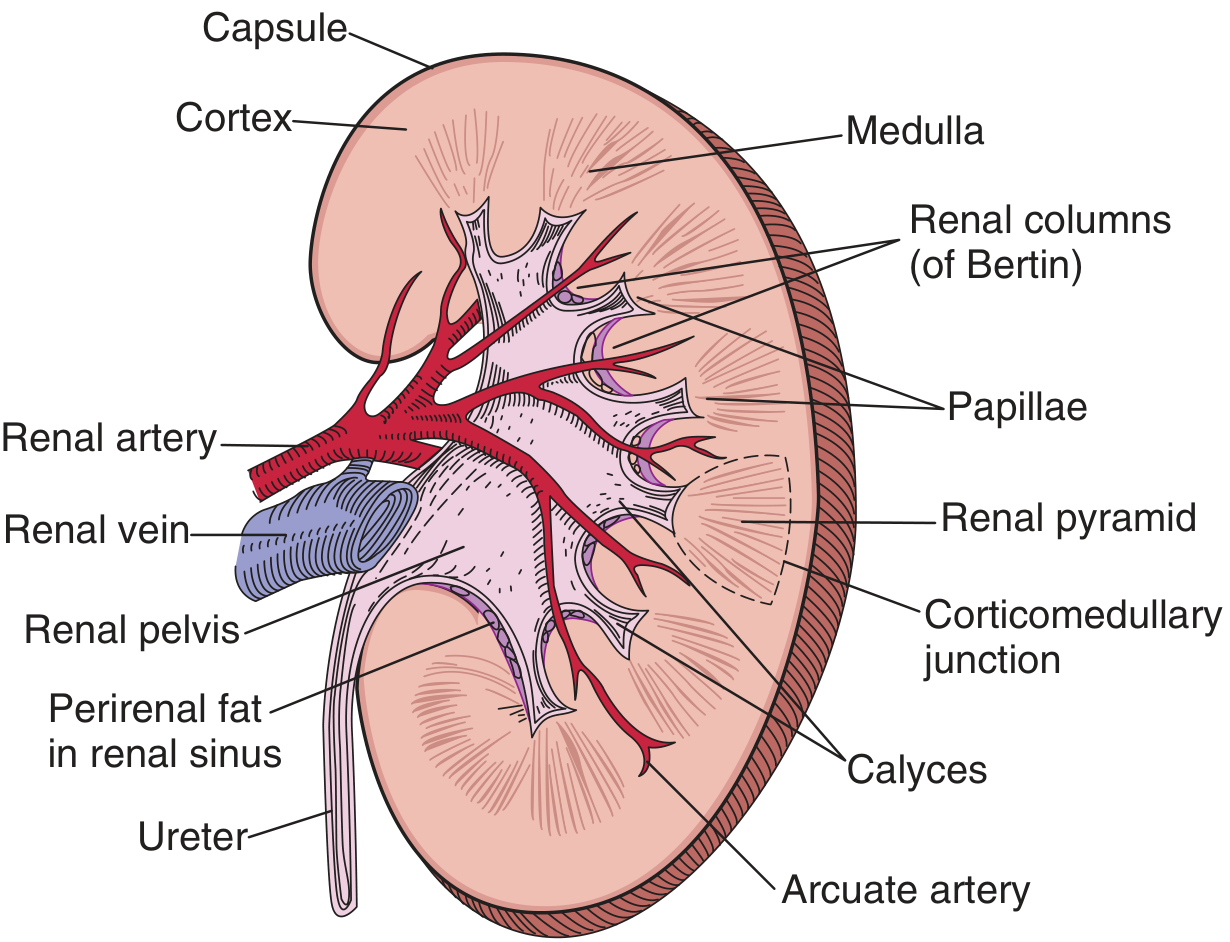
Fig. 2.4 - Cut surface of a bisected kidney showing major anatomical structures. (Brenner and Rector's The Kidney)
Collecting System
The renal pelvis is the expanded funnel of the upper urinary tract. It branches into 2-3 major calyces, which subdivide into 8-14 minor calyces, each receiving urine from one papilla. The pelvis tapers at the ureteropelvic junction (UPJ) to continue as the ureter.
Renal Vasculature
The renal artery arises from the aorta at L1-L2. The right renal artery passes posterior to the IVC. Each artery divides at the hilum into:
- Anterior division - supplies ~2/3 of the kidney; gives 4 segmental branches: apical, upper, middle, lower
- Posterior division - supplies ~1/3; gives the posterior segmental artery (usually the first branch, arising before the hilum)
These are end arteries - no collateral anastomoses between segments, so occlusion causes segmental infarction.
Intrarenal hierarchy: Segmental → Lobar → Interlobar (run in cortical columns) → Arcuate (at corticomedullary junction) → Interlobular → Afferent arterioles → Glomeruli → Efferent arterioles → Peritubular capillaries / Vasa recta
Surgical landmark: The longitudinal avascular plane (line of Brodel) runs just posterior to the lateral convexity, between anterior and posterior segmental arterial territories. Incisions here minimize blood loss (used in anatrophic nephrolithotomy and preferred for percutaneous access).
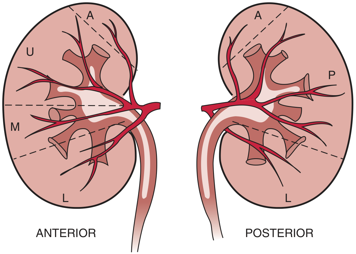
Fig. 2.1 - Vascular segmentation of the human kidney, anterior (left) and posterior (right). (Brenner and Rector's The Kidney)
Accessory renal arteries occur in 25-28% of patients and represent the sole supply to a segment (commonly the lower pole). They are important to identify before donor nephrectomy.
The Nephron
The nephron (~900,000-1 million per kidney) is the functional unit, comprising:
- Renal corpuscle (glomerulus + Bowman's capsule)
- Proximal convoluted tubule
- Loop of Henle (thin descending, thin ascending, thick ascending limbs)
- Distal convoluted tubule
- Connecting tubule → drains into collecting duct
The collecting duct system (derived from the ureteric bud, not metanephric blastema) is not strictly part of the nephron but is critical for final urine concentration.
2. URETERS
Course and Segments
The ureter is a ~25-30 cm muscular tube running from the renal pelvis to the bladder. For radiologic purposes, it is divided into three segments:
- Proximal - from UPJ to the upper border of the sacroiliac joint
- Middle - over the sacroiliac joint
- Distal - from lower border of the sacroiliac joint to the bladder
The ureter crosses over the bifurcation of the common iliac artery at the pelvic brim - a key landmark. It then curves medially in the pelvis to enter the posterolateral base of the bladder.
Key Surgical Relations
- In males: The ureter passes beneath the vas deferens ("water under the bridge"), lying near the tips of the seminal vesicles before entering the bladder
- In females: The ureter passes beneath the uterine artery ("water under the bridge") within the cardinal ligament - crucial in hysterectomy
Points of Physiological Narrowing
Three sites where stones most commonly obstruct:
- UPJ (ureteropelvic junction)
- Pelvic brim (crossing over iliac vessels)
- Ureterovesical junction (intramural ureter, most common site)
Microscopic Anatomy
The ureteral wall has three layers:
- Transitional epithelium (urothelium) with underlying lamina propria
- Smooth muscle (helical inner + outer longitudinal, becomes three layers distally)
- Adventitia with the vascular supply
The Waldeyer's muscular sheath surrounds the intramural ureter and is continuous with the trigonal smooth muscle.
Ureteral Duplication
- Incomplete (partial): Two ureteric buds fuse before entering the bladder (Y-shaped)
- Complete duplication: Two separate ureteric orifices in the bladder. The Weigert-Meyer rule applies: the upper pole ureter inserts inferomedially, the lower pole ureter inserts superolaterally. The upper pole ureter is prone to ectopic insertion and ureterocele; the lower pole ureter is prone to vesicoureteral reflux.
3. BLADDER
Position & Gross Anatomy
The bladder is an extraperitoneal organ in the anterior pelvis. When empty it sits behind the pubic symphysis; when full, the peritoneum reflects off the dome and the bladder rises into the abdomen.
Key spaces and relations:
- Space of Retzius (retropubic space): loose areolar tissue between the bladder and pubic bone anteriorly - opened in retropubic surgery
- Rectovesical pouch (males): peritoneal reflection between rectum and posterior bladder
- Vesicouterine pouch (females): between anterior uterus and posterior bladder
The bladder is divided into:
- Dome (body) - above the ureteral orifices; distensible
- Base - trigone + bladder neck; architecturally distinct
The trigone is the triangular region between the two ureteral orifices and the internal urethral meatus. It has a different embryological origin (from the mesonephric duct) and two muscle layers:
- Superficial trigone - continuous with the ureteral musculature
- Deep trigone - continuous with the detrusor
Wall Layers (from lumen outward)
- Urothelium (transitional epithelium, ~7 cell layers; apical "umbrella cells" are multinucleated)
- Lamina propria
- Muscularis propria (detrusor) - interlacing smooth muscle bundles in 3 layers
- Adventitia / perivesical fat
A complete ring of smooth muscle at the bladder neck (internal urethral sphincter) is present in males (facilitates antegrade ejaculation via sympathetic innervation), but not in females.
Blood Supply
- Superior vesical artery (from umbilical artery ← internal iliac)
- Inferior vesical artery (direct branch of anterior internal iliac; vaginal artery equivalent in females)
- Venous drainage: vesical venous plexus → internal iliac vein
Bladder & Outlet Anatomy (Diagram)
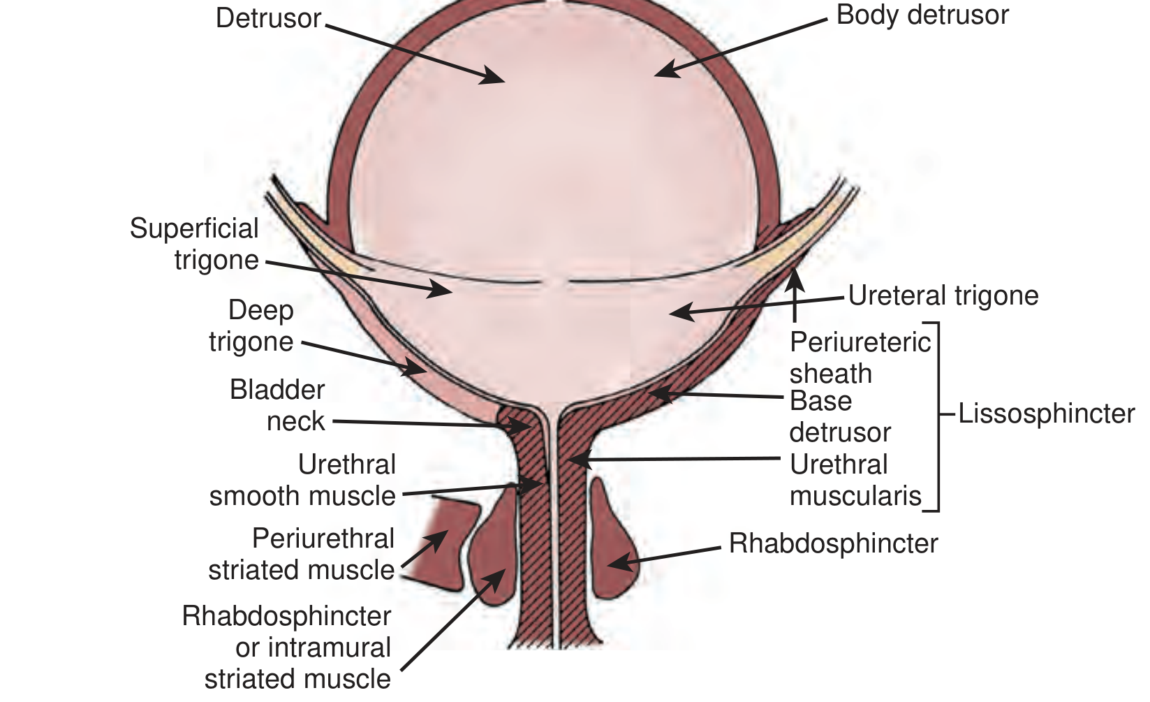
Fig. 110.1 - Bladder and outlet anatomy. (Campbell Walsh Wein Urology)
4. URETHRA
Male Urethra (~20 cm)
Seven anatomical subdivisions (International Consultation on Urological Diseases 2010 classification):
- Urethral meatus - slit-like external opening, slightly ventral on the glans tip
- Fossa navicularis - dilated within the glans; lined by stratified squamous epithelium
- Penile (pendulous) urethra - within the corpus spongiosum through the shaft
- Bulbous urethra - in the perineum; eccentrically placed within the spongiosum proximally
- Membranous urethra - traverses the urogenital diaphragm; contains the external urethral sphincter (rhabdosphincter) - the only striated voluntary sphincter
- Prostatic urethra - traverses the prostate; the verumontanum lies on the posterior wall where the ejaculatory ducts open; the urethra angles anteriorly at the verumontanum
- Bladder neck - the internal (smooth muscle) sphincter
The anterior urethra (fossa navicularis + pendulous + bulbous) and posterior urethra (membranous + prostatic + bladder neck) is the older, still-common clinical terminology.
Female Urethra (~3-4 cm)
A musculofascial tube from the bladder neck to the external meatus, suspended by the urethropelvic ligament (fused endopelvic and periurethral fascia). Layers include:
- Urothelium (proximal) transitioning to non-keratinized squamous epithelium (distal)
- Vascular lamina propria containing periurethral glands (tubuloalveolar; most prominent in the distal 2/3; drain into distal 1/3)
- Skene's glands - the largest and most distal, drain lateral to the meatus
- Smooth muscle (inner longitudinal, outer circular) and skeletal muscle (most prominent in the middle third; U-shaped, deficient dorsally)
Innervation: pudendal nerve (S2-S4). Lymphatics from proximal urethra drain to external/internal iliac nodes; from distal urethra to inguinal nodes.
Urethral Sphincter Diagram
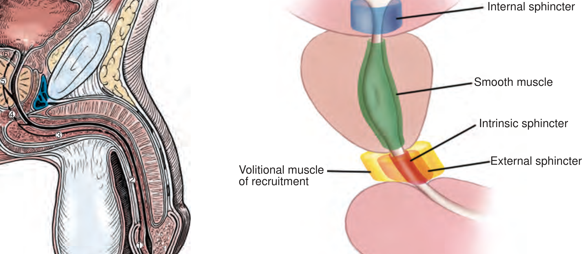
Sagittal male urethra and sphincter anatomy. Numbered segments: 1=fossa navicularis, 2=penile urethra, 3=bulbous urethra, 4=membranous urethra, 5=prostatic urethra, 6=bladder neck. (Campbell Walsh Wein Urology)
5. PROSTATE
Zonal Architecture (McNeal)
| Zone | % of gland | Clinical significance |
|---|---|---|
| Peripheral zone (PZ) | ~70% | Site of ~70% of adenocarcinomas; palpable on DRE |
| Transition zone (TZ) | ~5-10% (grows with BPH) | Benign prostatic hyperplasia originates here |
| Central zone (CZ) | ~25% | Surrounds ejaculatory ducts; rare cancers |
| Anterior fibromuscular stroma | No glands | Purely fibromuscular |
| Periurethral zone | Small | Surrounds proximal urethra |
Relations
The prostate lies between the bladder neck superiorly and the urogenital diaphragm inferiorly, anterior to the rectum (enabling TRUS imaging). The urethra traverses its entire length. The verumontanum marks the posterior wall of the mid-prostatic urethra where:
- Ejaculatory ducts open
- Prostatic utricle (Müllerian remnant) lies
- The urethra angles anteriorly
The seminal vesicles lie posterolateral at the base of the prostate, measuring ~4.5-5.5 cm in length. Each converges with its ipsilateral vas deferens to form an ejaculatory duct, which traverses the prostate to open at the verumontanum. The vasa deferentia course just above the seminal vesicles in the transverse plane.
Blood Supply & Innervation
Blood supply from the inferior vesical artery (prostatic branch). The neurovascular bundles (NVB) of Walsh run posterolaterally at the 5 and 7 o'clock positions - critical to preserve during radical prostatectomy to maintain erectile function.
6. MALE REPRODUCTIVE ANATOMY
Testis
- Paired organs in the scrotum; right commonly hangs lower in ~85% of men
- Normal size: 4-5 cm long, 3 cm wide, 2.5 cm deep; volume 15-25 mL
- Layers (outside to inside): Tunica vaginalis (visceral) → Tunica albuginea (tough fibromuscular capsule, contains smooth muscle) → Tunica vasculosa → Parenchyma
- The tunica albuginea invaginates to form the mediastinum testis and sends septa creating 200-300 cone-shaped lobules
- Each lobule contains 1-4 seminiferous tubules (~1 m long each if uncoiled)
- Sertoli cells - line seminiferous tubules; form the blood-testis barrier via tight junctions
- Leydig cells - interstitial; produce testosterone
- Rete testis → 12-20 efferent ductules → caput epididymis
Arterial supply (3 sources):
- Testicular (internal spermatic) artery - from abdominal aorta; main supply
- Deferential artery - from inferior vesical artery
- Cremasteric (external spermatic) artery - from inferior epigastric artery
Lymphatic drainage: Para-aortic and interaortocaval lymph nodes (at the level of the renal vessels, reflecting embryologic origin). This is why testicular tumors first spread to retroperitoneal nodes, NOT inguinal nodes.
Epididymis
Comma-shaped, attached to the posterolateral testis. ~3-4 m long if uncoiled. Three regions:
- Caput (head) - formed by 8-12 efferent ductules from the rete testis
- Corpus (body) - single tightly coiled duct
- Cauda (tail) - continuous with the vas deferens; sperm storage and maturation
Vas Deferens (Ductus Deferens)
Thick-walled muscular tube (~45 cm) running from the cauda epididymis through the inguinal canal, crossing over the ureter in the pelvis, then joining the seminal vesicle duct to form the ejaculatory duct. The wall contains a prominent thick smooth muscle coat (palpable as a hard cord in the spermatic cord).
Spermatic Cord Contents (mnemonic: Pampiniform plexus, Artery, Nerves, Ductus deferens, Lymphatics, I(nferior) cremaster, Coverings)
- Testicular artery + deferential artery + cremasteric artery
- Pampiniform venous plexus (drains to testicular vein; right → IVC, left → left renal vein)
- Vas deferens
- Lymphatics
- Genital branch of the genitofemoral nerve + ilioinguinal nerve
- Cremasteric muscle fibers
Penis
Three erectile bodies:
- Two corpora cavernosa - surrounded by the thick tunica albuginea; communicate through an incompletely septate midline. Proximally they diverge as the crura, attaching to the ischiopubic rami
- Corpus spongiosum - ventral; contains the urethra; expands distally as the glans penis and proximally as the bulb; its tunica albuginea is thinner
Fascial layers (superficial to deep):
- Skin
- Dartos fascia (areolar, no fat; carries superficial vessels/nerves)
- Buck fascia (deep, tough; contains the deep dorsal vein, paired dorsal arteries, and dorsal nerves on the dorsum; splits ventrally around the corpus spongiosum)
- Tunica albuginea
The glans is covered by the prepuce (foreskin). The meatus is a vertical slit, slightly ventral.
Summary Table
| Structure | Key Dimensions | Blood Supply | Lymphatics |
|---|---|---|---|
| Kidney | 11-12 cm; 125-170 g | Renal artery (aorta L1-L2) | Para-aortic nodes |
| Ureter | ~25-30 cm | Branches from renal, gonadal, aorta, iliac, vesical arteries | Para-aortic, iliac, pelvic nodes |
| Bladder | 300-500 mL capacity | Superior + inferior vesical (internal iliac) | External/internal iliac nodes |
| Female urethra | 3-4 cm | Inferior vesical / vaginal artery | Iliac + inguinal nodes |
| Male urethra | ~20 cm | Urethral artery (internal pudendal) | Superficial inguinal (distal), deep inguinal, iliac (proximal) |
| Prostate | ~20 g normal | Inferior vesical artery | Internal iliac, obturator nodes |
| Testis | 4-5 cm; 15-25 mL | Testicular + deferential + cremasteric arteries | Para-aortic / interaortocaval |
Sources:
- Campbell Walsh Wein Urology, 3-Volume Set - Chapters 34, 45, 46, 60, 73, 74, 83
- Brenner and Rector's The Kidney, 2-Volume Set - Chapter 2 (Anatomy of the Kidney)
- Smith and Tanagho's General Urology, 19th Edition - Chapter on erectile anatomyThat covers the full urology anatomy overview with illustrations. Here's a quick orientation to what was covered:
6 major regions covered:
- Kidneys - retroperitoneal position, gross structure (cortex/medulla/pyramids/calyces), vascular segmentation (5 end-arterial segments, line of Brodel), and the nephron unit
- Ureters - 3-segment radiologic classification, 3 physiological narrowings (stones get stuck here), key surgical relations (vas deferens in males, uterine artery in females), and the Weigert-Meyer rule for duplex systems
- Bladder - extraperitoneal position, trigone anatomy, detrusor layers, blood supply from the internal iliac artery
- Urethra - the 7-segment male urethra (with the rhabdosphincter at the membranous segment) and the 3-4 cm female urethra with Skene's glands
- Prostate - McNeal's zonal anatomy (peripheral zone = cancer, transition zone = BPH), neurovascular bundles at 5 & 7 o'clock
- Male reproductive tract - testis layers and cell types (Sertoli/Leydig), epididymis (caput/corpus/cauda), vas deferens, spermatic cord contents, and penile erectile bodies (Buck fascia, corpora cavernosa, corpus spongiosum)
Let me know if you'd like to go deeper on any specific region - for example the nephron microanatomy, pelvic floor, lymphatic drainage in more detail, or female pelvic anatomy.
This is a shared conversation. Sign in to Orris to start your own chat.