
steps,principle,procedure,modifications,examples of gram staining,afb staining
Reading File
Now let me search the medical library for both Gram staining and AFB staining simultaneously.
Finding Sources
Finding Sources
Finding Sources
Finding Sources
Reading File
Reading File
Finding Sources
Finding Sources
Reading File
Reading File
Reading File
Reading File
Reading File
Reading File
Reading File
Reading File
I now have excellent content from multiple authoritative textbooks. Let me fetch the key diagrams for a visual reference.
Loading Image
Loading Image
I now have all the information needed for a comprehensive answer. Let me compile it.
Gram Staining and Acid-Fast (AFB) Staining
GRAM STAINING
Principle
The Gram stain is a differential stain that separates bacteria into two major classes - Gram-positive and Gram-negative - based on differences in cell wall structure.
- Gram-positive bacteria have a thick peptidoglycan layer that traps the crystal violet-iodine complex after decolorization, retaining the purple color.
- Gram-negative bacteria have a thin peptidoglycan layer plus an outer lipopolysaccharide membrane. The decolorizer (alcohol/acetone) dissolves the outer membrane and washes the dye from the thin peptidoglycan layer, leaving the cells colorless until counterstained red with safranin.
The key mnemonic: P - PURPLE - POSITIVE.
(Medical Microbiology 9e; Jawetz Melnick & Adelbergs Medical Microbiology 28E)
Steps / Procedure
| Step | Reagent | Action | G+ Result | G- Result |
|---|---|---|---|---|
| 1 | Crystal violet (primary stain) | Flood smear for 1 min | Purple | Purple |
| 2 | Gram's iodine (mordant) | Forms CV-iodine complex; flood 1 min | Dark purple | Dark purple |
| 3 | Decolorizer (95% alcohol or acetone) | Wash briefly (~15-30 sec) | Retains purple | Decolorized (colorless) |
| 4 | Safranin (counterstain) | Flood 30-60 sec | Still purple | Red/pink |
Final result:
- Gram-positive = purple/violet
- Gram-negative = red/pink
This process takes less than 10 minutes. (Medical Microbiology 9e)
Visual - Gram Stain Steps
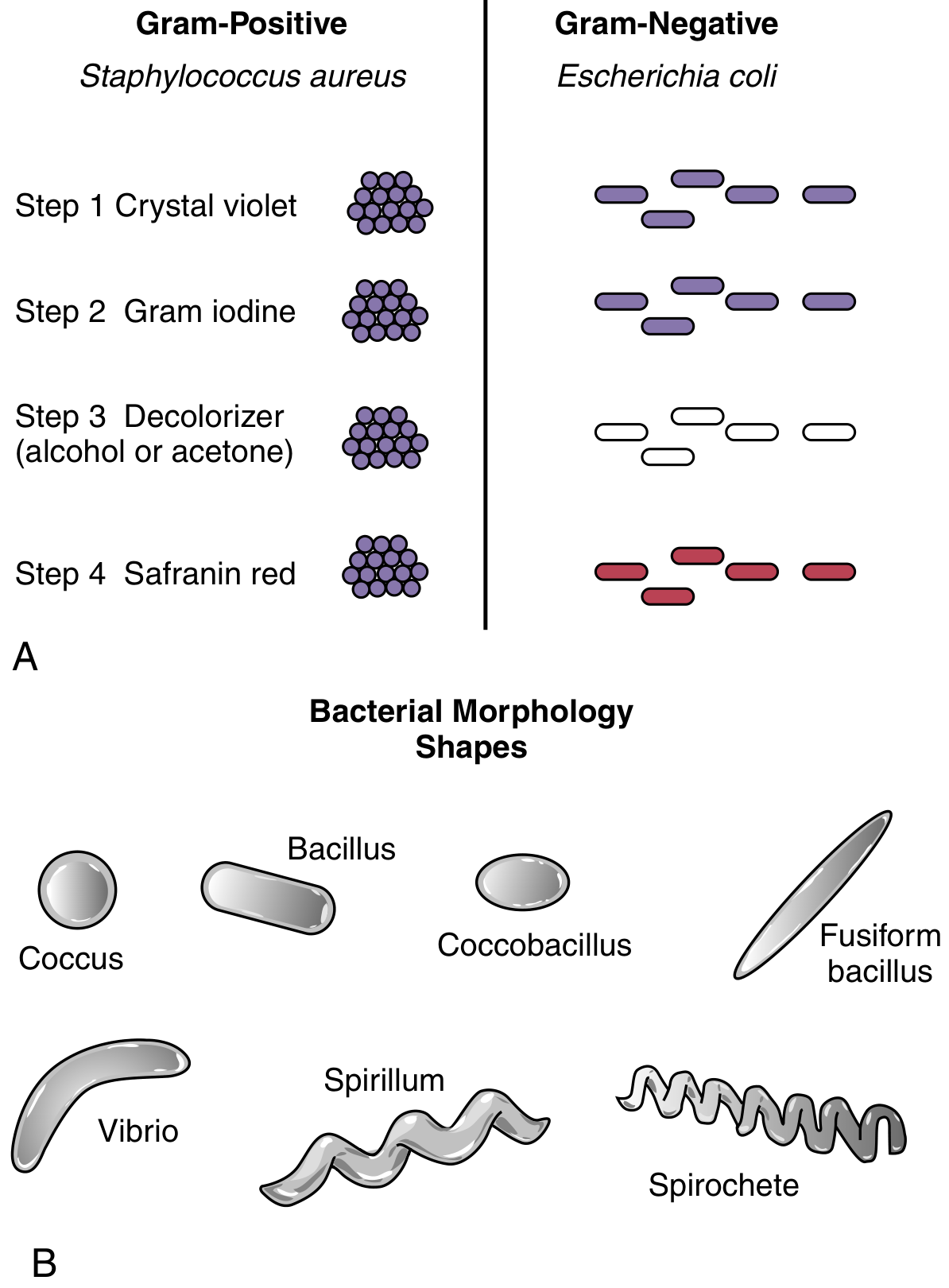
Modifications of Gram Staining
- Burke's modification - Uses sodium bicarbonate solution to enhance crystal violet uptake.
- Kopeloff's modification - Replaces iodine with picric acid; useful for brain tissue.
- Brown and Brenn modification - Used for Gram staining in tissue sections (histopathology); differentiates between Gram-positive and Gram-negative bacteria in formalin-fixed paraffin-embedded tissue.
- Hucker's modification - Uses crystal violet with ammonium oxalate; gives sharper staining of bacteria.
- Gram-Twort stain - Used in histopathology with neutral red and fast green as counterstains.
- Weigert modification - Uses safranin + neutral red as counterstain for tissue sections.
Examples of Gram-Positive and Gram-Negative Organisms
| Gram-Positive (Purple) | Gram-Negative (Red/Pink) |
|---|---|
| Staphylococcus aureus (cocci in clusters) | Escherichia coli (bacilli) |
| Streptococcus pyogenes (cocci in chains) | Klebsiella pneumoniae (bacilli) |
| Bacillus anthracis (large rods) | Neisseria meningitidis (diplococci) |
| Clostridium perfringens (rods) | Pseudomonas aeruginosa (rods) |
| Listeria monocytogenes (rods) | Haemophilus influenzae (coccobacilli) |
| Corynebacterium diphtheriae (pleomorphic rods) | Salmonella typhi (rods) |
Organisms That Stain Poorly or Are Missed on Gram Stain
- Mycobacteria - waxy lipid-rich cell wall resists crystal violet penetration; appear as "ghosts" or colorless bacilli
- Mycoplasma - no cell wall
- Treponema, Leptospira, Borrelia - too thin to visualize
- Chlamydia, Rickettsia - obligate intracellular; not seen on routine smear
- Legionella - stains very faintly Gram-negative; better detected by silver stain
ACID-FAST (AFB) STAINING
Principle
Mycobacteria and related organisms have a cell wall rich in mycolic acids (long-chain fatty acids) - this hydrophobic waxy wall:
- Resists penetration of routine dyes (including crystal violet in Gram stain)
- Once a dye penetrates (using heat/phenol), it forms stable complexes with mycolic acids
- These complexes resist decolorization by concentrated mineral acid + alcohol (acid-fast)
- Non-acid-fast organisms lose the primary stain and pick up the counterstain
(Sherris & Ryan's Medical Microbiology 8e; Tietz Textbook of Laboratory Medicine 7e)
Classic AFB Stain: Ziehl-Neelsen (ZN) - Hot Method
Reagents needed:
- Carbol-fuchsin (primary stain)
- 3% acid-alcohol (20% H2SO4 in 95% ethanol) - decolorizer
- Methylene blue or malachite green (counterstain)
Steps:
| Step | Action | Duration |
|---|---|---|
| 1 | Prepare smear; heat-fix | - |
| 2 | Flood with carbol-fuchsin; heat gently until steam rises (DO NOT BOIL) | 5 min |
| 3 | Cool; rinse with water | - |
| 4 | Decolorize with 3% acid-alcohol | 3-5 min |
| 5 | Rinse with water | - |
| 6 | Counterstain with methylene blue | 30-60 sec |
| 7 | Rinse, dry, examine under oil immersion (100x) | - |
Result:
- AFB (mycobacteria) = red/pink, beaded rods on a blue background
- Non-AFB = blue
AFB Stain Visual
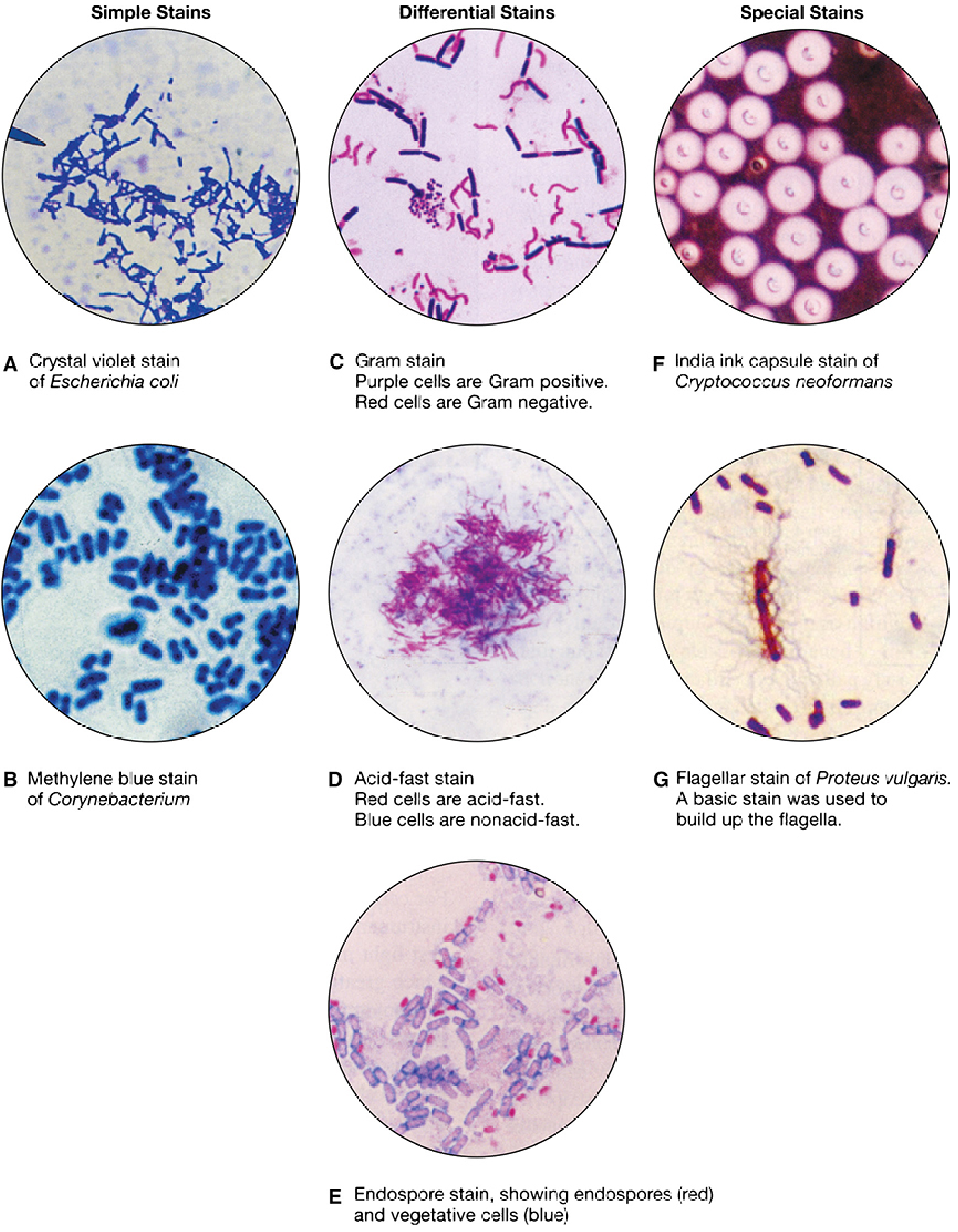
(Panel D shows the acid-fast stain: red cells are acid-fast, blue cells are non-acid-fast)
Modifications of AFB Staining
| Modification | Key Difference | Use |
|---|---|---|
| Ziehl-Neelsen (ZN) - classic | Hot method; heat applied during carbol-fuchsin step | Standard; M. tuberculosis, most mycobacteria |
| Kinyoun's cold stain | No heat; uses higher phenol concentration (4%) for permeabilization; "cold ZN" | Same principle as ZN; lab convenience; less risk of aerosol |
| Auramine-rhodamine (fluorochrome) | Fluorescent dyes (auramine O + rhodamine B); viewed under fluorescence microscope; acid-fast organisms fluoresce yellow-orange against black background | Method of choice per WHO; ~20% more sensitive than carbol-fuchsin; allows rapid low-magnification screening |
| Fite stain | Weaker decolorizer (1% H2SO4); paraffin sections pre-treated with peanut oil to restore mycolic acids | Histopathology of formalin-fixed tissue; detects M. leprae, Nocardia |
| Modified Kinyoun (modified acid-fast) | Even weaker acid (0.5-1% H2SO4) as decolorizer | Detects partially acid-fast organisms - Nocardia, Cryptosporidium, Isospora, Cyclospora |
| Truant fluorochrome stain | Auramine O alone with phenol | Variant of fluorochrome method |
(Medical Microbiology 9e; Tietz Textbook of Laboratory Medicine 7e; Quick Compendium of Clinical Pathology 5e)
Examples of AFB-Positive Organisms
| Organism | Classification | Disease |
|---|---|---|
| Mycobacterium tuberculosis | Strongly acid-fast | Tuberculosis |
| M. leprae | Weakly acid-fast (Fite stain preferred) | Leprosy |
| M. avium-intracellulare (MAC) | Strongly acid-fast | Disseminated infection in HIV |
| M. kansasii | Strongly acid-fast | Pulmonary disease |
| Nocardia spp. | Weakly/partially acid-fast (modified Kinyoun) | Nocardiosis |
| Cryptosporidium parvum | Partially acid-fast oocysts | Diarrhea (immunocompromised) |
| Cyclospora cayetanensis | Partially acid-fast oocysts | Diarrhea |
| Isospora belli | Partially acid-fast oocysts | Diarrhea (HIV) |
| Rhodococcus equi | Partially acid-fast | Pulmonary infection (HIV) |
Key Comparison: Gram Stain vs AFB Stain
| Feature | Gram Stain | AFB/Ziehl-Neelsen |
|---|---|---|
| Primary stain | Crystal violet | Carbol-fuchsin |
| Mordant | Gram's iodine | Heat (ZN) or phenol (Kinyoun) |
| Decolorizer | 95% ethanol or acetone | Acid-alcohol (3% HCl-alcohol) |
| Counterstain | Safranin (red) | Methylene blue (blue) |
| Positive result color | Purple | Red/pink beaded rods |
| Negative result color | Red | Blue |
| Principle | Peptidoglycan thickness | Mycolic acid retention |
| Mycobacteria detected? | No (appears as ghost) | Yes |
| Time | < 10 min | ~20-30 min |
Sources: Jawetz Melnick & Adelbergs Medical Microbiology 28E | Sherris & Ryan's Medical Microbiology 8e | Medical Microbiology 9e (Murray) | Tietz Textbook of Laboratory Medicine 7e | Henry's Clinical Diagnosis and Management by Laboratory Methods | Quick Compendium of Clinical Pathology 5e
This is a shared conversation. Sign in to Orris to start your own chat.