
All nerve system and their course in head and neck chapter
cranial nerves head and neck course diagram overview
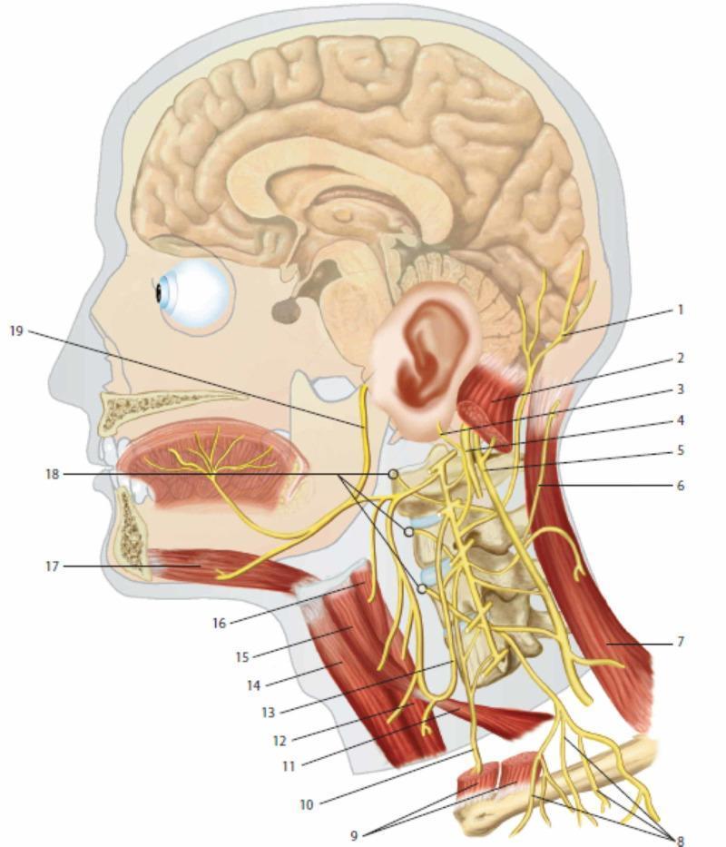
This medical anatomical diagram provides a lateral view of the human head and neck, focusing on the cervical plexus and its integration with cranial nerves. Key neural structures include the lesser occipital nerve (1), greater auricular nerve (3), and transverse cutaneous nerve of the neck (6), demonstrating the sensory distribution of the cervical plexus. The phrenic nerve (10) is shown descending inferiorly anterior to the prevertebral muscles (18). Cranial nerves X (vagus, 4), XI (accessory, 5), and XII (hypoglossal, 19) are illustrated to show their spatial relationships with cervical spinal branches. Muscular landmarks include the sternocleidomastoid (2, 9), trapezius (7), and infrahyoid muscles such as the omohyoid (11, 15) and thyrohyoid (16). The ansa cervicalis (13) is depicted innervating the sub-hyoid musculature. The diagram also illustrates the sagittal section of the brain and oral cavity, highlighting the innervation of the tongue and the relationship between the cervical spine vertebrae and emerging nerve roots. This resource is intended for medical education regarding the peripheral nervous system and regional anatomy of the neck.
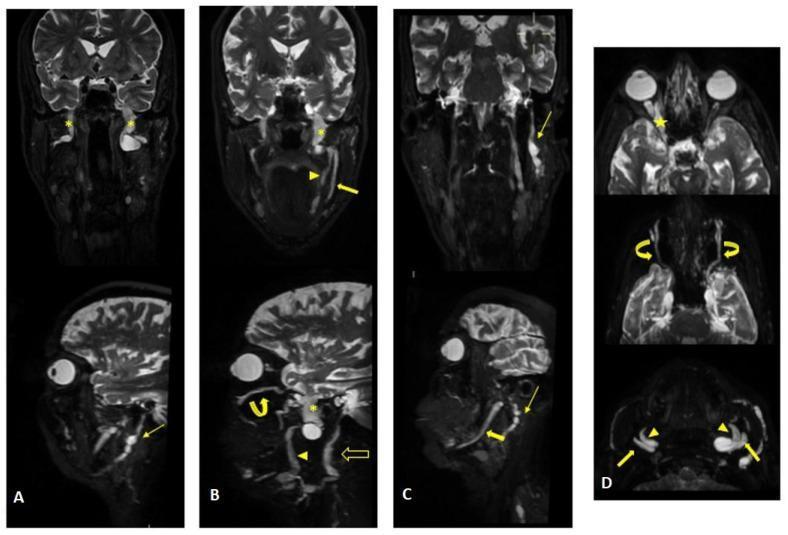
This diagnostic image set consists of multiplanar reconstructions from a 3D MR neurography study of the head and neck. The images demonstrate significant pathological hypertrophy and diffuse thickening of multiple cranial nerves along their extracranial pathways. Key findings include marked enlargement of the trigeminal nerve (CN V) branches: the ophthalmic (V1, yellow star), maxillary (V2, curved arrows), and mandibular (V3, asterisks) divisions, with involvement extending to the lingual (arrowheads) and alveolar (thick arrows) nerves. The left facial nerve (CN VII) exhibits pathological thickening within its intraparotid course (thin arrows), and the hypoglossal nerve (CN XII) shows asymmetric enlargement (open arrow). The thickened nerve segments display heterogeneous signal intensity with visible cystic-like components. Such diffuse, multi-cranial nerve hypertrophy is a distinctive feature often associated with chronic inflammatory demyelinating polyneuropathy (CIDP) or hereditary neuropathies like Charcot-Marie-Tooth disease. The educational focus is on identifying extracranial cranial nerve anatomy and recognizing signs of neurofibromatosis or hypertrophic neuropathies via MR neurography.
This historical anatomical diagram illustrates the principles of phrenology as developed by Franz Joseph Gall. The image depicts a right-profile view of a human head and neck in a classic etching style. The cranial region is subdivided into numerous independent, numbered, and labeled compartments, representing the pseudo-scientific theory of cortical localization where specific cognitive functions, personality traits, and behaviors are mapped to distinct areas of the brain and overlying skull. The labels are in German, featuring terms such as 'Willenskraft' (willpower), 'Selbstvertrau' (self-confidence), and 'Kinderliebe' (love for children). The spatial distribution covers the frontal, parietal, temporal, and occipital regions, suggesting that different faculties like 'Frohsinn' (cheerfulness) or 'Mut' (courage) are localized to specific topographic coordinates. At the base of the neck, the philosophical inscription 'Erkenne Dich selbst' (Know thyself) is visible. While phrenology is now considered an obsolete theory, this diagram is an important precursor in medical history for the development of modern neuroscientific concepts regarding functional brain localization and eloquent cortical areas.
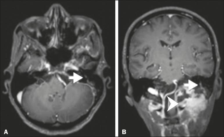
Diagnostic imaging consisting of two post-contrast T1-weighted MRI scans of the head and skull base. Image A is an axial section demonstrating abnormal linear enhancement along the intracranial and intratemporal course of the left facial nerve (Cranial Nerve VII), indicated by a white arrow. Image B is a coronal section showing the same perineural spread (PNS) of malignancy along the facial nerve (arrow) and a primary or recurred soft tissue neoplasm in the infratemporal/parapharyngeal region, marked by a white arrowhead. The imaging illustrates perineural tumor invasion originating from squamous cell carcinoma, highlighting the clinical significance of recognizing continuous nerve enhancement as a sign of disease progression. Key structures visible include the brainstem, cerebellum, and the temporal bone anatomy. This material serves as an educational tool for neuroradiology and oncology, specifically for identifying patterns of head and neck cancer spread along cranial nerves.
Nerves of the Head and Neck - Complete Chapter
Overview: The 12 Cranial Nerves
| CN | Name | Type | Foramen of Exit |
|---|---|---|---|
| I | Olfactory | Sensory | Cribriform plate |
| II | Optic | Sensory | Optic canal |
| III | Oculomotor | Motor (+ parasympathetic) | Superior orbital fissure |
| IV | Trochlear | Motor | Superior orbital fissure |
| V | Trigeminal | Mixed | V1: SOF; V2: Foramen rotundum; V3: Foramen ovale |
| VI | Abducent | Motor | Superior orbital fissure |
| VII | Facial | Mixed | Internal acoustic meatus → stylomastoid foramen |
| VIII | Vestibulocochlear | Sensory | Internal acoustic meatus |
| IX | Glossopharyngeal | Mixed | Jugular foramen |
| X | Vagus | Mixed | Jugular foramen |
| XI | Accessory | Motor | Jugular foramen |
| XII | Hypoglossal | Motor | Hypoglossal canal |
CN I - Olfactory Nerve
CN II - Optic Nerve
- The optic nerve leaves the orbit through the optic canal, accompanied by the ophthalmic artery.
- The two optic nerves meet at the optic chiasm, where fibers from the nasal half of each retina decussate.
- From the chiasm, optic tracts pass to the lateral geniculate nuclei, then via optic radiations to the primary visual cortex (occipital lobe).
CN III - Oculomotor Nerve
- Emerges from the anterior surface of the midbrain (between the midbrain and pons).
- Passes forward in the lateral wall of the cavernous sinus.
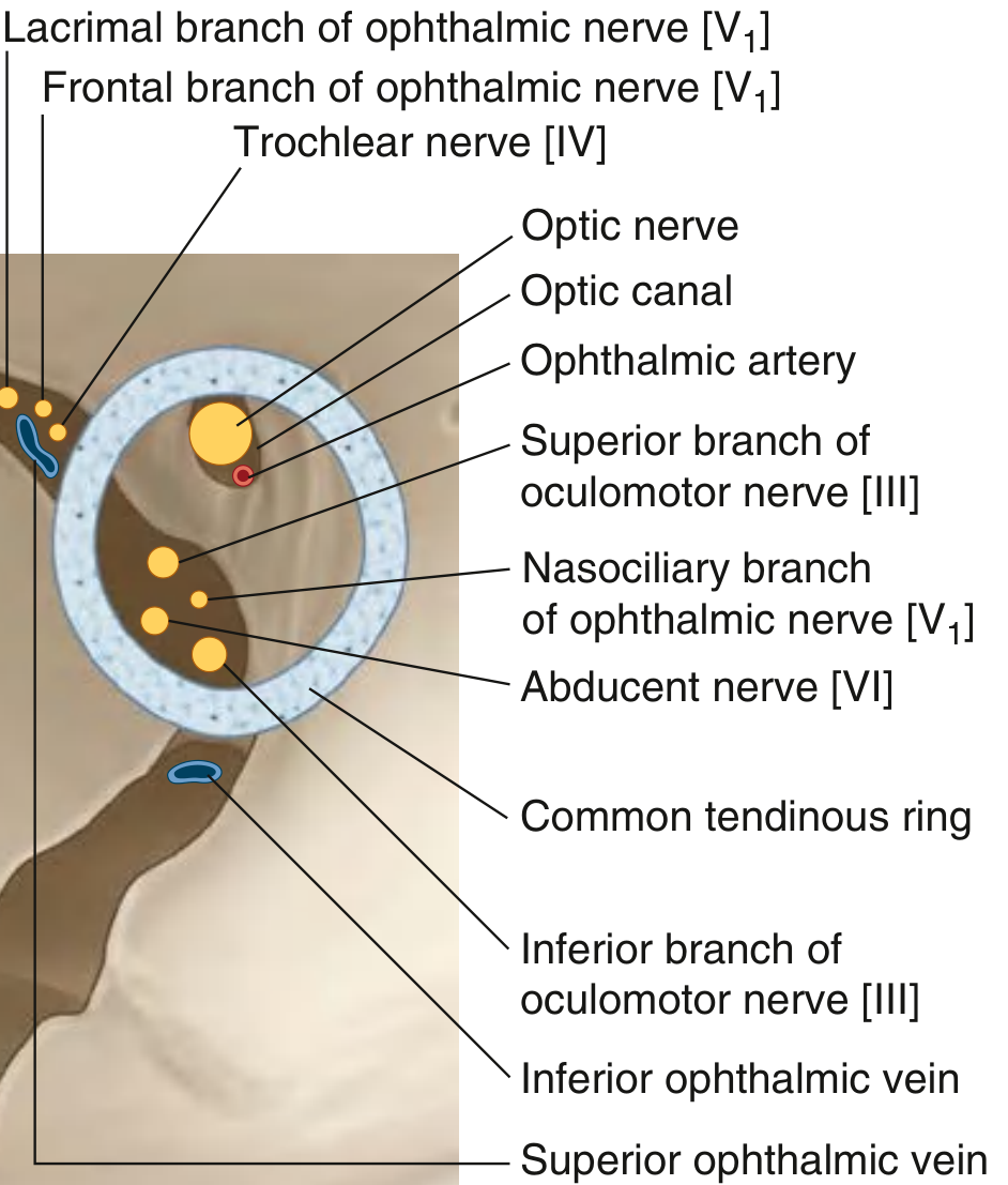
- Divides into superior and inferior branches just before entering the orbit through the superior orbital fissure (within the common tendinous ring).
- Superior branch: passes upward over the lateral side of the optic nerve → innervates superior rectus and levator palpebrae superioris.
- Inferior branch (3 sub-branches):
- To medial rectus (passes below the optic nerve medially)
- To inferior rectus (descends directly)
- To inferior oblique (runs forward along the orbital floor) - this branch also gives off the root to the ciliary ganglion (parasympathetic preganglionic fibers).
CN IV - Trochlear Nerve
- The only cranial nerve to emerge from the dorsal surface of the brainstem (posterior midbrain).
- Wraps around the midbrain, crosses to the opposite side, and passes forward in the lateral wall of the cavernous sinus (inferior to CN III).
- Enters the orbit through the superior orbital fissure outside the common tendinous ring.
- Crosses over to innervate the superior oblique muscle (the only extraocular muscle it supplies).
CN V - Trigeminal Nerve
V1 - Ophthalmic Nerve
- Leaves the trigeminal ganglion and passes forward in the lateral wall of the cavernous sinus (inferior to CN IV and CN III).
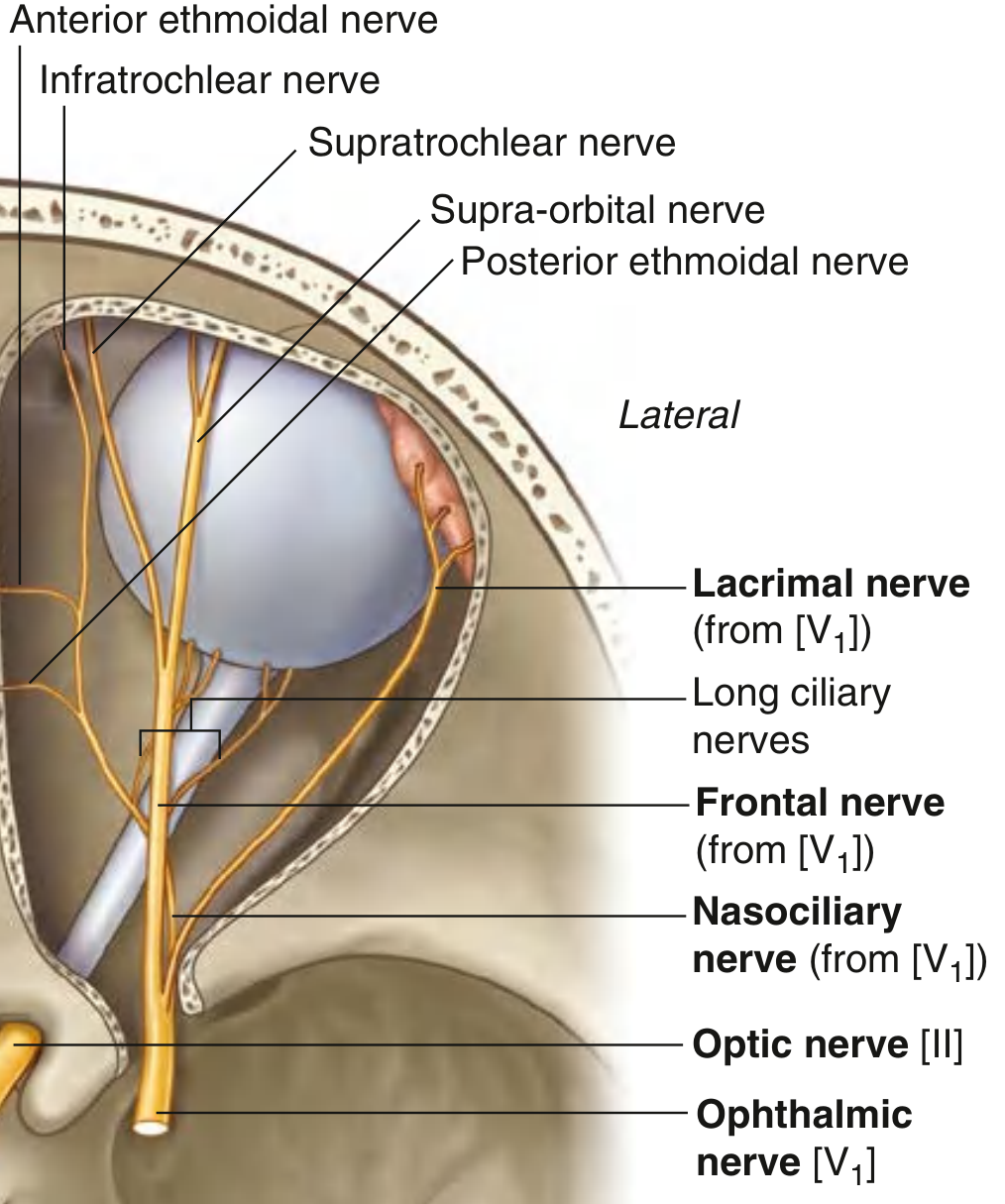
- Divides just before entering the orbit into 3 branches: nasociliary, frontal, and lacrimal.
- All three enter the orbit through the superior orbital fissure (frontal and lacrimal outside the common tendinous ring; nasociliary within it).
- Lacrimal nerve (smallest): Passes along the upper border of the lateral rectus muscle → supplies lacrimal gland, conjunctiva, and lateral upper eyelid. Receives a communicating branch from the zygomaticotemporal nerve carrying postganglionic parasympathetic fibers for the lacrimal gland.
- Frontal nerve (largest): Passes between levator palpebrae superioris and the periorbita. Divides midway into:
- Supratrochlear nerve: anteromedial direction, above the trochlea → skin of medial forehead and upper eyelid
- Supraorbital nerve: exits via supraorbital notch/foramen → skin of forehead and scalp (anterior to lambdoid suture)
- Nasociliary nerve: Crosses the optic nerve to reach the medial orbital wall; gives off:
- Long ciliary nerves → cornea, iris (also carry sympathetic fibers)
- Branch to the ciliary ganglion (sensory root)
- Anterior and posterior ethmoidal nerves → ethmoidal air cells, anterior cranial fossa dura, nasal cavity, external nose (as external nasal nerve)
- Infratrochlear nerve → medial eyelids, adjacent skin and conjunctiva
V2 - Maxillary Nerve
- Leaves the trigeminal ganglion and passes through the foramen rotundum into the pterygopalatine fossa.
- Continues as the infra-orbital nerve through the inferior orbital fissure → infra-orbital groove → infra-orbital canal → exits via the infra-orbital foramen onto the face.
- Meningeal branch (middle meningeal nerve): supplies dura of the middle cranial fossa
- Zygomatic nerve: enters orbit via inferior orbital fissure → divides into:
- Zygomaticotemporal nerve: exits zygoma → skin of temporal region; carries parasympathetic fibers (from pterygopalatine ganglion) via a communicating branch to the lacrimal nerve
- Zygomaticofacial nerve: skin over the zygomatic prominence
- Ganglionic branches (pterygopalatine nerves): sensory fibers that pass through the pterygopalatine ganglion (without synapsing) and distribute as:
- Nasal branches (lateral and medial walls of nasal cavity)
- Palatine nerves (greater and lesser): hard and soft palate
- Nasopalatine nerve: nasal septum and anterior hard palate
- Pharyngeal branch: roof of the nasopharynx
- Posterior superior alveolar nerve: descends into infratemporal fossa → enters posterior surface of maxilla → upper molar teeth
- Infra-orbital nerve terminal branches: middle superior alveolar (premolars), anterior superior alveolar (incisors and canine), infraorbital (skin of cheek, lower eyelid, upper lip, side of nose)
V3 - Mandibular Nerve
- Leaves the cranial cavity through foramen ovale into the infratemporal fossa.
- Divides into a small anterior trunk (mostly motor) and a large posterior trunk (mostly sensory).
- Nerve to medial pterygoid: descends medially to supply medial pterygoid; small branches also supply tensor veli palatini and tensor tympani
- Buccal nerve (sensory): passes laterally between heads of lateral pterygoid → through tendon of temporalis → cheek (skin and mucosa, buccal gingiva of lower molars)
- Masseteric nerve (motor): passes through mandibular notch → masseter muscle
- Deep temporal nerves (motor, usually 2): curve around infratemporal crest → supply temporalis from deep surface
- Nerve to lateral pterygoid (motor): enters directly
- Auriculotemporal nerve (sensory): encircles the middle meningeal artery (2 roots), passes posterior to the neck of the mandible, ascends behind the TMJ, and crosses the zygomatic arch → skin of temple, auricle (including external acoustic meatus), and parotid gland (carries postganglionic parasympathetic fibers from otic ganglion to parotid)
- Lingual nerve (sensory): descends medial to the lateral pterygoid, then between the medial pterygoid and the mandibular ramus, curves below the mucous membrane of the floor of the mouth → general sensation to the anterior 2/3 of tongue, floor of mouth, and lower gingiva. Joined by the chorda tympani (from CN VII) in the infratemporal fossa, which adds taste from anterior 2/3 tongue and preganglionic parasympathetic fibers for submandibular and sublingual glands.
- Inferior alveolar nerve (mixed): descends medial to lateral pterygoid, then between sphenomandibular ligament and mandible → enters mandibular foramen → passes through mandibular canal → supplies all lower teeth (inferior dental plexus); terminal branch exits through the mental foramen as the mental nerve (skin and mucosa of lower lip and chin). Before entering the mandibular foramen gives off the nerve to mylohyoid (motor to mylohyoid and anterior belly of digastric).
- Meningeal branch: ascends through foramen spinosum with the middle meningeal artery → sensory to dura of middle cranial fossa
CN VI - Abducent Nerve
- Emerges from the pontomedullary junction at the inferior border of the pons.
- Has the longest intracranial course of any cranial nerve, making it vulnerable to raised intracranial pressure.
- Passes through the cavernous sinus (here it lies within the sinus, not just in the wall - unlike other nerves) in close relation to the internal carotid artery.
- Enters the orbit through the superior orbital fissure within the common tendinous ring.
- Innervates the lateral rectus muscle (abduction of the eye).
CN VII - Facial Nerve
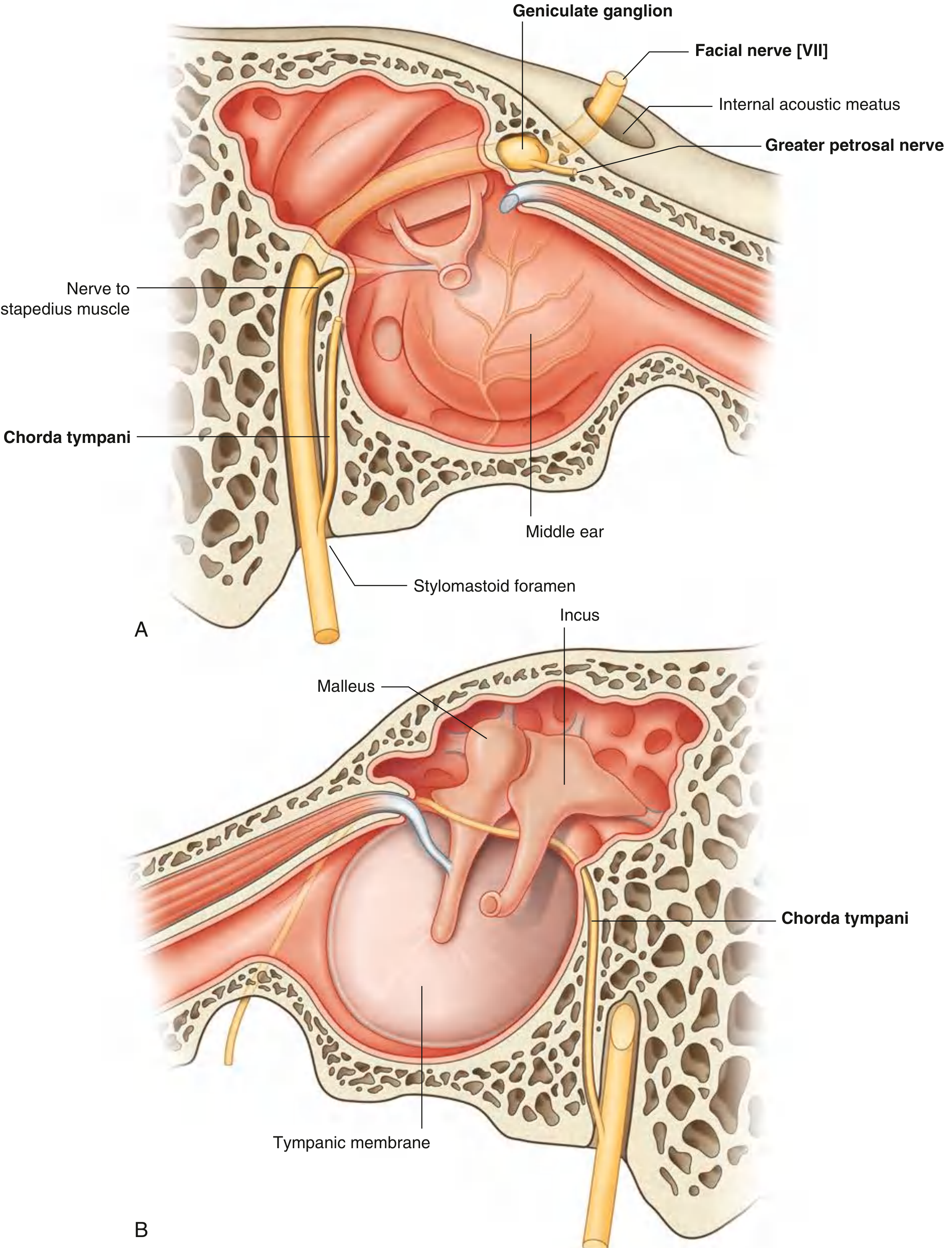
- Enters the internal acoustic meatus in the petrous part of the temporal bone, accompanied by CN VIII and the labyrinthine artery.
- Travels through the facial canal:
- Passes laterally between the internal and middle ears
- Enlarges at the geniculate ganglion (sensory ganglion) and bends posteriorly and laterally
- Turns sharply downward in almost vertical direction
- Exits the skull through the stylomastoid foramen
- Greater petrosal nerve (from geniculate ganglion): carries preganglionic parasympathetic fibers to the pterygopalatine ganglion (for lacrimal gland, nasal and palatine glands) + some taste fibers from soft palate
- Nerve to stapedius: small branch innervating the stapedius muscle in the middle ear (dampens ossicular vibration - hyperacusis when damaged)
- Chorda tympani: arises just before exit from stylomastoid foramen → ascends into middle ear (passes between malleus and incus near the tympanic membrane) → exits through petrotympanic fissure → joins the lingual nerve in the infratemporal fossa → carries:
- Taste from anterior 2/3 of tongue
- Preganglionic parasympathetics to submandibular ganglion → submandibular and sublingual salivary glands
- Enters the parotid gland and divides into its terminal branches within the gland.
- Five groups of terminal motor branches (branches to muscles of facial expression):
- Temporal - frontalis, orbicularis oculi (upper), corrugator supercilii
- Zygomatic - orbicularis oculi (lower), zygomaticus
- Buccal - buccinator, orbicularis oris, muscles of upper lip
- Marginal mandibular - muscles of lower lip, mentalis
- Cervical - platysma
- Also gives off the posterior auricular nerve (just after exiting stylomastoid foramen) → occipitalis and auricular muscles; and branches to the posterior belly of digastric and stylohyoid.
CN VIII - Vestibulocochlear Nerve
- Arises from the inner ear and travels through the internal acoustic meatus alongside CN VII and the labyrinthine artery.
- Has two functional divisions:
- Cochlear nerve (hearing): from the spiral organ (of Corti) in the cochlea → cochlear nuclei in the pons
- Vestibular nerve (balance): from the semicircular canals, utricle, and saccule → vestibular nuclei in the pons and medulla; some fibers go directly to the cerebellum
CN IX - Glossopharyngeal Nerve
- Exits the cranial cavity through the jugular foramen.
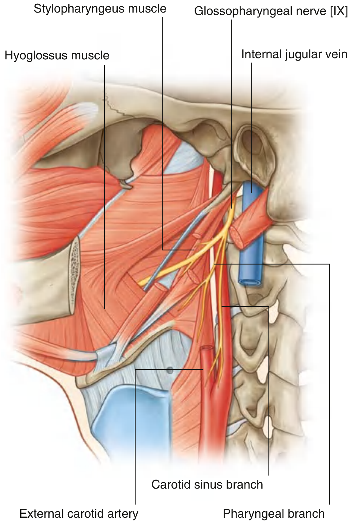
- Begins its descent between the internal carotid artery and the internal jugular vein, lying deep to the styloid process and its associated muscles.
- Passes forward between the internal and external carotid arteries, curves around the lateral border of the stylopharyngeus muscle.
- Continues anteriorly, deep to the hyoglossus muscle, to reach the base of the tongue and the area of the palatine tonsil.
- Tympanic nerve (Jacobson's nerve): re-enters the skull via the inferior tympanic canaliculus → tympanic plexus in the middle ear → lesser petrosal nerve → otic ganglion (preganglionic parasympathetics for parotid gland via auriculotemporal nerve)
- Branch to stylopharyngeus (only motor branch) - the only muscle CN IX innervates
- Carotid sinus branch (nerve of Hering): to the carotid sinus (baroreceptor) and carotid body (chemoreceptor)
- Pharyngeal branches: contribute to pharyngeal plexus → sensory to the pharynx
- Tonsillar branches: sensory to palatine tonsil and adjacent soft palate
- Lingual branches: taste and general sensation to posterior 1/3 of tongue (and vallate papillae)
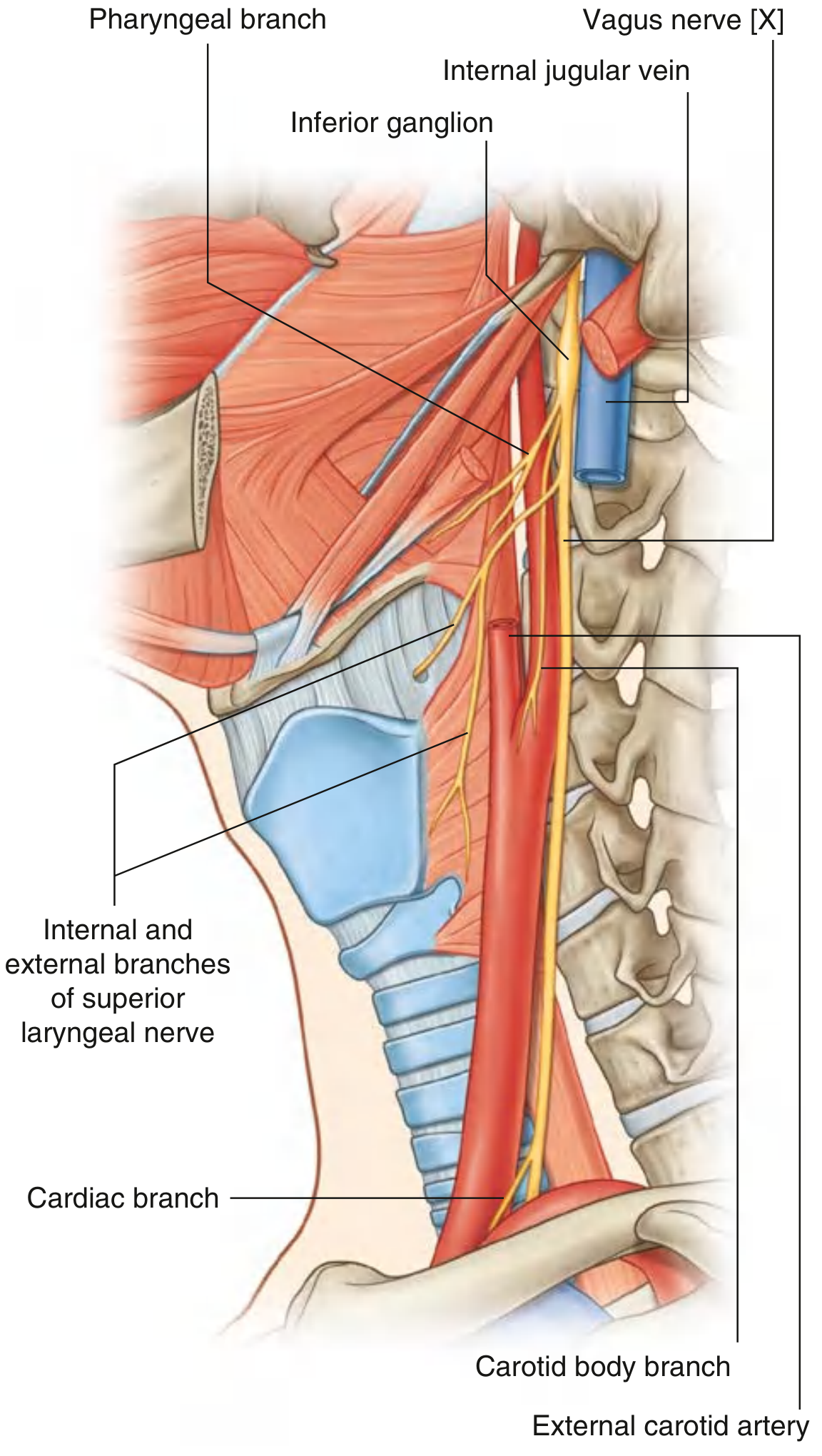
CN X - Vagus Nerve
- Exits the cranial cavity through the jugular foramen between CN IX and CN XI.
- Outside the skull, it enters the carotid sheath and descends through the neck enclosed within this structure - medial to the internal jugular vein, posterior to the internal carotid artery (and then the common carotid artery).
- Has two sensory ganglia:
- Superior ganglion (jugular ganglion): in the jugular foramen
- Inferior ganglion (nodose ganglion): just below the skull base
- Meningeal branch: from superior ganglion → dura mater of posterior cranial fossa
- Auricular branch (Arnold's nerve): from superior ganglion → exits through mastoid canaliculus → external acoustic meatus and adjacent auricle (touching this area can trigger a cough reflex or bradycardia)
- Pharyngeal branches: contribute to pharyngeal plexus → motor to pharyngeal constrictors and soft palate muscles (except tensor veli palatini = CN V3)
- Branch to carotid body
- Superior laryngeal nerve: branches from the inferior ganglion → divides into:
- Internal laryngeal nerve (sensory): passes through thyrohyoid membrane → sensory to laryngeal mucosa above the vocal folds
- External laryngeal nerve (motor): descends alongside the superior thyroid artery → innervates cricothyroid muscle (the only intrinsic laryngeal muscle NOT supplied by the recurrent laryngeal nerve)
- Recurrent laryngeal nerve (RLN): loops around the subclavian artery (right) or aortic arch (left) and ascends in the tracheoesophageal groove → enters larynx posterior to the cricothyroid joint → motor to all intrinsic laryngeal muscles EXCEPT cricothyroid; sensory below the vocal folds
- Cardiac branches (superior and inferior cervical cardiac branches)
CN XI - Accessory Nerve
- Cranial root: from the nucleus ambiguus in the medulla; fibers join the vagus and are distributed via the vagus (some consider this part of CN X)
- Spinal root (the "true" accessory nerve): fibers from anterior horn cells C1-C5/6 of the spinal cord; ascends through the foramen magnum
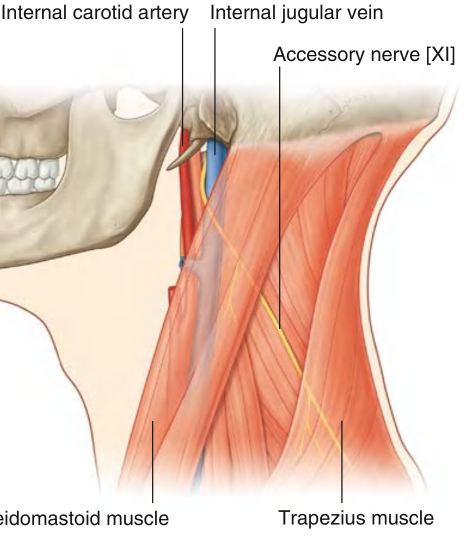
- Exits the cranial cavity through the jugular foramen (most posterior of the three CN IX, X, XI nerves exiting there).
- Begins its descent medial to the internal jugular vein, then emerges from between the internal jugular vein and internal carotid artery to cross the lateral surface of the internal jugular vein.
- Descends through the posterior triangle of the neck (crossing from the posterior border of the sternocleidomastoid to the anterior border of the trapezius), where it lies in the roof of the posterior triangle deep to the investing fascia.
- Gives off no branches as it passes through the anterior triangle.
- Motor innervation: sternocleidomastoid and trapezius muscles.
CN XII - Hypoglossal Nerve
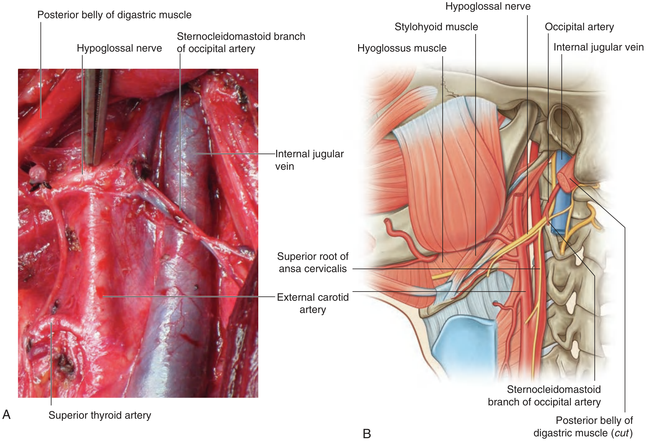
- Leaves the cranial cavity through the hypoglossal canal (in the occipital bone), medial to the internal jugular vein and internal carotid artery immediately outside the skull.
- As it descends, passes outward between the internal jugular vein and internal carotid artery.
- Passes forward, hooking around the occipital artery.
- Crosses the lateral surfaces of the internal and external carotid arteries and the lingual artery.
- Continues deep to the posterior belly of the digastric and stylohyoid muscles.
- Passes over the surface of the hyoglossus muscle and disappears deep to the mylohyoid muscle to enter the tongue.
- Gives off no branches as it passes through the anterior triangle of the neck.
- Motor innervation: all intrinsic and extrinsic muscles of the tongue EXCEPT the palatoglossus (innervated by CN X/pharyngeal plexus).
Cervical Plexus (Neck Nerves)
| Nerve | Origin | Course & Distribution |
|---|---|---|
| Lesser occipital nerve | C2 | Ascends along posterior border of SCM → skin of posterior scalp |
| Greater auricular nerve | C2, C3 | Crosses SCM → skin of auricle, over parotid, and mastoid region |
| Transverse cervical nerve | C2, C3 | Emerges from posterior border of SCM near midpoint → crosses SCM anteriorly → cutaneous innervation of anterior neck |
| Supraclavicular nerves | C3, C4 | Emerge beneath SCM → descend across clavicle → skin of lower neck, shoulder, and upper chest |
| Phrenic nerve | C3, C4, C5 | Descends on anterior surface of anterior scalene → thorax → motor to diaphragm, sensory to pericardium, mediastinal pleura, and diaphragmatic pleura |
| Ansa cervicalis | C1-C3 | Loop of fibers (superior root = C1 fibers with CN XII; inferior root = C2, C3) → motor to infrahyoid "strap" muscles (sternohyoid, sternothyroid, omohyoid) |
Autonomic Nerves of the Head and Neck
Sympathetic Supply
- Origin: T1-T2 lateral horn (ciliospinal center of Budge) → synapse in the superior cervical ganglion
- Postganglionic fibers travel along the internal carotid artery (internal carotid plexus) and external carotid artery branches to reach the head
- Functions in the head: dilator pupillae (pupil dilation), superior tarsal muscle (partial elevation of upper lid), inferior tarsal muscle, sweat glands of face, blood vessels
- Horner's syndrome: interruption at any point of this 3-neuron pathway (hypothalamus → ciliospinal center → superior cervical ganglion → end organ) causes ptosis, miosis, anhidrosis (if below the superior cervical ganglion), and enophthalmos (apparent)
Four Parasympathetic Ganglia in the Head
| Ganglion | CN carrying preganglionic fibers | Postganglionic target |
|---|---|---|
| Ciliary ganglion | CN III (via branch to inferior oblique) | Sphincter pupillae (miosis) and ciliary muscle (accommodation) via short ciliary nerves |
| Pterygopalatine ganglion | CN VII (via greater petrosal nerve) | Lacrimal gland, nasal glands, palatine glands |
| Submandibular ganglion | CN VII (via chorda tympani and lingual nerve) | Submandibular and sublingual salivary glands |
| Otic ganglion | CN IX (via tympanic nerve and lesser petrosal nerve) | Parotid salivary gland (via auriculotemporal nerve) |
Key Clinical Correlations
| Lesion | Result |
|---|---|
| CN III palsy | "Down and out" eye, ptosis, dilated pupil (mydriasis) |
| CN IV palsy | Vertical diplopia, head tilt toward opposite side |
| CN V (trigeminal neuralgia) | Severe lancinating pain in one or more divisions of the face |
| CN VI palsy | Medial deviation of eye at rest, inability to abduct |
| CN VII (Bell's palsy - LMN) | Complete unilateral facial paralysis (including forehead) |
| CN VII (UMN lesion) | Lower facial paralysis only (forehead spared due to bilateral cortical representation) |
| CN IX lesion | Loss of gag reflex, loss of taste and sensation posterior 1/3 tongue, loss of parotid secretion |
| CN X (vagal lesion) | Hoarseness (RLN), dysphagia, loss of gag reflex, tachycardia |
| CN XI (accessory palsy) | Weakness of shoulder shrug (trapezius) and head rotation (SCM) |
| CN XII palsy | Tongue deviates toward the side of the lesion |
| Horner's syndrome | Ptosis, miosis, anhidrosis, enophthalmos |