
Que 1. Draw and label the blood supply of the heart. Briefly describe the histopathological changes in myocardial infarction. Marks 20 (8+12) Que 2. Describe the types of hypersensitivity reactions with its examples. How will you proceed for conducting medicolegal autopsy in a case of death due to anaphylactic shock? Marks 20 (5+3+12) Que 3. Discuss the pathophysiology, gross and microscopic autopsy findings, and biochemical markers of DIC. Describe the pathological features of maternal death from HELLP syndrome. Marks 20 (3+3+3+3+8) Que 4. Write short notes on a) What is the moment of death? Discuss brain stem death criteria. b) What do you understand by Disaster Victim Identification (DVI)? Describe in detail, how does the virtual autopsy help in this concept? c) Describe the histopathology of wound healing. Add a note on immunohistochemistry for the determination of the age of the wound. d) Pathophysiology of shock with its medico-legal importance
coronary arteries blood supply of heart diagram anterior view
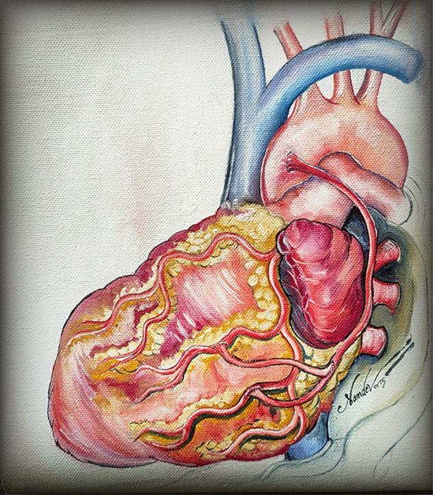
This medical illustration depicts the external anatomy of a human heart with a focus on surgical revascularization. The artwork shows the heart in an anterior view, highlighting the epicardial surface with prominent fatty tissue (yellow) and myocardium (red/pink). Key vascular structures include the superior vena cava (blue), the aortic arch with its branching vessels, and the pulmonary trunk. The illustration specifically demonstrates a 'baby Y' arterial anastomosis technique, where supplementary arterial conduits are grafted onto a parent bypass limb. Red-colored arteries, representing coronary bypass grafts, are shown in a branching pattern, with a proximal anastomosis connecting a secondary limb to the main conduit to supply multiple target vessels, such as the obtuse marginal or diagonal arteries. This anatomical diagram serves to educate on total arterial revascularization strategies in coronary artery bypass grafting (CABG) surgery, emphasizing the distribution and pattern of surgical anastomoses to restore myocardial blood flow.
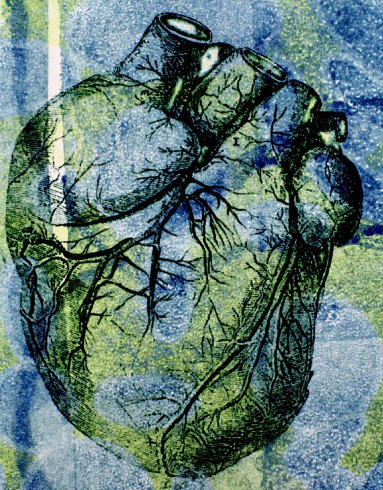
This stylized anatomical illustration depicts a human heart in an anterior view, emphasizing its gross external morphology and coronary vasculature. The organ exhibits its characteristic ovoid shape with superiorly positioned great vessels, including cross-sectional views of the aorta and pulmonary trunk. The surface anatomy highlights the intricate branching patterns of the coronary arteries and veins, demonstrating their distribution across the myocardium toward the apex. The image uses an artistic, high-contrast color palette of green, blue, and yellow to delineate anatomical boundaries and the etched texture of the cardiac tissue. Educationally, the diagram serves to illustrate the relationship between the major cardiac chambers and the superficial vascular network, particularly in the context of cardiovascular health and hemodynamic pressures. The visualization emphasizes the complexity of the organ's blood supply, which is critical for understanding conditions like hypertension and coronary artery disease.
Disaster Victim Identification DVI process role of virtual autopsy CT scan
HELLP syndrome maternal death autopsy pathological findings liver subcapsular hematoma
"disseminated intravascular coagulation" AND biomarkers
https://link.springer.com/chapter/10.1007/978-1-59259-786-4_…
Forensic Medicine & Pathology - Model Answers
Q1. Blood Supply of the Heart & Histopathology of Myocardial Infarction (8 + 12)
A. Blood Supply of the Heart (8 marks)
- Short trunk (0.5-2 cm) running behind the pulmonary trunk, then divides into:
- Left Anterior Descending (LAD) - runs in the anterior interventricular groove to the apex; gives off diagonal branches (to anterolateral LV wall) and septal perforators (anterior 2/3 of interventricular septum, bundle branches). Supplies anterior wall of LV, anterior septum, and apex.
- Left Circumflex (LCx) - runs in the left AV (coronary) groove; gives obtuse marginal branches to the lateral/posterior LV wall. In ~40% supplies the SA nodal artery.
- Runs in the right AV groove; gives:
- Right marginal (acute marginal) branch - to the right ventricle
- SA nodal artery (in ~60%)
- AV nodal artery (in ~90% - "right dominant" circulation)
- Posterior descending/interventricular artery (PDA) in the posterior interventricular groove - supplies posterior 1/3 of septum and posteroinferior LV wall (right dominance ~85%; left dominance ~8%; co-dominance ~7%)
B. Histopathological Changes in Myocardial Infarction (12 marks)
| Time since onset | Gross appearance | Light microscopy |
|---|---|---|
| 0-4 hours | Usually none visible | None by light microscopy (only EM: mitochondrial swelling, glycogen loss) |
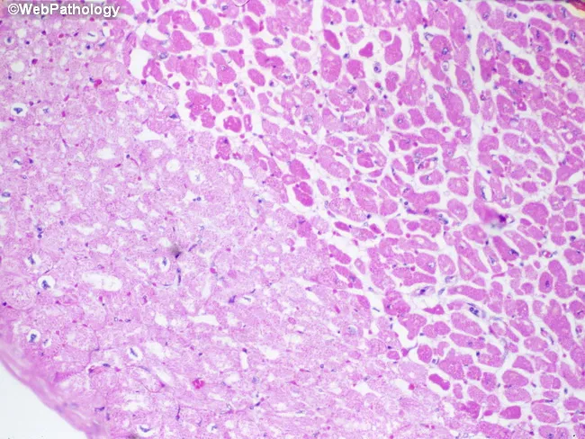
| 4-12 hours | Occasionally dark mottling | Early coagulative necrosis, edema, hemorrhage, wavy fibers, contraction band necrosis at margins |
| 12-24 hours | Dark mottling | Continuing coagulative necrosis; nuclei pyknotic/lost; hypereosinophilia of myocytes; early neutrophilic infiltrate begins |
| 1-3 days | Mottling with yellow-tan infarct center | Coagulative necrosis with loss of nuclei and striations; brisk interstitial neutrophilic infiltrate |
| 3-7 days | Hyperemic border, yellow-tan, soft center (maximal risk of rupture) | Beginning disintegration of dead myofibers with dying neutrophils; early phagocytosis of dead cells at infarct border by macrophages |
| 7-10 days | Maximally yellow-tan, soft, depressed red-tan margins | Well-developed phagocytosis by macrophages; early formation of fibrovascular granulation tissue at margins |
| 10-14 days | Red-gray depressed borders | Well-established granulation tissue with new blood vessels and collagen deposition |
| 2-8 weeks | Gray-white scar, progresses from border toward core | Increased collagen deposition with decreased cellularity |
| >2 months | Dense, white, collagenous scar (complete) | Dense acellular collagenous scar |
Q2. Hypersensitivity Reactions & Medicolegal Autopsy in Anaphylactic Death (5 + 3 + 12)
A. Classification of Hypersensitivity Reactions (Gell and Coombs) (5 marks)
| Type | Mechanism |
|---|---|
| Type I (Immediate/Anaphylactic) | Antigen cross-links IgE on mast cells/basophils → degranulation → release of histamine, leukotrienes, prostaglandins → vasodilation, increased vascular permeability, smooth muscle contraction. Occurs within minutes. |
| Type II (Antibody-mediated/Cytotoxic) | IgG/IgM antibodies bind antigens on cell surface → complement activation, opsonization/phagocytosis, ADCC, or receptor dysfunction. |
| Type III (Immune complex-mediated) | Antigen-antibody immune complexes deposit in vessel walls/tissues → complement activation → neutrophilic infiltration and tissue damage (vasculitis). |
| Type IV (Delayed-type/Cell-mediated) | T-cell mediated (CD4+ Th1 via macrophage activation, or CD8+ cytotoxic T-cell killing) - no antibody involved; peaks at 24-72 hours. |
B. Examples (3 marks)
- Type I: Anaphylaxis (drugs, insect stings, food), bronchial asthma, allergic rhinitis (hay fever), urticaria, angioedema
- Type II: Autoimmune hemolytic anemia, Goodpasture syndrome, hemolytic disease of the newborn (Rh incompatibility), hemolytic transfusion reactions, Graves' disease, myasthenia gravis
- Type III: Serum sickness, systemic lupus erythematosus, post-streptococcal glomerulonephritis, Arthus reaction, polyarteritis nodosa
- Type IV: Tuberculin (Mantoux) reaction, contact dermatitis (poison ivy, nickel), tuberculosis granuloma, graft rejection, Type 1 diabetes mellitus
C. Medicolegal Autopsy in Death Due to Anaphylactic Shock (12 marks)
- Circumstances of collapse, timing relative to drug/injection/sting/food, known atopy or prior allergic history, medical records of the terminal event, drugs administered by treating doctors (adrenaline, antihistamines given?).
- Search meticulously for the injection/sting site - excise it with a 5 cm margin of surrounding skin and subcutaneous tissue, photograph it, and preserve for histology/antigen analysis.
- Look for local swelling; edema of face, eyelids, lips, and conjunctivae.
- Signs of asphyxia: subconjunctival hemorrhages, cyanosis of lips/nail beds, froth at mouth/nostrils.
- Generalized petechial hemorrhages in skin (due to histamine-mediated vasodilation and increased permeability).
- Laryngeal/glottic and epiglottic edema causing airway obstruction - this must be photographed immediately (from above) as it subsides quickly after death.
- Tracheobronchial tree: frothy fluid/mucus.
- Lungs: heavier than normal, markedly hyperinflated/distended (acute emphysema from bronchospasm), with edema and congestion.
- Generalized visceral congestion; occasionally urticaria of serosal surfaces.
- Heart: right ventricular dilatation may be seen; look for any co-existing cardiac pathology to exclude a natural cause.
- Larynx/trachea: submucosal edema, eosinophilic infiltration, mast cell degranulation.
- Lungs: mucus plugging, bronchial smooth muscle contraction, eosinophils in bronchial walls.
- Skin at injection/sting site: perivascular eosinophilic and mast cell infiltrate.
- Preserve blood (femoral vein) for serum tryptase (mast cell marker - remains elevated for several hours post-mortem, most reliable biochemical evidence of anaphylaxis) and total/specific IgE.
- Preserve viscera in saturated saline (not formalin) for toxicological analysis to rule out a drug/poison cause, and to identify the causative antigen.
- Skin biopsy from injection/sting site for histological/immunological confirmation.
- Review the ante-mortem clinical records and any skin prick/RAST test results if the person survived long enough to have them performed.
- Correlate history + external/internal findings + raised serum tryptase/IgE + exclusion of other causes (natural disease, poisoning, trauma) to certify death as due to anaphylactic shock, mentioning the probable allergen if identified.
Q3. DIC and HELLP Syndrome (3+3+3+3+8)
A. Pathophysiology of DIC (3 marks)
B. Gross Autopsy Findings (3 marks)
- Widespread, disproportionate bleeding at venepuncture/injection/surgical sites, mucosal surfaces, and serosae
- Petechiae and ecchymoses in skin and internal organs
- Multiple organ infarcts - kidneys ("shock kidney"/cortical necrosis), adrenals (hemorrhagic infarction - Waterhouse-Friderichsen pattern in sepsis), lungs, brain, pituitary (Sheehan syndrome in obstetric cases)
- Hemorrhage into serous cavities, gastrointestinal tract, and lungs
C. Microscopic Autopsy Findings (3 marks)
- Fibrin/platelet microthrombi in capillaries, arterioles and venules of kidney glomeruli, lung, brain and adrenal (best demonstrated before postmortem fibrinolysis sets in - autopsy should be prompt)
- Microinfarcts with ischemic necrosis in affected organs
- Microangiopathic hemolytic changes (fragmented RBCs/schistocytes on peripheral smear/histology)
- Note: postmortem fibrinolysis can make fibrin difficult to demonstrate histologically if autopsy is delayed - lungs, liver, kidney, and adrenals should specifically be examined.
D. Biochemical Markers (3 marks)
- Thrombocytopenia (progressive fall in platelet count)
- Prolonged PT and APTT
- Decreased fibrinogen
- Elevated D-dimer and fibrin degradation products (FDPs)
- Schistocytes/helmet cells on peripheral smear
- ISTH scoring system combines platelet count, D-dimer, PT prolongation, and fibrinogen level (score ≥5 = overt DIC)
E. Pathological Features of Maternal Death from HELLP Syndrome (8 marks)
- Petechiae and hemorrhagic suffusions in conjunctivae, skin, and serosal/mucosal surfaces of internal organs
- Cerebral edema
- Signs of acute respiratory distress syndrome (ARDS)
- Peripheral (lower limb) edema, hydropericardium
- Splenic hyperemia and "shock kidneys"
- Gross: liver of rigid consistency, yellow-brown cut surface with confluent hemorrhagic (hemorrhagic infarct-like) foci, occasionally subcapsular hematoma (typically over the anterosuperior right lobe) or frank hepatic rupture - the most feared, often fatal complication
- Microscopic: periportal hepatocellular necrosis, hemorrhages sharply demarcated from surrounding normal parenchyma by a fibrin network, and leukostasis in liver sinusoids
- Glomeruli enlarged and appear "bloodless" due to obliteration of capillary lumina by swollen, vacuolated, occasionally foamy endocapillary (endothelial) cells - characteristic glomerular capillary endotheliosis, similar to but often more marked than in pre-eclampsia alone
Q4. Short Notes
a) Moment of Death & Brain Stem Death Criteria
- No spontaneous respiration - apnea test (disconnect ventilator after pre-oxygenation; no respiratory effort despite PaCO2 rising above 60 mmHg)
- Fixed, dilated pupils, unreactive to light
- Absent corneal reflex
- Absent oculo-vestibular reflex (no eye movement on caloric testing with ice-cold water)
- Absent oculocephalic (doll's eye) reflex
- Absent gag/pharyngeal and tracheal (cough) reflex
- No motor response in cranial nerve distribution to painful stimuli
b) Disaster Victim Identification (DVI) and Role of Virtual Autopsy
- Scene examination and recovery of remains
- Post-mortem (PM) data collection (dental, fingerprint, DNA, anthropological, radiological, personal effects)
- Ante-mortem (AM) data collection from families/records
- Reconciliation - comparing AM and PM data (primary identifiers: DNA, fingerprints, odontology; secondary: medical/anthropological findings, personal effects)
- Identification confirmation by a reconciliation board and issue of a death certificate
- Rapid, non-destructive triage of large numbers of bodies/fragmented remains, particularly valuable when bodies are commingled, decomposed, burnt, or religiously/culturally sensitive to invasive dissection
- Detection of radio-opaque personal identifiers: surgical implants, prostheses, pacemakers (with serial numbers), dental fillings/restorations, and foreign bodies useful for AM-PM dental/radiological comparison
- 3D skeletal reconstruction for anthropological assessment of age, sex, stature, and ante-mortem skeletal trauma/old fractures/disease that can be matched against medical records
- Detects hazardous material (explosive fragments, shrapnel) before handling, improving mortuary staff safety
- Provides a permanent, repeatable digital record that can be reviewed by multiple experts without further disturbing the remains, and used in later legal proceedings
- Speeds up throughput dramatically in high-volume disasters, allowing conventional autopsy to be reserved for cases where CT findings are inconclusive
- Limitation: cannot replace histology, toxicology or definitive cause-of-death determination in medicolegally contentious cases - used as an adjunct, not a full replacement, for conventional autopsy.
c) Histopathology of Wound Healing & Immunohistochemistry for Age of Wound
| Phase | Time | Histology |
|---|---|---|
| Inflammatory (hemostasis + inflammation) | 0-3 days | Fibrin-platelet clot; margination and infiltration of neutrophils (peak 24-48 h) clearing debris/bacteria; then macrophages (from day 2-3) which phagocytose debris and secrete growth factors |
| Proliferative | 3 days-3 weeks | Fibroblast proliferation and migration; angiogenesis with formation of granulation tissue (loose connective tissue with new capillaries); re-epithelialization from wound margins; myofibroblasts appear and cause wound contraction |
| Remodeling/Maturation | 3 weeks-months | Type III collagen replaced by stronger type I collagen; decreasing cellularity and vascularity; progressive gain in tensile strength; scar formation |
- Fibronectin - appears very early (within 1-4 hours), one of the earliest markers of vital reaction at wound margins
- P-selectin/E-selectin and other adhesion molecules - within hours, marking early leukocyte margination
- Myeloperoxidase / CD15, CD11b - neutrophil markers, useful in first 1-3 days
- CD68 (macrophage marker) - increases from day 2-3, peaks around day 5-7
- Vimentin/alpha-smooth muscle actin (myofibroblast marker) - appears from day 3-5, increasing through granulation tissue formation
- Factor VIII/CD31/CD34 (endothelial markers) - mark neoangiogenesis from day 3 onward
- Collagen type III vs type I ratio and tenascin - useful for dating wounds of several days to weeks
- Ki-67 (proliferation marker) in epidermal cells at wound margin - marks re-epithelialization activity
d) Pathophysiology of Shock and its Medicolegal Importance
- Hypovolemic - reduced blood/plasma volume (hemorrhage, burns, dehydration) → decreased preload → decreased cardiac output
- Cardiogenic - pump failure (MI, arrhythmia, tamponade) → decreased cardiac output despite adequate volume
- Obstructive - mechanical obstruction to flow (tension pneumothorax, massive PE, tamponade)
- Distributive - profound peripheral vasodilation/maldistribution of flow:
- Septic - endotoxin/cytokine-mediated vasodilation and endothelial injury with DIC
- Anaphylactic - IgE-mediated histamine release causing vasodilation, increased permeability, bronchospasm
- Neurogenic - loss of sympathetic vasomotor tone (spinal cord injury, deep anesthesia)
- Shock (traumatic, hemorrhagic, septic, anaphylactic, neurogenic) is a common immediate cause of death in trauma cases and must be specifically identified at autopsy and correlated with antemortem clinical course
- Distinguishes deaths due to primary injury versus deaths due to a complication (shock) of that injury, which affects causation and the legal chain linking injury to death
- Determines the interval between injury and death, relevant in cases with intervening medical treatment (novus actus interveniens) and for opinions on survivability/negligence
- Autopsy diagnosis of shock (visceral congestion, petechiae, shock organs) supports or refutes claims of natural disease versus violence as the true cause of death
- In poisoning, transfusion reactions, and anaphylaxis, recognizing shock pathology at autopsy is essential to correctly certify manner and cause of death and to identify any third-party liability (e.g., negligent transfusion, drug administration, or failure to treat).
Que 1. Define Euthanasia. Discuss various types of Euthanasia and its religious and current legal status in India. Describe the 2018 Supreme Court guidelines issued in this context and the later 2023 amendments, and discuss the reasons behind these amendments. Marks 20 (2+6+8+4) Que 2. What is surrogacy? Describe in detail the need and the important features of the Surrogacy Regulation Act, 2021. Write a note on the medicolegal implications of surrogacy. Marks 20 (2+10+8) Que 3. Describe various sections of BNS and BNSS applicable to routine medicolegal practice in India. Discuss major changes in comparison to earlier relevant provisions of IPC and CRPC. Marks 20 (6+6+4+4) Que 4. Write short notes on a) Describe the procedure of Narco Analysis in detail. Discuss the legal status of Narco Analysis in India with relevant court judgments. (4+6) b) Define and classify torture. Enumerate features of torture and discuss the role of a forensic expert in the examination of a victim of torture. c) Discuss the morphological changes occurring over the symphyseal face of the hip bone (Innominate) and its relevance in determining age. (2+3+5) d) Discuss the role of consent and its components in research involving human participants. (6+4)
Supreme Court 2023 amendment living will guidelines euthanasia Common Cause procedure simplified
BNS BNSS sections relevant forensic medicine medicolegal changes from IPC CrPC 2023
https://ijfmts.com/archive/volume/9/issue/2/article/2367
Selvi v State of Karnataka 2010 Supreme Court narco analysis polygraph legal status judgment
https://en.wikipedia.org/wiki/Surrogacy_(Regulation)_Act,_20…
Medical Jurisprudence & Forensic Medicine - Model Answers
Q1. Euthanasia (2+6+8+4)
A. Definition (2 marks)
B. Types, Religious Views, and Current Legal Status in India (6 marks)
- Active/Positive euthanasia - a deliberate act of commission to hasten death (e.g. administering a lethal drug dose). Illegal in India (amounts to culpable homicide/murder).
- Passive/Negative euthanasia - an act of omission; withholding or withdrawing extraordinary life-sustaining treatment (ventilator, feeding tube, CPR), allowing natural death to occur. Legal in India under specific safeguards.
- Voluntary - with the patient's own informed consent/request
- Involuntary - performed against the expressed wishes of the patient (illegal everywhere - amounts to murder)
- Non-voluntary - patient unable to express wishes (irreversible coma, severely defective neonate); decision taken by surrogate/family/court
C. 2018 Supreme Court Guidelines (8 marks)
- Execution of the living will: Can only be executed by an adult of sound and healthy mind, acting voluntarily, based on informed consent, free of coercion/inducement, expressed in clear and unambiguous terms.
- Attestation: The document must be signed by the executor in the presence of two attesting witnesses and countersigned by a Judicial Magistrate of First Class (JMFC), who preserves a copy and forwards one to the Registry of the district and to the family physician (if any).
- Content: It should specify the circumstances in which treatment may be withdrawn, and name a guardian/close relative authorized to give consent for withdrawal when the executor becomes incompetent.
- When the directive is to be enforced: The treating physician, on being satisfied that the patient is suffering from a terminal illness/permanent vegetative state with no hope of cure, must inform the hospital, which constitutes a Medical Board.
- Two-tier Medical Board scrutiny:
- Primary Medical Board - constituted by the hospital, headed by the treating physician, with at least three experts from general medicine/cardiology/neurology/nephrology/psychiatry/oncology, each with 20 years' experience, to certify the diagnosis and prognosis.
- Secondary/District-level Medical Board - constituted by the District Collector, chaired by the Chief District Medical Officer with three more expert doctors, to independently review and countersign the Primary Board's decision.
- Judicial oversight: The decision of the Boards is informed to the JMFC, who visits the patient and may endorse or authorize withdrawal of treatment.
- In the absence of an advance directive, similar Board-based safeguards apply, with the family/next of kin approaching the High Court if there is disagreement.
D. 2023 Amendments and Reasons (4 marks)
- The Advance Directive no longer needs attestation/countersignature by a JMFC - it can now be signed before a notary or Gazetted Officer, who also maintains a register.
- The experience requirement for Medical Board members was reduced from 20 years to 5 years, widening the pool of eligible doctors, especially in smaller hospitals/districts.
- Both the primary and secondary hospital-constituted Medical Boards retained, but the earlier requirement of District Collector-appointed Boards was eased - the secondary board is now also constituted by the hospital itself (chaired by a different nominated head of department along with the hospital medical superintendent), removing dependence on collectorate administration.
- A preferable time limit of 48 hours was fixed for both Boards to render their opinion, avoiding delay.
- Requiring a 20-year-experienced specialist board and District Collector-level involvement caused long delays, defeating the purpose of a "dignified" death and prolonging patient/family suffering.
- Small and rural hospitals often lacked doctors with 20 years' experience, making the process unworkable outside large tertiary centres.
- Mandatory JMFC countersignature was seen as an unnecessary bureaucratic/judicial hurdle discouraging people from executing living wills at all.
- The medical fraternity (through ISCCM, backed by the Vidhi Centre for Legal Policy and the End-of-Life Care in India Task Force) petitioned the Court, arguing the guidelines needed to balance patient autonomy/dignity with administrative feasibility.
- The Court accepted that simplification would make the right to a dignified death more accessible and effective in practice, without diluting the safeguards against misuse.
Q2. Surrogacy and the Surrogacy (Regulation) Act, 2021 (2+10+8)
A. Definition (2 marks)
- Gestational (host) surrogacy - the embryo is created using the gametes of the intending couple (or donor gametes) and implanted in the surrogate, who has no genetic link to the child - this is the only form legally permitted in India.
- Genetic (traditional/partial) surrogacy - the surrogate's own ovum is used, making her the biological mother - not permitted under Indian law.
B. Need and Key Features of the Surrogacy (Regulation) Act, 2021 (10 marks)
- Exploitation and commodification of poor/vulnerable women as "surrogacy for hire"
- Absence of legal protection, medical insurance, or informed consent safeguards for surrogates
- Ambiguous parentage/citizenship disputes in cross-border surrogacy (e.g., Baby Manji Yamada case, Jan Balaz case)
- Trafficking of women and children, and abandonment of surrogate-born children (including those with disabilities)
- No central registry/regulation of ART clinics and surrogacy agents
- Prohibits commercial surrogacy entirely; permits only altruistic gestational surrogacy - the surrogate may receive only medical expenses and insurance coverage, no monetary compensation/reward.
- Eligibility of intending couple: Indian citizens, legally married for at least 5 years, wife aged 23-50 years and husband aged 26-55 years, with no surviving biological/adopted/surrogate child (exception: if the existing child is mentally/physically challenged or suffers a life-threatening disorder/fatal illness).
- Certificates required: "Certificate of essentiality" (proof of infertility, court order re: parentage/custody, insurance) and "certificate of eligibility" issued by the appropriate authority before surrogacy can proceed.
- Eligibility of surrogate mother: Must be a close relative of the intending couple, a married woman having a child of her own, aged 25-35 years, medically and psychologically fit, and may act as a surrogate only once in her lifetime.
- Single women: A widow or divorced woman aged 35-45 years ("intending woman") is also permitted to avail gestational surrogacy using donor gametes.
- 2023/2024 amendment: The original Rules required both gametes to come from the intending couple. Following representations about couples with genuine medical conditions, the Rules were amended (2024) to permit use of a donor egg or donor sperm where either the intending husband or wife has a medical indication necessitating it - provided the child retains a genetic link to at least one intending parent (donor gametes for both partners simultaneously remains prohibited).
- Regulatory bodies: Constitution of the National Surrogacy Board and State Surrogacy Boards, and registration of all surrogacy clinics/ART banks with the appropriate authority.
- Insurance: Mandatory medical insurance coverage for the surrogate for 36 months, covering postpartum complications.
- Offences and penalties: Commercial surrogacy, exploitation/abandonment of the surrogate or child, sale of human embryo/gametes, and trafficking are punishable with imprisonment (up to 10 years) and fine.
- Enacted alongside the complementary Assisted Reproductive Technology (Regulation) Act, 2021, which separately regulates ART clinics and banks.
C. Medicolegal Implications of Surrogacy (8 marks)
- Establishment of legal parentage/maternity: The intending parent(s), not the gestational surrogate, are recorded as the legal parents on the birth certificate - requires clear documentation and, in disputed cases, DNA/genetic testing to establish parentage.
- Informed consent: The surrogate's consent must be free, informed, and revocable up to a defined stage; issues of coercion by family/agents remain a medicolegal concern despite the "close relative" restriction.
- Custody disputes: Death, divorce, or refusal of the intending couple to accept the child (especially if born with a disability) raises complex custody and abandonment issues; the Act mandates the intending couple accept the child irrespective of any abnormality.
- Health risks to the surrogate: Complications of pregnancy/delivery, multiple gestation, and the need for adequate insurance and medical care - the treating doctor's medicolegal duty of care extends to the surrogate as a patient in her own right.
- Determining death during pregnancy: If the surrogate or an intending parent dies during the process, legal succession, custody, and citizenship of the child require judicial determination.
- Cross-border/citizenship issues (historically significant, now curtailed by restricting eligibility to Indian citizens) - relevant to residual disputes and NRI intending parents.
- Exploitation and trafficking: Doctors and clinics have a duty to verify eligibility certificates, guard against commercial transactions disguised as "altruistic" arrangements, and report violations.
- Role of the forensic/medical expert: Verification of age, marital status, medical/psychological fitness of the surrogate, documentation of informed consent, and testifying in disputes over parentage, consent, or alleged exploitation.
Q3. BNS and BNSS in Medicolegal Practice (6+6+4+4)
A. Important BNS Sections in Medicolegal Practice (6 marks)
| Old IPC | Subject | New BNS |
|---|---|---|
| S.299/300 | Culpable homicide / Murder | S.100/101 |
| S.302 | Punishment for murder | S.103(1) - new S.103(2) adds enhanced punishment for murder by 5+ persons on grounds of race, caste, sex, place of birth, language etc. (mob lynching) |
| S.304 | Culpable homicide not amounting to murder | S.105 |
| S.304A | Death by negligence | S.106 - now differentiates rash/negligent act by a registered medical practitioner (max 2 years + fine) from other persons (max 5 years + fine); escape from scene increases punishment |
| S.306 | Abetment of suicide | S.108 |
| S.307 | Attempt to murder | S.109 |
| S.320 | Grievous hurt (definition) | S.116-117 |
| S.323/325/326 | Hurt / Grievous hurt / by dangerous weapons | S.115/117/118 |
| S.330/331 | Hurt/grievous hurt to extort confession | S.120/121 |
| S.375/376 | Rape and punishment | S.63/64 |
| S.82/83 | Doli incapax (child <7 / 7-12) | S.20/21 |
| S.84 | Act of person of unsound mind | S.22 |
| S.85/86 | Intoxication | S.23/24 |
| S.87/88 | Acts done by consent | S.25/26 |
| S.309 | Attempt to suicide | Largely decriminalized; retained narrowly (S.226, attempt to suicide to compel/restrain a public servant) |
B. Important BNSS Sections in Medicolegal Practice (6 marks)
| Old CrPC | Subject | New BNSS |
|---|---|---|
| S.53 | Examination of arrested/accused person by medical practitioner at police request | S.51 |
| S.53A | Examination of person accused of rape | S.52 |
| S.54 | Examination of arrested person by medical officer (records injuries, approximate time) | S.53 (BNSS drops the earlier requirement of repeat examination every 48 hours of detention) |
| S.164A | Medical examination of the victim of rape | S.184 |
| S.174 | Police inquest into unnatural/suspicious deaths | S.194 |
| S.175 | Power to summon witnesses for inquest | S.195 |
| S.176 | Magisterial inquiry into custodial deaths/disappearances | S.196 - now also covers deaths/disappearance or rape in police custody with mandatory magisterial inquiry |
| S.161/164 | Statements/confessions to police | ~S.180/183 |
| S.164A | - | New S.176(3): mandates a forensic expert's visit to the crime scene and videography of the investigation process (mobile/electronic device) for all offences punishable with 7+ years imprisonment |
C. Major Changes from IPC (4 marks)
- Consolidation: 511 IPC sections reduced/reorganized into 358 BNS sections; several overlapping provisions merged.
- Doctor-specific liability (S.106): For the first time, medical negligence causing death by a registered medical practitioner is treated distinctly (lower maximum sentence of 2 years vs 5 years for others), addressing longstanding concerns about doctors being clubbed with ordinary offenders under S.304A - though initial mandatory imprisonment provisions caused significant controversy within the medical community before this differentiation was clarified.
- New offences: Organized crime, terrorism, and mob lynching (aggravated murder) formally defined for the first time as substantive offences, relevant to forensic documentation of custodial/group-violence deaths.
- Community service introduced as an alternative punishment for certain minor offences.
- Decriminalization/modification of certain colonial-era provisions (e.g., adultery already struck down; attempt to suicide narrowed).
D. Major Changes from CrPC (4 marks)
- Expanded procedural code: 484 CrPC sections expanded to 531 BNSS sections across 39 chapters.
- Mandatory forensic investigation (S.176(3)): For offences punishable by 7+ years, a forensic expert must visit the crime scene and collect evidence, with video documentation - a major forensic-medicine-relevant addition absent in the CrPC.
- Timelines introduced: Statutory time limits for investigation, filing of charge sheets, and judgment delivery (e.g., judgment within 30 days of conclusion of arguments) to reduce delay - relevant to timely medicolegal report submission and testimony.
- Use of technology: Provision for audio-video recording of search, seizure, and statements; electronic service of summons.
- Custodial examination: BNSS S.53 no longer explicitly mandates the earlier CrPC requirement of medical re-examination every 48 hours during detention - a change flagged by forensic/human-rights commentators as reducing a key safeguard against custodial torture.
- Sentencing discretion (S.25 BNSS vs S.427 CrPC): Courts must now expressly record reasons on whether multiple sentences run concurrently or consecutively rather than a default presumption, and the age threshold for exemption from personal attendance before an investigating officer reduced from 65 to 60 years.
Q4. Short Notes
a) Narco Analysis - Procedure and Legal Status (4+6)
- Also called the "truth serum" test. A barbiturate - classically sodium pentothal (thiopental sodium) or sodium amytal - is administered slowly intravenously in a controlled hospital/forensic setting.
- Pre-test: Physical and psychiatric fitness assessment of the subject; ideally informed consent obtained; team includes an anaesthesiologist, physician, psychiatrist, forensic expert, and videographer; resuscitation equipment kept ready.
- The drug is titrated to induce a hypnotic, disinhibited, semi-conscious "twilight" state between full consciousness and sleep, theoretically depressing higher cortical (inhibitory) control and reducing the subject's ability to fabricate/lie, while leaving speech centres relatively active.
- Pre-formulated questions are asked by the interrogator during this window; vital parameters (BP, pulse, SpO2, ECG) are continuously monitored due to risks of respiratory depression, hypotension, or anaphylaxis.
- The entire session is video-recorded; the transcript/recording, not a "confession" per se, is what is preserved as a record.
- Landmark case: Selvi & Ors. v State of Karnataka (2010) - a 3-judge Supreme Court bench held that compulsory/involuntary administration of narco-analysis, polygraph, and Brain Electrical Activation Profile (BEAP)/brain-mapping tests violates:
- Article 20(3) - the right against self-incrimination (compelling a person to be a witness against himself), and
- Article 21 - personal liberty, mental privacy, and the right against "cruel, inhuman or degrading treatment."
- The Court held these tests can only be conducted with the subject's free, voluntary, and informed consent, given without threat, inducement, or coercion, ideally with legal counsel access.
- Even where validly consented to, the results/statements of narco-analysis do not by themselves constitute a "confession" and are not admissible as direct evidence of guilt; however, any physical/independent evidence subsequently discovered as a result of a lead from the test may be admissible under Section 27 of the Evidence Act (now Section 23 BSA), subject to safeguards.
- Reaffirmed in later cases (e.g., Amlesh Kumar v State of Bihar), which clarified the converse also holds - an accused has no absolute right to demand a narco-analysis test on himself either.
- National Human Rights Commission Guidelines (2000) similarly require prior informed consent, legal representation, and that the test be conducted only at government hospitals with judicial oversight.
b) Torture - Definition, Classification, Features, and Role of the Forensic Expert
- By consent: (a) Self-inflicted/consensual (rare, e.g. ritual practices) and (b) Inflicted by others without consent - further divided into torture of an innocent person versus a person accused of a crime (criminals/terrorists/detainees).
- By nature: Physical torture and Psychological/mental torture.
- By perpetrator: Criminal/terrorist groups; police/security forces during detention/interrogation; military on war captives; domestic/household torture.
- Take a detailed, careful history in a private, non-threatening setting, correlating the account of events with objective findings (as per Istanbul Protocol principles).
- Complete physical examination, systematically documenting all injuries (site, size, shape, colour/age, pattern-matching to alleged weapons e.g. tram-line bruises), noting patterned injuries suggestive of repeated/positional restraint.
- Ancillary investigations: radiography and bone scintigraphy to detect old, healed fractures not visible externally; ophthalmologic and ENT examination (e.g., tympanic membrane rupture from telefono); psychological/psychiatric assessment for PTSD and mental sequelae.
- Photographic documentation with scale markers, maintaining chain of custody for potential legal/human-rights proceedings.
- Provide a written medicolegal opinion correlating findings with the alleged history, explicitly noting consistency, inconsistency, or non-specificity of findings (since with delayed examination, e.g. months later, cutaneous injuries may have healed leaving no trace unless full-thickness skin breach caused scarring).
- Ensure treatment and psychological rehabilitation, without re-traumatizing the victim by repeated recounting of events.
- Maintain strict professional independence/impartiality and refuse to participate in or certify torture, per the Geneva Declaration and Tokyo Declaration obligations.
c) Morphological Changes of the Symphyseal Face (Pubic Symphysis) and Age Determination (2+3+5)
- Young adults (teens-20s): Symphyseal face shows deep, well-marked transverse ridges and furrows running across the entire face; no defined rim; billowing surface.
- 20s-30s: Ridges/furrows begin to fill in and flatten; ossific nodules appear and coalesce to begin forming a rim, first on the dorsal margin, later the ventral margin (the ventral rampart forms last, sometimes not completing until the 30s-40s).
- 30s-40s: Rim becomes essentially complete, oval outline forms; face becomes finely to coarsely granular; no lipping.
- 40s onward: Progressive rim lipping, formation of bony ligamentous outgrowths at the margins, and increasing rarefaction (porosity) of the face.
- Older age (50s+): Marked erosion, pitting, and irregular ossific outgrowths; ventral margin becomes irregular; the face may show a coarsely eroded, friable appearance with disintegration of the rim.
- The pubic symphysis is regarded as one of the most reliable single skeletal sites for adult age estimation (alongside the auricular surface and sternal rib ends), especially superior to cranial suture closure.
- Todd's method (10 phases) and the McKern-Stewart method (component scoring, originally for male military personnel) were early standard techniques; the Suchey-Brooks method (6 sex-specific phases, validated on large modern populations, using reference casts) is now the most widely used and taught.
- It is particularly accurate and useful in young to middle-aged adults (age estimates within about ±5 years up to the 30s-40s), which is medicolegally valuable for identifying unidentified skeletal remains, disaster victims, and in determining whether remains belong to a juvenile or adult.
- Accuracy diminishes with advancing age - later phases (particularly Suchey-Brooks Phase V-VI) span very wide and overlapping age ranges (e.g., Phase V may span 25-83 years), limiting precision in older individuals.
- For medicolegal practice, it is used in combination with other age indicators (auricular surface morphology, sternal rib end changes, dental wear, radiographic epiphyseal fusion) to arrive at a composite, corroborated age estimate, important in disaster victim identification, unidentified body cases, and any legal proceeding where the age of skeletal remains is in question (e.g., determining minority/majority status).
d) Consent and its Components in Human Research (6+4)
- Disclosure - full information on the purpose, procedures, expected duration, foreseeable risks and discomforts, potential benefits, alternative procedures/treatments available, and confidentiality safeguards.
- Comprehension - information must be conveyed in simple, understandable language (participant's own language/literacy level), allowing adequate time for questions.
- Voluntariness - participation must be free of coercion, undue influence, or inducement (financial or otherwise); the participant must have a genuine, unfettered right to refuse or withdraw at any time without penalty or loss of benefits to which they are otherwise entitled.
- Competence - the participant must have the legal and mental capacity to understand and decide; where competence is lacking (minors, unconscious patients, persons with mental illness/intellectual disability), consent must be obtained from a legally acceptable representative (LAR), with assent additionally sought from the participant where possible.
- Documentation - a signed and dated written Informed Consent Form (ICF), witnessed appropriately; for clinical trials in India, audio-visual recording of the consent process is mandatory (New Drugs and Clinical Trials Rules, 2019).
- Continuing/ongoing consent - particularly for long-duration studies, re-consent as protocols change or new risks emerge.
- Rooted in the Nuremberg Code (1947), Declaration of Helsinki, and the Belmont Report (1979) principles of respect for persons, beneficence, and justice.
- In India, governed by the ICMR National Ethical Guidelines for Biomedical and Health Research on Human Participants (2017) and the New Drugs and Clinical Trials Rules, 2019.
- Mandatory prior approval of the research protocol and consent process by an Institutional Ethics Committee (IEC) - constituted with an odd number of members (commonly 9-11), including a legal expert, a social scientist/lay/lady member, and a chairperson from outside the institution, to independently safeguard participant welfare.
- Special additional protections required for vulnerable populations (children, prisoners, economically/educationally disadvantaged persons, pregnant women) to prevent exploitation.
- Medicolegal importance: Absence of valid, documented consent can expose the researcher and institution to liability for assault/battery, professional misconduct, and civil/criminal claims, and undermines the ethical and scientific validity of the entire research project; robust consent processes remain central to both participant protection and defensible research conduct.
_______________________________________________________________ Que 1. Mention various ways in which a person can be injured during a bomb blast. Describe the autopsy protocol and autopsy findings in a case of death due to a bomb explosion. Marks 20 (8+4+8) Que 2. How will you conduct a fetal autopsy and give opinion about the age of the fetus? Discuss the fallacies of the hydrostatic test. Marks 20 (8+6+6) Que 3. Discuss the concept of Poison Information Centre and Poison Control Centre. Mention the basic modalities of placing these facilities. Marks 20 (8+6+6) Que 4. Write short notes on a) Define poison. Enumerate and describe the procedure of bedside tests used in the diagnosis of common household poisons. (2+8) b) Explain the mechanism of action, toxicokinetics, signs and symptoms, management, and post-mortem findings in a case of carbon monoxide poisoning. (2+2+2+2+2) c) Discuss the interpretation of clinical and laboratory examination findings in a case of alleged sexual assault survivor. (5+5) d) Describe the various date rape drugs, their mechanism of action, and fatal dose. What are the various detection technologies for date rape drugs? (3+3+4)
Poison Information Centre India NPIC AIIMS modalities setting up poison control centre
date rape drugs GHB ketamine flunitrazepam detection technology hair testing LC-MS fatal dose
forensic medical examination sexual assault survivor interpretation of findings genital injury significance
Forensic Medicine & Toxicology - Model Answers
Q1. Bomb Blast Injuries and Autopsy (8+4+8)
A. Ways a Person Can Be Injured in a Bomb Blast (8 marks)
- Disruptive effect - if the victim is in contact with/carrying the bomb, the body may be blown into fragments scattered over an area up to ~200 m radius; a bomb exploding on the ground may cause traumatic amputation of the lower legs; if the victim was bending over it, severe damage to face, arms and front of chest results.
- Burns - explosive gas temperature can exceed 2000°C, causing flash burns of exposed skin and clothing/nearby objects; areas under tight clothing (collar, bra, waistband, socks) are often spared ("clothing protection pattern").
- Air blast (shock wave) injury - Primary blast injury - a high-pressure shock wave (~1120 ft/sec) followed by a negative-pressure suction wave causes barotrauma at air-fluid interfaces: rupture of tympanic membranes, pulmonary contusion/hemorrhage ("blast lung"), and rupture of hollow viscera (bowel more often affected than lung); fractures of legs/vertebral column from the pressure wave; death can occur with no external injury.
- Flying missiles - Secondary blast injury - bomb fragments or nearby debris (glass, gravel, wood, brick) driven through the air produce a mixture of bruises, abrasions, and puncture lacerations on the skin ("Marshall's triad" - diagnostic of bomb injury), usually <1 cm each, ragged, sometimes containing embedded foreign material; explosive dust may cause uniform "tattooing" of skin.
- Falling masonry/structural collapse - Tertiary blast injury - when a building collapses, victims sustain blunt trauma and may die of traumatic/crush asphyxia; body may also be thrown against objects.
- Toxic fumes/gases - Quaternary blast injury - explosion in a confined space generates carbon monoxide causing asphyxial death; quaternary injury also includes thermal, inhalational, and chemical/biological/radiological contamination effects.
- Incendiary bombs (napalm, phosphorus/magnesium-based, Molotov cocktails) - produce extensive burns (~1000°C) as the primary mechanism.
B. Autopsy Protocol in Bomb Blast Death (4 marks)
- Scene investigation and history: Type of explosive device, confined vs open space, position of victim relative to blast, number of casualties.
- Whole-body radiography (X-ray) before autopsy - mandatory to localize embedded metal fragments, shrapnel, and parts of the detonating mechanism before dissection, and to identify occult fractures.
- Careful external examination and documentation - photograph and map the distribution/pattern of Marshall's triad injuries, burns, and tattooing to reconstruct the victim's position relative to the bomb.
- Systematic internal examination with special attention to ears (tympanic membrane), lungs, and bowel (sites of primary blast barotrauma), and retrieval of all foreign material/fragments for forensic ballistic/explosive residue analysis.
- Identification procedures - in cases of severe fragmentation, DNA profiling, dental records, fingerprints, and anthropological methods are required (may overlap with DVI protocols in mass-casualty blasts).
- Toxicological sampling - blood for carboxyhemoglobin (COHb) if confined-space explosion suspected; tissue and swab samples for explosive residue analysis (nitrates/nitrites).
C. Autopsy Findings (8 marks)
- External: Disruption/mutilation or traumatic amputation of parts (if in contact with bomb); flash burns with sparing under tight clothing; multiple small bruises, abrasions, and puncture-lacerations (Marshall's triad) often containing foreign debris; skin tattooing by explosive dust; pattern of injury indicating position (carrying/bending over the device).
- Ears: Rupture of tympanic membrane(s) - a sensitive marker of significant blast overpressure exposure.
- Lungs: "Blast lung" - patchy or diffuse subpleural and interstitial hemorrhage, pulmonary contusion, and edema; occasionally frank laceration; pneumothorax/air embolism from alveolar-vascular fistulae.
- Gastrointestinal tract: Contusion, perforation, or rupture of bowel loops (gas-filled hollow viscus is especially vulnerable to blast overpressure).
- Skeletal system: Fractures of long bones (especially lower limbs) and vertebral column from the pressure wave or from tertiary impact/structural collapse.
- Embedded foreign material: Metal fragments, shrapnel, pieces of the detonator/casing recovered from wounds - crucial physical evidence for the investigating agency.
- Signs of asphyxia (traumatic/crush asphyxia) if death occurred due to building collapse or CO if the explosion occurred in a confined space (cherry-red lividity, raised COHb on blood analysis).
- Absence of external injury with internal blast injury is well recognized - death can occur purely from primary blast lung/bowel injury without any visible external wound, so a negative external exam does not rule out blast as cause of death.
Q2. Fetal Autopsy, Age Estimation, and Fallacies of the Hydrostatic Test (8+6+6)
A. Conducting a Fetal Autopsy (8 marks)
- Anthropometric measurements: Crown-heel length (CHL), crown-rump length (CRL), head circumference, chest/abdominal circumference, foot length, and body weight - all key indicators of gestational maturity.
- Examine skin for vernix caseosa, lanugo hair, nail development, presence of maceration (skin slippage, discoloration - indicates intrauterine death before delivery) or putrefaction.
- Umbilical cord: length, number of vessels (2 arteries + 1 vein normally), presence of Wharton's jelly, mode of separation/ligation (cut with a sharp/blunt instrument, torn, or naturally separated), signs of drying/mummification (dries and shrivels only after live birth with atmospheric exposure).
- Placenta: weight, completeness, infarcts, retroplacental clot (abruption), signs of infection.
- Look for external congenital anomalies, birth injury (caput succedaneum, cephalhematoma, fractures).
- Respiratory system: Note lung volume/expansion, color, texture; perform hydrostatic and other tests for live birth (see below).
- Gastrointestinal tract: Presence of air in stomach/intestines (Breslau's test - air progressively reaches further down the gut the longer respiration/survival continued).
- Cardiovascular system: Check patency/closure status of foramen ovale, ductus arteriosus, and ductus venosus (progressive closure occurs after birth, aiding in estimating survival time).
- Skeletal ossification centers: Presence/absence and size of centers in calcaneum, talus, and (near term) the distal femoral epiphysis - important both for age estimation and viability assessment.
- Full radiographic examination and, where needed, histopathology of lung, umbilical cord, and placenta.
B. Opinion on the Age of the Fetus (6 marks)
| Parameter | Approx. finding at full term (~40 weeks) |
|---|---|
| Crown-heel length | 48-52 cm |
| Crown-rump length | 28-32 cm |
| Head circumference | 30-35 cm |
| Body weight | 2500-3300 g (female ~100 g less) |
| Nails | Reach/project beyond fingertips |
| Testes | Descended into scrotum (male) |
| Skin | Pink, vernix mainly in skin folds, lanugo scanty (mainly shoulders) |
- Haase's rule (approximation of length by lunar month): for the first 5 months, length (cm) = month²; from the 6th to 10th month, length (cm) = month × 5.
- Ossification centers are a key adjunct: calcaneum ossifies by the 5th lunar month, talus by the 7th month, and the distal (lower) femoral epiphysis appears close to term (~36 weeks) - its presence is strong evidence the fetus was near or at full term, and is specifically used in stillbirth/viability opinions.
- A fetus is considered viable after 210 days (7 lunar months) of intrauterine life (occasionally survivable after 180 days/6 months, but usually immature); an immature infant is defined as one weighing 2000 g or less regardless of gestational duration.
- Degree of maturity (viable/non-viable, mature/immature) is assessed by combining CHL/CRL, weight, and ossification status rather than any single parameter.
C. Fallacies of the Hydrostatic Test (6 marks)
- Diseases causing consolidation/collapse: acute pulmonary edema, pneumonia, congenital syphilis
- Atelectasis - air fails to reach alveoli due to feeble respiration, air is completely reabsorbed by continuing circulation after respiration ceased (as in asphyxia), or excessive air is expelled on expiration due to lung elastic recoil, or obstruction by alveolar duct membrane
- Drowning at birth (e.g., delivery into a toilet bowl/bucket of water)
- Putrefactive gases - though fetal lungs are relatively resistant to putrefaction (less blood content), gas bubbles can still form and cause floating (however, such gas bubbles are typically large, uneven, and shift/reduce on pressure, unlike the fine uniform bubbles of true aeration)
- Artificial inflation - mouth-to-mouth resuscitation or tube insufflation may partially inflate lungs and introduce air into the stomach, mimicking respiration
- Alcoholic fixation of the tissue can alter buoyancy
- Fetus less than 180 days' gestation
- Fetus is a "monster" (major congenital malformation)
- Fetus is macerated or mummified
- Umbilical cord already separated/cicatrized (implying survival beyond the immediate perinatal period, making the test moot)
- Stomach contains milk (clear evidence of feeding/survival regardless of lung findings)
- If the entire thoracic contents float together, the test is regarded as more reliable, but sectional (piecemeal) floating requires cautious interpretation
Q3. Poison Information Centre and Poison Control Centre (8+6+6)
A. Concept (8 marks)
- 24x7, 365-day telephone helpline service providing product-specific information: signs/symptoms, pre-hospital first aid, supportive/symptomatic care, and antidote availability/dosage
- Maintain standardized information cards/protocols for common poisons (organophosphates, carbamates, organochlorines, aluminium phosphide, corrosives, tricyclic antidepressants, benzodiazepines, methanol, carbon monoxide, paracetamol, opioids, plant poisons like Datura and yellow oleander, scorpion sting, cyanide, etc.)
- Toxicovigilance and surveillance - recording poisoning patterns/epidemiology in the region to guide public health/preventive policy
- Research on antidotes, environmental/biological monitoring of chemicals, and toxicokinetics
- Teaching and training programs for healthcare workers on poisoning prevention and management
- Development of contingency/disaster plans for chemical mass-casualty incidents
- Maintenance of a National/Regional Antidote Bank stocking scarce antidotes for emergency dispatch
B. Basic Modalities of Setting Up a Poison Information/Control Centre (6+6 marks)
- Institutional affiliation - ideally attached to a medical college/tertiary hospital with departments of Pharmacology, Forensic Medicine/Toxicology, and Emergency Medicine for multidisciplinary expertise.
- Staffing - qualified "Poison Information Consultants" (pharmacologists/toxicologists/physicians) available round-the-clock, supported by trained call-handling staff, with access to specialist backup (nephrology for dialysis, critical care, etc.).
- Communication infrastructure - dedicated toll-free telephone lines, computerized database/software for rapid retrieval of poison-specific data, and (increasingly) web/app-based query systems.
- Reference resources - current toxicology textbooks/manuals, an indexed card system or database of individual poisons/products (trade names, active ingredients, LD50, clinical course), and access to poison analysis literature updates.
- Laboratory backup (for a full PCC) - a clinical toxicology laboratory capable of rapid qualitative/quantitative analysis of common poisons in biological samples.
- Antidote bank - a stocked, regularly audited inventory of essential and scarce antidotes (atropine, pralidoxime, N-acetylcysteine, naloxone, digoxin-specific antibody, methylene blue, sodium nitrite/thiosulfate, fomepizole, etc.) with a mechanism for emergency dispatch to peripheral hospitals.
- Standard intake protocol - the consultant records an exact history (product name/composition, route, amount, time since exposure, patient's clinical status) on a structured proforma for every call, enabling later data evaluation and follow-up.
- Provision of standardized management protocols - readymade information cards on signs/symptoms, prehospital first aid, hospital treatment steps, and antidote dosage schedules for common poisons.
- Follow-up and case tracking for outcome data and audit.
- Toxicovigilance reporting system feeding into regional/national poisoning surveillance and prevention programs.
- Linkages/networking with other national and regional poison centres, hospitals, and public health/disaster-management authorities for coordinated response to mass poisoning/chemical disaster events.
- Public awareness and training programs on poisoning prevention (e.g., safe storage of household chemicals, pesticides) as an outreach function.
Q4. Short Notes
a) Poison Definition and Bedside Tests for Common Household Poisons (2+8)
| Poison | Bedside test | Principle/Finding |
|---|---|---|
| Organophosphate/Carbamate | (i) Characteristic garlic-like odor of breath/vomitus/clothing (ii) Atropine challenge test - give IV atropine 1-2 mg; failure to produce signs of atropinization (dry mouth, tachycardia, mydriasis) supports OP poisoning | Cholinesterase inhibition blunts anticholinergic response to test-dose atropine |
| Aluminium/Zinc phosphide (rodenticide) | Silver nitrate paper test - a filter paper moistened with silver nitrate held over gastric contents/vomitus turns black | Liberated phosphine gas reduces silver nitrate to black metallic silver; garlic-like odor also present |
| Corrosive acids/alkalis | Litmus paper test on vomitus/gastric aspirate - turns red with acids, blue with alkalis; pH testing | Direct chemical reaction confirming acidic or alkaline corrosive |
| Cyanide | (i) Bitter almond odor of breath (not detectable by all examiners) (ii) Prussian blue test on gastric contents - ferrous sulfate + NaOH added, then acidified; blue color confirms cyanide | Forms ferric ferrocyanide (Prussian blue) complex |
| Salicylates | Ferric chloride (Trinder's) test on urine - purple/violet color | Ferric ions form a colored complex with salicylate |
| Iron salts | Deferoxamine challenge/urine color test - urine turns "vin rosé" (pink-red) after deferoxamine | Chelation of free iron forms ferrioxamine, excreted as colored urine |
| Opioids | Naloxone challenge test - IV naloxone reverses miosis, respiratory depression, and sedation within minutes | Competitive opioid receptor antagonism |
| Carbon monoxide | (i) Blood appears bright pink/cherry red on visual inspection (ii) Ammonium hydroxide/NaOH dilution test - blood diluted with water + NaOH turns straw-yellow (<20% COHb) or pink (>20% COHb), while normal blood turns brown | COHb resists denaturation by alkali unlike normal oxyhemoglobin |
| Barbiturates | Zwikker's test - cobalt-based reagent produces a violet/purple color with barbiturate-containing urine/gastric contents | Cobalt-barbiturate complex formation |
| Methanol/Ethylene glycol (antifreeze) | Fluorescence of vomitus/urine under Wood's (UV) lamp if a fluorescent dye is present in the commercial product | Detects the added fluorescent tracer dye in many antifreeze formulations |
b) Carbon Monoxide Poisoning (2+2+2+2+2)
c) Interpretation of Clinical and Laboratory Findings in Alleged Sexual Assault (5+5)
- General/systemic examination: Document demeanor, any evidence of restraint, intoxication, or physical struggle (bruises, defense wounds on forearms/hands, bite marks, strangulation signs - facial/periorbital/conjunctival petechiae).
- Genital/anogenital examination: Common sites of injury in females are the labia minora and posterior vagina/posterior fourchette; examine labia majora/minora, clitoral hood, periurethral area, hymen, fossa navicularis, and perineum systematically; in males, examine the external genitalia and perianal region.
- Absence of injury does not exclude assault - genital/anogenital injury is found in only about 40% of cases of alleged rape in females; a normal examination is common, especially with lubrication, elapsed time, or if only digital/oral contact occurred, and must never be used alone to disprove an allegation.
- Colposcopy is particularly sensitive for detecting subtle genital trauma (magnification of minute injuries not visible to the naked eye), and a Wood's lamp may help detect semen or foreign debris fluorescing on skin (though not entirely specific).
- Findings must always be interpreted in the context of history - recency of injury, consensual sexual activity, prior surgery/childbirth, and menstrual status can all alter appearance and must be factored in before opining on "recent" trauma.
- All findings should be documented with body/anogenital diagrams and photographs (with consent), regardless of whether injury is found ("no visible trauma noted" is itself a valid, important finding).
- Trace evidence collection: Swabs from vaginal, cervical, anal, and oral sites (as per history) for semen/spermatozoa (acid phosphatase, PSA/p30, and microscopic sperm identification) and for DNA profiling - critical for identifying the assailant; timing of collection matters as spermatozoa motility/detectability declines over hours to days.
- STI testing: Recommended routinely in children/adolescents (can be near-conclusive evidence of assault); in adults, testing is offered based on symptoms/risk or patient request, balanced against rape-shield concerns about using pre-existing STI status to discredit the survivor; prophylactic treatment is generally given regardless of test results.
- Toxicology screening for drug-facilitated sexual assault (DFSA) when history suggests impaired consciousness/amnesia - blood (within ~4-24 hours) and urine (up to 72-96 hours) samples collected with strict chain-of-custody, tested for ethanol, benzodiazepines, GHB, ketamine, cannabinoids, and other common agents.
- Pregnancy testing where relevant, and baseline serology (HIV, hepatitis B/C, syphilis) for medicolegal and treatment purposes.
- All samples must be handled per standardized forensic evidence-collection kits, correctly labeled, refrigerated, and accompanied by unbroken chain-of-custody documentation to ensure admissibility in court; negative laboratory findings (e.g., no semen detected) do not exclude assault, particularly if a condom was used, ejaculation did not occur, or samples were collected late.
d) Date Rape Drugs - Types, Mechanism, Fatal Dose, and Detection (3+3+4)
- Gamma-hydroxybutyrate (GHB)/GBL - a GABA-B receptor agonist causing rapid-onset sedation, amnesia, and muscle relaxation; endogenous CNS depressant analogue.
- Flunitrazepam (Rohypnol) and other benzodiazepines (midazolam, triazolam) - potentiate GABA-A receptor activity, causing profound sedation, anterograde amnesia, and muscle relaxation.
- Ketamine - an NMDA receptor antagonist producing dissociative anesthesia, analgesia, and amnesia.
- Ethanol - remains the most commonly used and detected agent, potentiating GABAergic transmission and impairing judgment/resistance/memory.
- Other agents reported: scopolamine (anticholinergic, causes amnesia/sedation), chloral hydrate, and various sedating antihistamines/prescription sedatives, all sharing the common effect of impairing consciousness, resistance, and memory formation.
- GHB - fatal dose reported in the range of ~50 mg/kg or higher; therapeutic/recreational doses (1-2.5 g) are close to doses causing respiratory depression, making the margin of safety narrow, especially with alcohol.
- Flunitrazepam - fatal dose is high in isolation (benzodiazepines alone rarely fatal), but co-ingestion with alcohol/opioids markedly lowers the lethal threshold via synergistic respiratory depression.
- Ketamine - fatal dose not precisely established in humans as a sole agent; risk arises mainly from combination with other CNS depressants and from aspiration/respiratory compromise during dissociation. (Because these drugs are typically administered surreptitiously in small covert doses to incapacitate rather than kill, death is more often a complication of aspiration, respiratory depression, or combination with alcohol/other depressants than from a defined "fatal dose" of the drug alone.)
- Blood analysis - most useful if collected within the first 4-6 hours post-ingestion (short half-life of most agents, especially GHB, which is detectable in blood/serum for only a few hours).
- Urine analysis - wider detection window than blood: GHB detectable up to ~8-12 hours; flunitrazepam and its major metabolite 7-aminoflunitrazepam detectable in urine for up to 3-5 days (immunoassay/EIA and confirmatory GC-MS).
- Gas chromatography-mass spectrometry (GC-MS) and liquid chromatography-tandem mass spectrometry (LC-MS/MS) are the forensic gold-standard confirmatory techniques, offering high sensitivity and specificity for GHB, ketamine, benzodiazepines, and their metabolites.
- Hair analysis - provides the longest retrospective detection window (weeks), useful when a victim presents late; segmental hair analysis can even help correlate drug exposure with the approximate time of the alleged assault, using GC-MS/MS or LC-MS/MS on hair segments.
- Point-of-care immunoassay (EIA) kits are used for rapid screening in the emergency setting, with confirmatory chromatography-mass spectrometry required for legal purposes.
- Rapid, expeditious sample collection (blood, urine, and ideally hair) within the recommended forensic protocol window (up to 72-96 hours) with strict chain-of-custody is essential, since many of these agents are rapidly metabolized/eliminated and may not be detectable if testing is delayed.
Que 1. Discuss the ethics, law and the rights of the accused in relation to brain mapping and narcoanalysis. Marks 20 (5+5+5) Que 2. What is a necrobiome? Discuss the role of the necrobiome in the estimation of the postmortem interval. Marks 20 (4+16) Que 3. What is DNA fingerprinting? Discuss various techniques and laboratory procedures used for DNA fingerprinting, and their uses/applications as per the DNA Technology Regulation Bill, 2019. Marks 20 (4+8+8) Que 4. Write short notes on a) Define Forensic Psychiatry. How do the concepts of Psychology help in criminal cases? (2+8) b) Describe in detail about superimposition technique. How does it help in identification in case of a mutilated body? (6+4) c) What is nanotechnology? Discuss uses of nanotechnology in forensic investigations. (2+8) d) Discuss medicolegal issues involved in the use of artificial intelligence and ChatGPT in medical practice. (5+5)
Selvi v State of Karnataka 2010 Supreme Court narcoanalysis brain mapping polygraph rights of accused Article 20(3)
DNA Technology Regulation Bill 2019 India provisions DNA profiling board uses applications
https://prsindia.org/billtrack/the-dna-technology-use-and-ap…
necrobiome postmortem interval estimation microbial succession review
https://www.fsigenetics.com/article/S1872-4973(18)30403-4/fu…
medicolegal issues ChatGPT artificial intelligence medical practice liability confidentiality India
https://pmc.ncbi.nlm.nih.gov/articles/PMC10486263/
Forensic Medicine & Medical Jurisprudence - Model Answers
Q1. Ethics, Law and Rights of the Accused in Brain Mapping and Narcoanalysis (5+5+5)
A. Ethical Issues (5 marks)
- Violation of autonomy and bodily integrity - the subject's mind and body are used as a source of evidence against their own will, reducing the person to a mere object of investigation rather than a rights-bearing individual.
- Doctor's dual loyalty conflict - the physician administering the drug/test serves the interest of the investigating agency rather than the patient's therapeutic welfare, breaching the fundamental medical ethic of acting in the patient's best interest (non-maleficence).
- Absence of true informed consent - consent obtained from a person in custody is inherently questionable given the power imbalance, fear of reprisal, and coercive custodial environment; "consent" here rarely meets the standard of free, voluntary, informed consent required in medical ethics.
- Risk of self-incrimination through an altered mental state - the subject cannot exercise judgment about what to disclose, unlike a normal interrogation, raising concerns about dignity and mental privacy ("mental privacy" as an extension of bodily privacy).
- Scientific unreliability - narcoanalysis can produce confabulation, false memories, or suggestible responses (the drugged subject may be more susceptible to leading questions), so using an unreliable technique to extract "truth" is ethically problematic when it may convict the innocent or fail to protect genuine rights.
- Risk of physical/psychological harm - sedative drugs carry risks of adverse reactions, and the psychological harm of forced disclosure of private thoughts can amount to a form of "mental torture."
- Declaration of Tokyo (WMA) principles - physicians must not participate in acts of torture or degrading treatment; critics have equated compulsory application of these tests to a form of psychological coercion inconsistent with the physician's ethical obligation to "do no harm" and to refuse any professional role in producing evidence through non-consensual bodily/mental intrusion.
B. Legal Position (5 marks)
- The Court held that involuntary administration of narcoanalysis, polygraph, and BEAP (brain mapping) tests violates Article 20(3) (right against self-incrimination) and Article 21 (right to life and personal liberty, including the right to privacy and substantive due process).
- Testimonial character: The Court reasoned that in a polygraph or brain-mapping test the subject makes a mental effort that leads to a physiological response, thereby imparting personal knowledge relevant to the facts in issue - this constitutes a "testimonial act" and therefore falls within the protective ambit of Article 20(3), even though the outward act is not a verbal statement.
- Scope of protection: Article 20(3), read with Section 161(2) of the Cr.P.C. (now corresponding provisions of BNSS 2023), protects not only the formally accused but also suspects and witnesses examined during investigation.
- No adverse inference: There is a rule against drawing adverse inferences from a person's refusal to undergo these tests or to answer questions.
- Right to counsel: The Court noted these tests negate the effective right to legal counsel, since counsel cannot advise the client contemporaneously while the tests are administered.
- Evidentiary value: Results obtained through compulsion are inadmissible as evidence. Even where the subject voluntarily consents, only information/material subsequently and independently discovered with the help of such a test (i.e., derivative/confirmatory physical evidence, per Section 27 Evidence Act principles) may be admissible - the test result/statement itself cannot be treated as a "confession."
- Mandatory safeguards laid down in Selvi (following NHRC 2000 guidelines) if the subject voluntarily consents:
- Consent should be recorded before a Judicial Magistrate.
- The subject must be informed of the physical/legal implications and given access to a lawyer.
- Magistrate must ensure consent is voluntary, given without coercion/inducement.
- The test must be conducted by an independent agency (e.g., a hospital) in the presence of the lawyer, with the entire process video-recorded.
- A copy of the Magistrate's order must be given to the accused/counsel.
- Guidelines under the Dhanaraju Committee (NHRC) regarding medical/psychological fitness of the subject prior to the test must be followed.
- Prior conflicting High Court decisions (e.g., Ramchandra Ram Reddy v. State of Maharashtra, which had upheld narcoanalysis with consent) were effectively overtaken by Selvi's constitutional ruling.
C. Rights of the Accused (5 marks)
- Right against self-incrimination (Article 20(3)) - no one accused of an offence can be compelled to be a witness against himself; this extends to the investigation stage and protects against compulsory subjection to these tests.
- Right to personal liberty and privacy (Article 21) - includes "mental privacy," bodily integrity, and protection from state intrusion into one's mind without consent; compulsory administration amounts to unwarranted intrusion into personal liberty, tantamount to "testimonial compulsion."
- Right to a fair trial and due process - includes the right to remain silent and the right not to have adverse inferences drawn from silence.
- Right to legal representation/counsel during any interrogation-related process, including the opportunity to be advised prior to giving consent.
- Right to refuse - the accused (or a suspect/witness) has an absolute right to refuse to undergo narcoanalysis/polygraph/BEAP; refusal cannot itself be treated as evidence of guilt.
- Right to bodily integrity and dignity (Article 21) - forcible administration of a chemical substance into the body is a form of "bodily intrusion" that requires the constitutional standard of "substantive due process," which mere administrative or investigative necessity does not satisfy.
- Right to informed, voluntary consent with judicial oversight - even where a person volunteers, strict procedural safeguards (Magistrate-recorded consent, presence of counsel, video recording, independent conducting agency) must be followed to protect against coerced or induced "consent" in custody.
- Protection of the resulting information - any statement made during the test cannot be used as a confession; only independently, subsequently verified physical evidence discovered as a result may be led in evidence.
Q2. Necrobiome and Postmortem Interval Estimation (4+16)
A. Definition of Necrobiome (4 marks)
- The thanatomicrobiome - the endogenous microbial community that proliferates within internal organs/blood after death (derived largely from the host's own gut, oral, and skin microbiota once immune surveillance ceases).
- The epinecrotic community - microorganisms and invertebrates on the exterior surface of the remains and in the surrounding soil/substrate influenced by the decomposing body.
- Insect and other arthropod communities (forming the traditional basis of forensic entomology) that arrive and succeed one another in a broadly predictable sequence.
B. Role of the Necrobiome in PMI Estimation (16 marks)
- Early stages (fresh to bloat): Dominated by facultative anaerobes and aerobic bacteria (e.g., Enterobacteriaceae, Clostridium spp. from the gut), with rapid bacterial proliferation as tissue autolysis begins.
- Bloat stage: Marks a transition point - anaerobic gut bacteria proliferate explosively, generating gases (putrefaction) that distend the body; internal organ microbiomes shift from an oxygen-limited to anaerobe-dominated profile.
- Active/advanced decay: As tissue ruptures and becomes exposed to the external environment, there is a marked shift from anaerobic to aerobic microbiomes (soil- and insect-associated taxa colonize), providing an objective marker to segment decomposition into pre-rupture and post-rupture phases.
- Skeletonization: Strict anaerobes and soil-derived, environmentally hardy organisms dominate as the substrate becomes nutrient-depleted; fungal communities often become more prominent in these later stages.
- Different body sites/organs (e.g., gut, oral cavity, skin, internal organs) show distinct, site-specific succession patterns, so multi-site sampling improves accuracy.
- High-throughput 16S rRNA gene sequencing (bacteria) and 18S rRNA/ITS sequencing (fungi) to characterize community composition at different postmortem time points.
- Machine-learning "microbial clock" models trained on succession data from known-PMI samples (largely from human/animal decomposition facilities) to predict PMI from a "snapshot" microbial profile of an unknown-PMI case; these can achieve estimation accuracy within a few days for early PMI in controlled studies.
- Multimodal integration - combining necrobiome/microbial sequencing data with metabolomic and volatile organic compound (VOC) profiles (biochemical products of decomposition) improves accuracy, since unimodal (microbial-only) models have insufficient information to comprehensively predict PMI at all stages.
- Complementary use with classical forensic entomology (insect/larval age and species succession) since insect necrobiome and body-associated microbiome interact (insect colonization alters and accelerates microbial succession, and vice versa).
- Provides objective, quantifiable, reproducible markers (sequencing data) compared to subjective visual staging or temperature-based algorithms (e.g., Henssge's nomogram), which lose accuracy after the early postmortem period.
- Useful in the late postmortem interval (advanced decay/skeletonization) where classical methods (rigor mortis, livor mortis, algor mortis) are no longer applicable and entomological evidence may be absent or degraded.
- Can be applied even when the body has been moved, buried, or is in water, since certain necrobiome signatures (e.g., soil/aquatic microbiome shifts) are environment-specific and can additionally provide clues to the scene/environment of death.
- Amenable to modern machine learning/AI-based predictive modelling, potentially improving precision as reference databases expand.
- Succession patterns are influenced by environmental variables (temperature, humidity, soil type, burial vs surface exposure, access to insects, geographic/climatic region), limiting generalizability of reference "clocks" developed in one setting to another.
- Reference/training datasets remain relatively limited in size and diversity (species, geography, cause of death, body condition), restricting current forensic validation and courtroom acceptance.
- Antemortem factors (antibiotic use, underlying disease, gut microbiome variation between individuals) can alter the baseline microbiota and confound succession patterns.
- Still largely a research-stage/adjunct tool rather than a court-validated standalone method; it is best used to corroborate, not replace, traditional PMI indicators (rigor/livor/algor mortis, gastric emptying, entomological evidence, and scene investigation).
Q3. DNA Fingerprinting, Techniques, Laboratory Procedures and Applications under the DNA Technology Regulation Bill, 2019 (4+8+8)
A. What is DNA Fingerprinting? (4 marks)
B. Techniques and Laboratory Procedures (8 marks)
- Biological samples (blood, semen, saliva, hair with root/bulb, bone, teeth, buccal swabs) collected from the crime scene, victim, and suspect(s), each with a separate reference/control sample.
- Blood: at least 1 mL (preferably 5 mL) collected into an EDTA tube (chelates metal ions, prevents clotting, and inhibits DNA-degrading enzymes/microorganisms); stored frozen in a plastic (not glass) container if any delay in transit to lab; samples clearly labelled to avoid mix-up with samples needed for other forensic tests.
- Dried bloodstains: sent intact on the surface if possible, or lifted with a water-moistened cotton swab (dried without heat) or scraped with a scalpel into a plastic container, keeping the sample cool throughout transit; accompanied by a control swab of the unstained surface.
- Strict chain-of-custody documentation maintained at every step to ensure legal admissibility.
- RFLP (Restriction Fragment Length Polymorphism) - the original technique; DNA is cut with restriction enzymes at specific sequences, and fragments of varying length (due to individual variation in the number of repeats between cut sites) are separated by gel electrophoresis, transferred to a membrane (Southern blotting), and visualized using labelled probes. Highly discriminating but requires large, high-quality DNA samples and is time-consuming - now largely superseded.
- PCR-based STR (Short Tandem Repeat) profiling - the current gold standard. Polymerase Chain Reaction amplifies specific short tandem repeat loci (e.g., the CODIS/Indian core loci) even from minute or degraded samples; amplified fragments are separated and sized by capillary electrophoresis with fluorescent labelling, generating an electropherogram profile of allele sizes at each locus. Advantages: requires very small quantities of DNA, works on degraded samples, rapid, highly discriminating when multiple loci are combined.
- Mitochondrial DNA (mtDNA) analysis - used when nuclear DNA is severely degraded (old skeletal remains, hair shafts without root); inherited maternally, useful for maternal lineage matching in mass disaster/skeletal identification, though less individually discriminating (shared by all maternal relatives).
- Y-STR (Y-chromosome STR) analysis - useful in sexual assault cases with mixed male/female samples (selectively amplifies the male DNA component) and paternal lineage tracing.
- SNP (Single Nucleotide Polymorphism) analysis - increasingly used for highly degraded samples and for phenotype/ancestry prediction ("DNA phenotyping").
- Next-generation sequencing (NGS) - emerging technology allowing simultaneous analysis of multiple STR/SNP markers with greater sensitivity for highly degraded/mixed samples.
C. Uses/Applications as per the DNA Technology (Use and Application) Regulation Bill, 2019 (8 marks)
- Criminal investigation - identification of offenders and exculpation of the innocent by matching crime-scene biological evidence with suspect profiles (index categories proposed: crime scene index, suspects'/undertrials' index, offenders' index).
- Identification of missing persons - matching DNA of unidentified/missing persons against a missing-persons index and family reference samples.
- Identification of human remains - particularly in mass disaster victim identification (DVI) - earthquakes, terrorist attacks, transportation accidents, and other mass fatalities, where conventional methods (fingerprints, visual identification) fail due to decomposition or fragmentation.
- Civil disputes - resolution of disputed paternity and maternity matters.
- Immigration matters - establishing familial/biological relationships for immigration purposes.
- Organ transplantation - verification of biological relationships relevant to organ donation eligibility.
- Medical research and diagnostics - ancillary uses under regulated conditions (though this remained one of the more contested provisions).
- DNA Regulatory Board (DRB): apex body to grant accreditation/certification to DNA laboratories, lay down standards and procedures, and oversee the DNA Data Bank(s); given wide subordinate powers (e.g., Sections 12(k), 23(d), 31(4), 34(f)) to specify additional purposes, sources, and privacy-protection recommendations.
- National and Regional DNA Data Banks to store DNA profiles under defined indices (crime scene, suspects, offenders, missing persons, unknown deceased persons).
- Provisions for removal/expunction of profiles on acquittal/discharge (varying safeguards debated).
- Consent requirement for DNA sample collection in most circumstances (with exceptions for specified serious offences), and restriction of use of samples/profiles strictly to the purposes specified in the Act.
- Confidentiality and privacy safeguards intended to prevent misuse of genetic information, though critiqued (by the Law Commission and civil liberties commentators) for insufficient data-protection mechanisms, especially in the absence of a comprehensive standalone data protection law at the time of drafting (address partly since by the Digital Personal Data Protection Act, 2023).
- The Bill lapsed with the dissolution of the Lok Sabha and, as of now, has not been re-enacted; DNA evidence in India therefore continues to be governed by existing provisions of the Bharatiya Sakshya Adhiniyam/Indian Evidence Act, the CrPC/BNSS (Sections on medical examination of accused, e.g., old S.53A/BNSS equivalent), and judicial precedent (courts have generally permitted DNA evidence as material/scientific evidence not attracting the same self-incrimination bar as narcoanalysis, since it is treated as physical rather than testimonial evidence).
Q4. Short Notes
a) Forensic Psychiatry and the Role of Psychology in Criminal Cases (2+8)
- Assessment of criminal responsibility/insanity defence - psychological/psychiatric evaluation determines whether the accused, at the time of the offence, was suffering from "unsoundness of mind" such that they could not understand the nature of the act or that it was wrong or contrary to law (McNaghten Rules, incorporated in Indian law under BNS S.22, formerly IPC S.84).
- Fitness/competency to stand trial - psychological assessment of the accused's present mental capacity to understand proceedings and instruct counsel, distinct from their mental state at the time of the offence.
- Assessment of intent (mens rea) and diminished responsibility - psychological evaluation helps distinguish full culpability from partial/diminished responsibility due to mental impairment, immaturity, or intoxication.
- Risk assessment and recidivism prediction - structured psychological risk-assessment tools inform bail, sentencing, and parole decisions, especially in violent or sexual offences.
- Eyewitness testimony and memory reliability - psychology examines factors affecting the accuracy of eyewitness identification and testimony (stress, weapon focus, suggestibility, cross-racial identification errors, memory decay), critical in evaluating the reliability of witness evidence.
- Criminal profiling - behavioral/psychological analysis of crime scene evidence to narrow the pool of likely offenders based on personality, motive, and behavioral patterns (used particularly in serial crime investigation).
- Assessment of confessions and interrogation-related vulnerabilities - psychology assists in evaluating the voluntariness of confessions and susceptibility to false confession, particularly in juveniles or the intellectually disabled.
- Detection of malingering and deception - psychological evaluation to distinguish genuine mental illness from feigned symptoms (malingering) raised as a legal defence.
- Victim psychology - understanding trauma responses (e.g., in sexual assault or domestic violence victims), which informs interpretation of victim behavior/testimony (delayed disclosure, freeze response) that might otherwise be misinterpreted by investigators or courts.
- Sentencing and rehabilitation planning - psychological reports guide courts in deciding between punitive and rehabilitative/treatment-oriented dispositions, particularly for juvenile offenders and those with mental illness.
b) Superimposition Technique and its Role in Identification of a Mutilated Body (6+4)
- The recovered skull is cleaned, oriented, and X-rayed/photographed in standard frontal and lateral views, matching the same camera angle, distance, and orientation as an available antemortem photograph of the person suspected to be the deceased.
- The antemortem photograph (or its negative) is enlarged/scaled to match the exact proportions of the skull photograph, using fixed anthropometric reference points (e.g., inter-orbital distance, gonion, nasion) for accurate scaling.
- The two images - the skull photograph/X-ray (or its transparency/negative) and the life photograph (positive) - are then superimposed, either photographically (double exposure) or, in modern practice, digitally/electronically using two video cameras (or digital image overlay software), one capturing the skull and one the photograph, merging the two signals in real time.
- The examiner assesses whether the contours of the face and the underlying skull tally - specifically, whether bony landmarks (orbital margins, zygomatic arches, nasal aperture, mandibular outline, dentition) align with the corresponding soft-tissue features in the photograph.
- 3D/CT-based superimposition and computer-assisted craniofacial reconstruction are increasingly used, offering more objective and reproducible comparison than manual photographic overlay.
- Useful when a body is severely decomposed, skeletonized, or mutilated beyond recognition by visual means but the skull is intact and reasonably preserved.
- Provides a means of identification when other methods (fingerprints, visual recognition, distinctive marks) are unavailable due to destruction of soft tissue.
- Can corroborate a tentative identity derived from circumstantial evidence (missing person report, location, associated artifacts) by matching antemortem photographs to the recovered skull.
- Often used alongside facial reconstruction (forensic sculptors modelling soft tissue depths onto the skull with clay, or computer-based reconstruction/age-progression software) when no antemortem photograph is available but a general likeness needs to be generated for public identification appeals.
- In modern medicolegal practice, superimposition is typically used as a screening/corroborative adjunct, with final confirmation of identity relying on DNA profiling, dental records, or fingerprints wherever those are available.
c) Nanotechnology and its Uses in Forensic Investigations (2+8)
- Enhanced fingerprint development - nanoparticle-based powders and fluorescent nanocrystals (e.g., quantum dots, gold/silver nanoparticles) can develop latent fingerprints on difficult surfaces (wet, textured, or previously "unworkable" substrates) with far greater sensitivity, contrast, and reduced background interference than traditional powders.
- Trace evidence detection - nanosensors can detect vanishingly small quantities of explosive residues, narcotics, and chemical/biological agents at crime scenes or checkpoints, using functionalized nanoparticles that produce a colorimetric or fluorescent signal on binding target molecules.
- DNA analysis enhancement - nanotechnology-based extraction and amplification platforms (e.g., nanopore sequencing, nanoparticle-assisted DNA extraction) improve sensitivity and speed of DNA profiling from minute or degraded biological samples, and enable portable, rapid on-site DNA analysis devices.
- Gunshot residue (GSR) and toxicology analysis - nanomaterial-based sensors improve detection limits for GSR particles and for trace toxicological analysis of poisons/drugs in biological fluids.
- Document and currency examination - nanoscale imaging and analysis (e.g., atomic force microscopy) can detect forged security features, ink composition differences, and subtle alterations in questioned documents/currency not visible by conventional microscopy.
- Nanotechnology-based biosensors - rapid, field-deployable biosensors for on-scene detection of biological fluids (blood, semen, saliva) and pathogens relevant to bioterrorism investigations.
- Improved trace fiber/paint/glass analysis - nanoscale spectroscopic techniques (e.g., surface-enhanced Raman spectroscopy using metallic nanoparticles) allow highly sensitive characterization and individualization of microscopic trace evidence such as fibers, paint chips, and glass fragments.
- Toxicology/drug detection portable devices - lab-on-chip and nanosensor-based portable kits allow rapid roadside/field screening for drugs of abuse and poisons, supplementing or expediting definitive laboratory (GC-MS/LC-MS) confirmation.
d) Medicolegal Issues in the Use of Artificial Intelligence and ChatGPT in Medical Practice (5+5)
- Ambiguous liability allocation - when an AI tool (including large language models like ChatGPT) contributes to a diagnostic or treatment error, it is unclear whether liability falls on the treating physician, the hospital/institution, the software developer, or the AI vendor; existing medical negligence law (Bolam/Bolitho-type "reasonable doctor" standard) was not designed with AI decision-support in mind.
- "Black box" explainability problem - many AI/ML systems (and generative AI models) cannot fully explain the reasoning behind their output, making it difficult to establish whether the physician's reliance on the AI recommendation met the expected standard of care, or to attribute fault when the output is wrong.
- Risk of AI "hallucination"/factual inaccuracy - general-purpose generative AI tools such as ChatGPT are known to generate plausible-sounding but factually incorrect medical information (fabricated drug dosages, non-existent studies/citations), creating a direct risk of harm if used uncritically in clinical decision-making without independent verification.
- Absence of dedicated regulatory framework - in India, there is no dedicated statute governing medical AI use; existing frameworks (Clinical Establishments Act, NMC Code of Ethics, CDSCO medical device regulations for AI-based diagnostic software) only partially address AI-specific concerns, and international regulatory approaches (FDA's evolving Software as a Medical Device framework) remain in flux, creating uncertainty about approval pathways and validation requirements before clinical deployment.
- Need for mandatory human oversight ("human-in-the-loop") - professional and ethical guidelines increasingly require that AI-generated recommendations be reviewed and independently validated by a qualified physician before being acted upon, with the physician retaining ultimate clinical and legal responsibility - AI should support, not replace, professional judgment.
- Informed consent - patients should ideally be informed when AI tools materially influence their diagnosis or treatment plan, and be given the opportunity to decline AI-assisted care where feasible; existing informed consent frameworks do not explicitly address disclosure of AI use.
- Patient confidentiality and data protection - inputting identifiable patient data into general-purpose AI platforms (like public ChatGPT interfaces) risks breaching doctor-patient confidentiality, since such data may be stored, used for model training, or exposed to third parties, potentially violating the Digital Personal Data Protection Act, 2023 and professional confidentiality obligations under the NMC Code of Medical Ethics.
- Cybersecurity risk - AI systems handling sensitive health data expand the attack surface for data breaches, necessitating robust institutional safeguards before adoption.
- Bias and equity concerns - AI models trained on non-representative datasets may produce biased or less accurate recommendations for underrepresented populations, raising medicolegal and ethical concerns about equitable care.
- Documentation and medico-legal record-keeping - AI-assisted decisions should be clearly documented in the medical record (including which AI tool was used and how its output was validated/acted upon), to support defensibility in the event of a subsequent negligence claim or medicolegal inquiry.
- Intellectual property and authorship issues - use of AI-generated content in medical records, research, or patient communication raises unresolved questions of authorship, accountability, and appropriate disclosure.
- Institutional policy requirement - hospitals/medical colleges are increasingly expected to formulate internal AI-use policies (approved tools, permissible use-cases, mandatory verification steps, and audit trails) pending the development of comprehensive national regulation specific to medical AI.