
make concise ppt for carpal tunnel syndrome
mkdir -p /tmp/workspace/cts-ppt
python3 << 'EOF' from pptx import Presentation from pptx.util import Inches, Pt from pptx.enum.text import PP_ALIGN, MSO_ANCHOR from pptx.dml.color import RGBColor # Create presentation prs = Presentation() prs.slide_width = Inches(10) prs.slide_height = Inches(7.5) # Define color scheme PRIMARY_COLOR = RGBColor(25, 118, 210) # Blue ACCENT_COLOR = RGBColor(244, 67, 54) # Red TEXT_COLOR = RGBColor(33, 33, 33) # Dark gray LIGHT_BG = RGBColor(240, 248, 255) # Light blue def add_title_slide(prs, title, subtitle): """Add a title slide""" slide = prs.slides.add_slide(prs.slide_layouts[6]) # Blank layout background = slide.background fill = background.fill fill.solid() fill.fore_color.rgb = PRIMARY_COLOR # Title title_box = slide.shapes.add_textbox(Inches(0.5), Inches(2.5), Inches(9), Inches(1.5)) title_frame = title_box.text_frame title_frame.word_wrap = True p = title_frame.paragraphs[0] p.text = title p.font.size = Pt(54) p.font.bold = True p.font.color.rgb = RGBColor(255, 255, 255) # Subtitle subtitle_box = slide.shapes.add_textbox(Inches(0.5), Inches(4.2), Inches(9), Inches(2)) subtitle_frame = subtitle_box.text_frame subtitle_frame.word_wrap = True p = subtitle_frame.paragraphs[0] p.text = subtitle p.font.size = Pt(28) p.font.color.rgb = RGBColor(255, 255, 255) return slide def add_content_slide(prs, title, content_list): """Add a content slide with bullet points""" slide = prs.slides.add_slide(prs.slide_layouts[6]) # Blank layout background = slide.background fill = background.fill fill.solid() fill.fore_color.rgb = RGBColor(255, 255, 255) # Title bar title_shape = slide.shapes.add_shape(1, Inches(0), Inches(0), Inches(10), Inches(1)) title_shape.fill.solid() title_shape.fill.fore_color.rgb = PRIMARY_COLOR title_shape.line.color.rgb = PRIMARY_COLOR # Title text title_frame = title_shape.text_frame title_frame.clear() p = title_frame.paragraphs[0] p.text = title p.font.size = Pt(40) p.font.bold = True p.font.color.rgb = RGBColor(255, 255, 255) p.alignment = PP_ALIGN.LEFT title_frame.margin_left = Inches(0.4) title_frame.margin_top = Inches(0.15) # Content content_box = slide.shapes.add_textbox(Inches(0.5), Inches(1.3), Inches(9), Inches(5.7)) text_frame = content_box.text_frame text_frame.word_wrap = True for i, item in enumerate(content_list): if i == 0: p = text_frame.paragraphs[0] else: p = text_frame.add_paragraph() p.text = item p.level = 0 p.font.size = Pt(18) p.font.color.rgb = TEXT_COLOR p.space_before = Pt(8) p.space_after = Pt(8) p.line_spacing = 1.3 return slide # Slide 1: Title add_title_slide(prs, "Carpal Tunnel Syndrome", "Clinical Overview & Management") # Slide 2: Definition & Epidemiology add_content_slide(prs, "Definition & Epidemiology", [ "• Most common nerve entrapment syndrome in primary care", "• Median nerve compressed in the carpal tunnel", "• Affects ~3% of adult American population", "• More common in women than men", "• Often occupational; associated with repetitive hand use" ]) # Slide 3: Anatomy add_content_slide(prs, "Carpal Tunnel Anatomy", [ "• Bounded by carpal bones (3 sides) and transverse carpal ligament (volar)", "• Contains 9 flexor tendons + median nerve", "• Pressure increase compresses the median nerve", "• Most common cause: tenosynovitis of hand flexors" ]) # Slide 4: Risk Factors & Etiologies add_content_slide(prs, "Risk Factors & Associated Conditions", [ "• Pregnancy & postpartum", "• Diabetes mellitus", "• Rheumatoid arthritis", "• Hypothyroidism, acromegaly, sarcoidosis", "• Amyloid infiltration", "• Repetitive hand/wrist motion" ]) # Slide 5: Clinical Presentation add_content_slide(prs, "Clinical Presentation", [ "• Numbness/paresthesias in radial 3½ digits (thumb, index, middle)", "• Pain may radiate to shoulder", "• Symptoms worse at night or with repetitive activity", "• May progress to thenar muscle atrophy & weakness", "• Decreased grip strength" ]) # Slide 6: Physical Examination add_content_slide(prs, "Physical Examination Findings", [ "• Thenar (thumb) muscle atrophy", "• Decreased sensation over radial 3½ digits", "• Tinel sign: tap over wrist flexor retinaculum → paresthesias", "• Carpal compression test: direct compression elicits symptoms (most sensitive)", "• Phalen maneuver: wrist flexion 90° → symptoms develop within 1 minute" ]) # Slide 7: Diagnosis add_content_slide(prs, "Diagnosis", [ "• Clinical diagnosis based on history & physical exam", "• Imaging NOT necessary for diagnosis", "• Electrodiagnostic testing (NCS/EMG) confirms diagnosis", "• Should be done before surgery (25% false-negative rate)", "• Check screening labs: glucose, TSH, RF, ANA for systemic causes" ]) # Slide 8: Conservative Treatment add_content_slide(prs, "Conservative Treatment (First-Line)", [ "• Activity modification & ergonomic changes", "• Wrist splinting (especially at night)", "• Nerve gliding exercises", "• NSAIDs (modest benefit, not better than placebo)", "• Corticosteroid injection (helpful adjunct, better than placebo)", "• Most patients improve; symptoms often recur within 1 year" ]) # Slide 9: Surgical Treatment add_content_slide(prs, "Surgical Treatment", [ "• Indicated when: recurrent symptoms despite conservative measures", "• Open release vs. endoscopic release", "• Equivalent long-term outcomes", "• Excellent results in most patients", "• Consider if: long wait times, patient preference, severe disease" ]) # Slide 10: Summary add_content_slide(prs, "Key Takeaways", [ "• Clinical diagnosis: median nerve distribution symptoms + exam findings", "• Screen for systemic causes (DM, RA, hypothyroidism, pregnancy)", "• Conservative management effective in most patients", "• Surgery indicated for recurrent/refractory symptoms", "• Early diagnosis prevents complications (muscle atrophy, functional loss)" ]) # Save presentation output_path = "/tmp/workspace/cts-ppt/Carpal_Tunnel_Syndrome.pptx" prs.save(output_path) print(f"✓ Presentation created: {output_path}") EOF
- Title slide
- Definition & Epidemiology - Most common nerve entrapment, 3% population prevalence
- Anatomy - Carpal tunnel structure, 9 flexor tendons + median nerve
- Risk Factors - Pregnancy, diabetes, RA, hypothyroidism, repetitive stress
- Clinical Presentation - Radial 3½ digit paresthesias, nocturnal symptoms, thenar atrophy
- Physical Exam - Tinel sign, carpal compression test, Phalen maneuver
- Diagnosis - Clinical exam + electrodiagnostic testing; screen for systemic causes
- Conservative Treatment - Splinting, exercises, NSAIDs, steroid injection
- Surgical Treatment - Open vs. endoscopic release for refractory cases
- Key Takeaways - Clinical summary and management priorities
Carpal Tunnel Syndrome
Presentation · PPTX
make ppt for radiology in this topic\
carpal tunnel ultrasound imaging median nerve
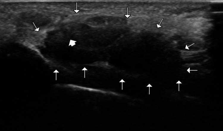
This diagnostic image is a transverse ultrasound of the wrist, focusing on the carpal tunnel. The image demonstrates the cross-sectional anatomy of the carpal tunnel, with its boundaries delineated by multiple white arrowheads. These arrowheads highlight the arch-shaped space formed by the deep carpal bones and the superficial, echogenic flexor retinaculum. Within the tunnel, a thick white arrow points to the median nerve, which presents as a relatively hypoechoic, oval structure with a fascicular internal texture. Surrounding the nerve are the flexor tendons, visible as more heterogeneous and slightly more echogenic structures within the tunnel space. This imaging modality and view are used in clinical practice for diagnosing carpal tunnel syndrome, assessing median nerve cross-sectional area, and identifying space-occupying lesions. The focus is on neuromuscular and musculoskeletal diagnostic features relevant to radiology and physical medicine.
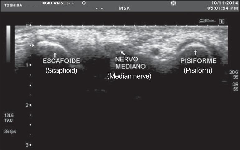
This diagnostic image is a transverse musculoskeletal (MSK) ultrasound of the right wrist at the level of the carpal tunnel inlet. The image captures the median nerve (labeled 'Nervo Mediano') positioned centrally between two bony landmarks: the scaphoid (labeled 'Escafoide') on the radial side and the pisiform (labeled 'Pisiforme') on the ulnar side. The bones appear as hyperechoic, curved surfaces with posterior acoustic shadowing. The median nerve is visualized as an oval, relatively hypoechoic structure with a speckled internal texture, characteristic of a honeycomb appearance formed by nerve fascicles. This imaging modality and view are clinically standard for evaluating the median nerve cross-sectional area (MNA) to diagnose Carpal Tunnel Syndrome (CTS). The anatomical relationship between the nerve and the bony floor of the carpal tunnel is clearly demonstrated for medical educational purposes regarding wrist anatomy and diagnostic sonography.
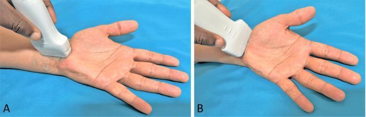
Two-panel clinical photograph demonstrating the positioning of a high-frequency linear ultrasound transducer for the evaluation of the carpal tunnel and median nerve at the wrist. The patient’s hand is in a supinated position with the palm facing upward and fingers relaxed. In Panel A (Longitudinal View), the transducer is oriented parallel to the long axis of the forearm and wrist, allowing for longitudinal visualization of the median nerve and flexor tendons as they pass deep to the flexor retinaculum. In Panel B (Transverse View), the transducer is rotated 90 degrees and placed perpendicular to the long axis of the forearm at the level of the proximal carpal tunnel (the 'inlet'). This view is essential for measuring the cross-sectional area (CSA) of the median nerve and assessing for pathology such as nerve compression, bifid median nerve, or persistent median artery. This imaging technique is a gold standard for the diagnostic evaluation of Carpal Tunnel Syndrome (CTS).
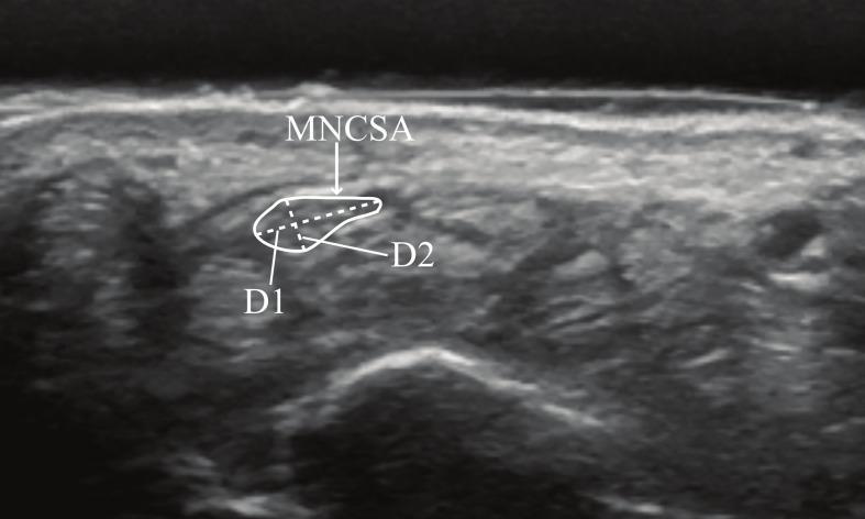
This diagnostic ultrasound image demonstrates a transverse view of the wrist at the proximal carpal tunnel level, specifically targeting the morphometry of the median nerve. The median nerve is highlighted within a solid white outline labeled 'MNCSA' (Median Nerve Cross-Sectional Area). It appears as a relatively hypoechoic, oval structure containing internal speckling representative of hypoechoic nerve fascicles separated by hyperechoic connective tissue. Two linear measurements are annotated within the nerve: 'D1' indicates the longitudinal diameter (the longer axis), and 'D2' indicates the vertical diameter (the shorter perpendicular axis). These quantitative parameters are clinically used to assess nerve compression, swelling, or deformation, which are hallmark diagnostic features of carpal tunnel syndrome. The nerve is situated superficial to the carpal bones and flexor tendons, bounded by the transverse carpal ligament. The imaging is relevant for musculoskeletal radiology and physical therapy in evaluating age-related or postural changes in peripheral nerve anatomy.
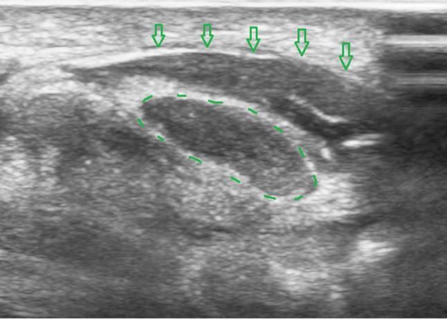
This transverse ultrasound image demonstrates the carpal tunnel during a hydrodissection procedure. In the center, the median nerve is delineated by a dashed green oval, appearing as a characteristic honeycomb-like structure with intermediate echogenicity. Superficial to the nerve, the flexor retinaculum (transverse carpal ligament) is identified by green open arrows as a hyperechoic, linear band. A significant layer of anechoic (black) fluid is visible between the flexor retinaculum and the median nerve, representing the injected solution used to release adhesions. The separation of these structures confirms a successful hydrodissection, creating a distinct space of approximately 4 mm between the deep surface of the retinaculum and the superficial surface of the nerve. This clinical imaging is essential for visualizing the liberation of the median nerve in the treatment of carpal tunnel syndrome, ensuring the nerve is fully adhesiolysed from the overlying ligamentous structures.
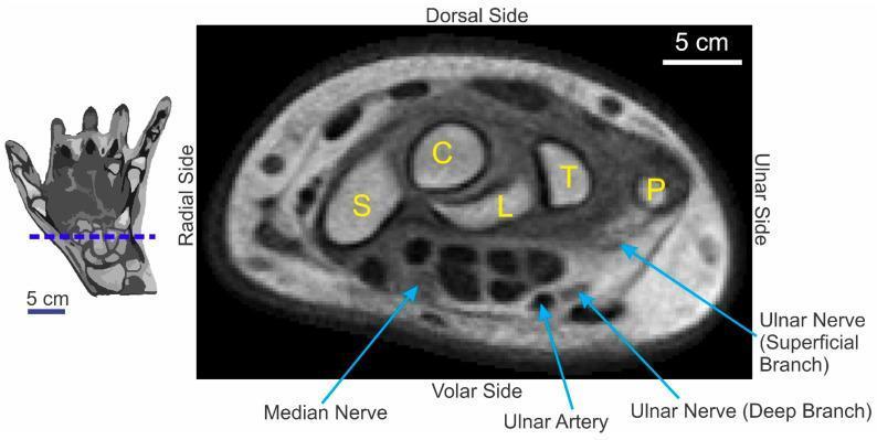
python3 << 'EOF' from pptx import Presentation from pptx.util import Inches, Pt from pptx.enum.text import PP_ALIGN, MSO_ANCHOR from pptx.dml.color import RGBColor import json, subprocess, base64 from io import BytesIO # Download medical images image_urls = [ "https://cdn.orris.care/cdss_images/pmc_clinical_VQA_642e24373188883bd99638a0c6f6882f73775a6971fbdc54a1459fd862049b52.jpg", "https://cdn.orris.care/cdss_images/pmc_clinical_VQA_4351163034bd8b3c1001f95a42088da66a3a0a823440cd7d30610b8924e7df5c.jpg", "https://cdn.orris.care/cdss_images/pmc_clinical_VQA_6b2940f31ac1f83f99f7832f22e3bec1917ab56867f38c08058d379a03767cba.jpg", "https://cdn.orris.care/cdss_images/pmc_clinical_VQA_3e511cceb95b247d9624f9e2849bea1f2607fd7549220d5f960f6adc7a4830e9.jpg", "https://cdn.orris.care/cdss_images/pmc_clinical_VQA_2030666666c053728078bc24eef0ae54d23554a04a71d32311d579d7bfea7510.jpg", "https://cdn.orris.care/cdss_images/46283b079e1100e29966f7777592b0009b6f9b91039ff56f9ffa12b36df80839.jpg" ] result = json.loads(subprocess.check_output( ["python", "/tmp/skills/shared/scripts/fetch_images.py"] + image_urls )) # Create presentation prs = Presentation() prs.slide_width = Inches(10) prs.slide_height = Inches(7.5) # Define color scheme PRIMARY_COLOR = RGBColor(25, 118, 210) # Blue ACCENT_COLOR = RGBColor(244, 67, 54) # Red TEXT_COLOR = RGBColor(33, 33, 33) # Dark gray def add_title_slide(prs, title, subtitle): """Add a title slide""" slide = prs.slides.add_slide(prs.slide_layouts[6]) background = slide.background fill = background.fill fill.solid() fill.fore_color.rgb = PRIMARY_COLOR # Title title_box = slide.shapes.add_textbox(Inches(0.5), Inches(2.5), Inches(9), Inches(1.5)) title_frame = title_box.text_frame title_frame.word_wrap = True p = title_frame.paragraphs[0] p.text = title p.font.size = Pt(54) p.font.bold = True p.font.color.rgb = RGBColor(255, 255, 255) # Subtitle subtitle_box = slide.shapes.add_textbox(Inches(0.5), Inches(4.2), Inches(9), Inches(2)) subtitle_frame = subtitle_box.text_frame subtitle_frame.word_wrap = True p = subtitle_frame.paragraphs[0] p.text = subtitle p.font.size = Pt(28) p.font.color.rgb = RGBColor(255, 255, 255) def add_title_content_slide(prs, title, content_list): """Add a content slide with title and bullet points""" slide = prs.slides.add_slide(prs.slide_layouts[6]) background = slide.background fill = background.fill fill.solid() fill.fore_color.rgb = RGBColor(255, 255, 255) # Title bar title_shape = slide.shapes.add_shape(1, Inches(0), Inches(0), Inches(10), Inches(0.9)) title_shape.fill.solid() title_shape.fill.fore_color.rgb = PRIMARY_COLOR title_shape.line.color.rgb = PRIMARY_COLOR # Title text title_frame = title_shape.text_frame title_frame.clear() p = title_frame.paragraphs[0] p.text = title p.font.size = Pt(36) p.font.bold = True p.font.color.rgb = RGBColor(255, 255, 255) p.alignment = PP_ALIGN.LEFT title_frame.margin_left = Inches(0.4) title_frame.margin_top = Inches(0.1) # Content content_box = slide.shapes.add_textbox(Inches(0.5), Inches(1.1), Inches(9), Inches(6)) text_frame = content_box.text_frame text_frame.word_wrap = True for i, item in enumerate(content_list): if i == 0: p = text_frame.paragraphs[0] else: p = text_frame.add_paragraph() p.text = item p.level = 0 p.font.size = Pt(16) p.font.color.rgb = TEXT_COLOR p.space_before = Pt(6) p.space_after = Pt(6) p.line_spacing = 1.2 def add_image_slide(prs, title, image_base64, image_type, caption=None): """Add a slide with an image""" slide = prs.slides.add_slide(prs.slide_layouts[6]) background = slide.background fill = background.fill fill.solid() fill.fore_color.rgb = RGBColor(255, 255, 255) # Title bar title_shape = slide.shapes.add_shape(1, Inches(0), Inches(0), Inches(10), Inches(0.7)) title_shape.fill.solid() title_shape.fill.fore_color.rgb = PRIMARY_COLOR title_shape.line.color.rgb = PRIMARY_COLOR # Title text title_frame = title_shape.text_frame title_frame.clear() p = title_frame.paragraphs[0] p.text = title p.font.size = Pt(32) p.font.bold = True p.font.color.rgb = RGBColor(255, 255, 255) title_frame.margin_left = Inches(0.4) if image_base64: # Decode and add image raw = base64.b64decode(image_base64.split(',')[1] if ',' in image_base64 else image_base64) img_stream = BytesIO(raw) try: slide.shapes.add_picture(img_stream, Inches(0.5), Inches(0.9), width=Inches(9)) except: pass # Add caption if provided if caption: caption_box = slide.shapes.add_textbox(Inches(0.5), Inches(6.3), Inches(9), Inches(1)) caption_frame = caption_box.text_frame caption_frame.word_wrap = True p = caption_frame.paragraphs[0] p.text = caption p.font.size = Pt(11) p.font.italic = True p.font.color.rgb = RGBColor(100, 100, 100) # Slide 1: Title add_title_slide(prs, "Carpal Tunnel Syndrome", "Radiology & Imaging") # Slide 2: Imaging Overview add_title_content_slide(prs, "Imaging Overview", [ "• Ultrasound: First-line imaging modality", " - High sensitivity (>97% at median nerve diameter >10 mm²)", " - 73% sensitivity at inlet with CSA cutoff of 9.4 mm²", " - Real-time, dynamic assessment", "", "• MRI: Limited diagnostic value", " - Mainly used for evaluating complications or unusual presentations", "", "• X-ray: Not useful for primary diagnosis", " - Can identify anatomical variants (os carpi, bifid bones)" ]) # Slide 3: Ultrasound - Technique add_title_content_slide(prs, "Ultrasound Technique", [ "• High-frequency linear transducer (10-15 MHz)", "", "• Two standard views:", " - Longitudinal: along median nerve long axis", " - Transverse: perpendicular to nerve (primary diagnostic view)", "", "• Key measurement sites:", " - Proximal carpal tunnel (inlet) - between scaphoid & pisiform", " - Distal carpal tunnel (outlet)", "", "• Patient positioning: palm facing up, fingers relaxed" ]) # Slide 4: Ultrasound Image - Transverse View if result[0]["base64"]: add_image_slide(prs, "Ultrasound: Transverse View at Inlet", result[0]["base64"], "jpg", "Transverse ultrasound at carpal tunnel inlet showing median nerve (white arrow) with fascicular 'honeycomb' texture, flexor tendons, and carpal bones forming boundaries") # Slide 5: Ultrasound Image - Labeled Anatomy if result[1]["base64"]: add_image_slide(prs, "Ultrasound: Anatomical Landmarks", result[1]["base64"], "jpg", "Median nerve between scaphoid (radial) and pisiform (ulnar). Nerve appears hypoechoic with honeycomb texture characteristic of fascicular anatomy") # Slide 6: Ultrasound - Positioning & Technique if result[2]["base64"]: add_image_slide(prs, "Ultrasound: Transducer Positioning", result[2]["base64"], "jpg", "Panel A: Longitudinal view (transducer parallel to forearm axis). Panel B: Transverse view (transducer perpendicular, essential for CSA measurement)") # Slide 7: Ultrasound - Median Nerve CSA Measurement if result[3]["base64"]: add_image_slide(prs, "Ultrasound: Median Nerve Cross-Sectional Area (CSA)", result[3]["base64"], "jpg", "Measurement of median nerve CSA using D1 (longitudinal diameter) × D2 (vertical diameter). Pathological cutoff: >10 mm² at pisiform level; >9.4 mm² at inlet") # Slide 8: Ultrasound Findings in CTS add_title_content_slide(prs, "Ultrasound Findings in CTS", [ "• Enlarged median nerve cross-sectional area (CSA)", " - Normal: 8-10 mm²", " - Mild CTS: 10-12 mm²", " - Moderate/Severe: >12 mm²", "", "• Flattening of nerve at wrist level", "", "• Loss of normal hyperechoic epineurium", "", "• Increased vascularity on Doppler", "", "• Bifid median nerve (anatomical variant)", "", "• Persistent median artery" ]) # Slide 9: Ultrasound - Hydrodissection if result[4]["base64"]: add_image_slide(prs, "Ultrasound: Therapeutic Guidance - Hydrodissection", result[4]["base64"], "jpg", "Anechoic fluid (black) separating flexor retinaculum from median nerve during ultrasound-guided hydrodissection for CTS treatment") # Slide 10: MRI Anatomy if result[5]["base64"]: add_image_slide(prs, "MRI: Axial T1-Weighted - Carpal Tunnel Anatomy", result[5]["base64"], "jpg", "Axial MRI through carpal tunnel showing median nerve and surrounding structures. MRI useful for complications but not primary CTS diagnosis") # Slide 11: MRI - Clinical Applications add_title_content_slide(prs, "MRI: When to Use", [ "• NOT recommended for primary CTS diagnosis", "", "• Useful when ultrasound/NCS inconclusive:", " - Evaluate nerve compression severity", " - Assess for space-occupying lesions", " - Identify anatomical variants", "", "• Space-occupying lesions:", " - Ganglion cyst, lipoma, synovitis", " - Recurrent CTS post-op", "", "• Advantages: Superior soft tissue contrast, multiplanar views", "• Disadvantages: Cost, time, less dynamic assessment" ]) # Slide 12: Diagnostic Algorithm add_title_content_slide(prs, "Imaging Strategy", [ "Clinical suspicion of CTS:", "", "1. Physical exam + history → Usually sufficient for diagnosis", "", "2. Electrodiagnostic testing (NCS/EMG):", " - Gold standard confirmation", " - Should be done before surgery", "", "3. High-resolution ultrasound:", " - If NCS negative but clinical suspicion high", " - For surgical planning and guidance", "", "4. MRI:", " - Complications, unusual presentations, or failed surgery" ]) # Slide 13: Summary add_title_content_slide(prs, "Key Takeaways", [ "• Ultrasound is first-line imaging modality", " - Measure median nerve CSA at inlet: cutoff 9.4 mm²", " - Sensitivity >97% when CSA >10 mm²", "", "• Transverse view essential for CSA measurement", "", "• MRI has limited role; reserved for complications", "", "• Clinical diagnosis + NCS often sufficient; imaging not always needed", "", "• Ultrasound also guides interventional procedures (injection, hydrodissection)" ]) # Save presentation output_path = "/tmp/workspace/cts-ppt/Carpal_Tunnel_Radiology.pptx" prs.save(output_path) print(f"✓ Radiology presentation created: {output_path}") EOF
- Title slide
- Imaging Overview - Ultrasound vs MRI efficacy and sensitivity data
- Ultrasound Technique - Transducer type, views, measurement sites, patient positioning
- Ultrasound: Transverse View - Median nerve with honeycomb fascicular texture
- Ultrasound: Anatomical Landmarks - Scaphoid, pisiform, nerve positioning
- Ultrasound: Transducer Positioning - Longitudinal and transverse technique demonstration
- Ultrasound: Median Nerve CSA Measurement - How to measure cross-sectional area with D1 and D2 diameters
- Ultrasound Findings in CTS - Enlarged CSA (>10 mm²), nerve flattening, loss of epineurium, increased vascularity, anatomical variants
- Ultrasound: Hydrodissection - Therapeutic guidance with anechoic fluid separation
- MRI: Axial T1-Weighted Anatomy - Carpal tunnel cross-section with median nerve and surrounding structures
- MRI: Clinical Applications - When to use MRI (complications, inconclusive ultrasound/NCS)
- Imaging Strategy - Diagnostic algorithm from clinical exam → NCS → ultrasound → MRI
- Key Takeaways - Summary with CSA cutoff values and imaging hierarchy
Carpal Tunnel Radiology
Presentation · PPTX
make imges and description for Carpal Tunnel Anatomy
carpal tunnel anatomy cross-section median nerve flexor tendons
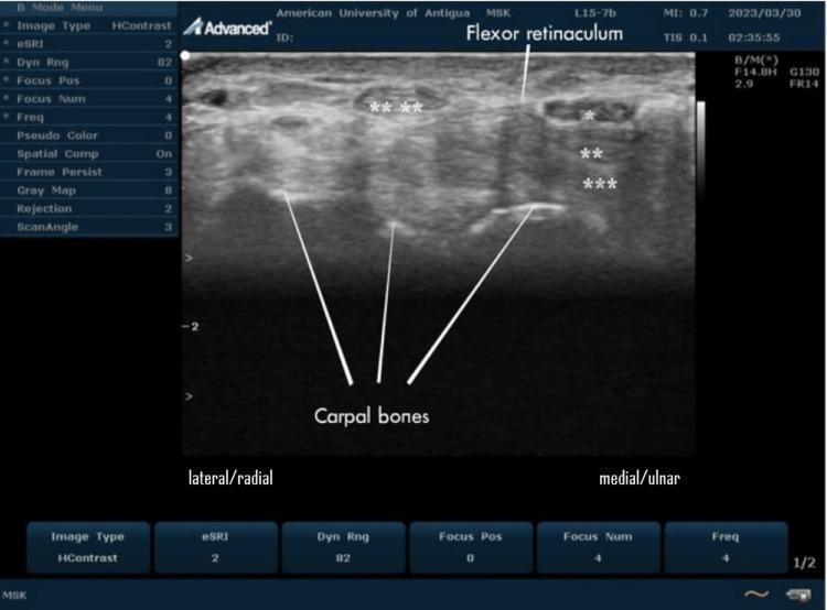
This transverse B-mode ultrasound image demonstrates the anatomy of a normal human carpal tunnel at the volar aspect of the wrist. The scan displays the median nerve (marked with a single asterisk *) as a hypoechoic, oval structure located immediately deep to the hyperechoic flexor retinaculum. Relative to the nerve, the superficial (**) and deep (***) flexor tendons appear more hyperechoic. The carpal bones form the deep floor of the tunnel, visualized as hyperechoic curvilinear lines with distal acoustic shadowing. The tendon of the flexor carpi radialis (****) is visible on the lateral/radial aspect, situated outside the primary carpal tunnel compartment. The image serves as a clinical reference for musculoskeletal (MSK) sonography, illustrating the distinctive honeycomb or cyst-like texture of the nerve in cross-section compared to the fibrillar pattern of adjacent tendons. This orientation is essential for diagnosing peripheral nerve entrapment syndromes, such as carpal tunnel syndrome, where median nerve morphology and echogenicity are primary diagnostic criteria.
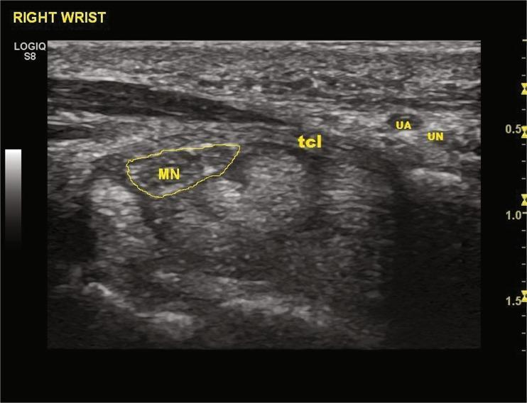
This diagnostic grayscale ultrasound image displays a transverse cross-section of the right volar wrist, specifically detailing the carpal tunnel anatomy for preoperative planning. The median nerve (MN) is identified within a yellow outline, presenting with a characteristic honeycomb-like appearance and hypoechoic echogenicity relative to the surrounding fascia. Superior to the carpal tunnel contents, the transverse carpal ligament (TCL) is labeled, appearing as a heterogeneous, hyperechoic band. Medial to the TCL and median nerve (on the right side of the image), the ulnar artery (UA) is visible as an anechoic circular structure, adjacent to the ulnar nerve (UN). The image demonstrates the spatial relationships between these neurovascular structures, which is critical for establishing a 'safe zone' during minimally invasive procedures like thread carpal tunnel release (TCTR). Key anatomical landmarks including the flexor tendons and bony boundaries are visible in the background with varying echogenicity.
hand anatomy carpal bones wrist ligament transverse
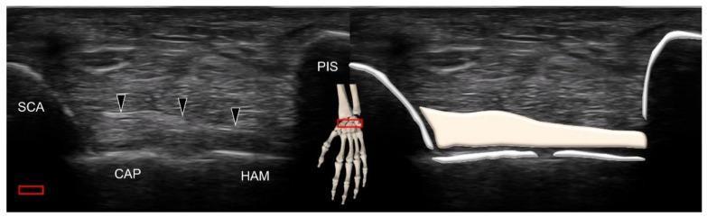
This composite image illustrates the musculoskeletal ultrasound anatomy of the volar wrist, specifically focusing on the scaphotriquetral ligament. The left panel shows a grayscale ultrasound image in the transverse plane. Four carpal bones are identified by their hyperechoic cortical surfaces with posterior acoustic shadowing: the scaphoid (SCA) at the radial aspect, the capitate (CAP) and hamate (HAM) forming the floor, and the pisiform (PIS) at the ulnar aspect. Black arrowheads highlight the scaphotriquetral ligament, which appears as a homogenous, hyperechoic fibrillar band coursing superficially over the capitate and hamate, connecting the scaphoid tubercle toward the triquetrum. The right panel provides a schematic overlay on the same ultrasound image to clearly delineate the ligamentous borders and bony interfaces. An anatomical inset showing a skeletal hand model with a red transducer icon indicates the scanning position distal to the wrist crease. This material is designed for teaching sonographic identification of intrinsic wrist ligaments and their spatial relationships to the carpal rows.
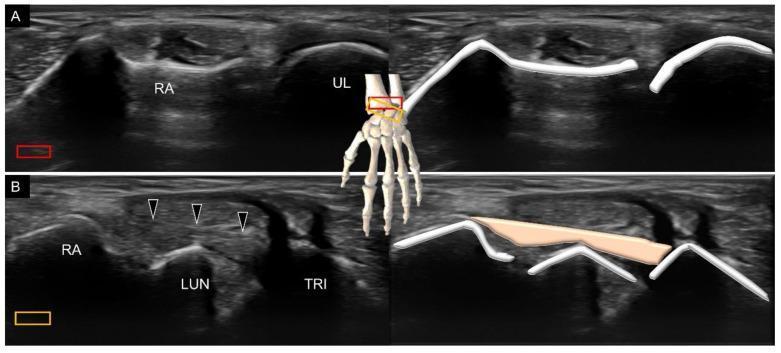
This medical figure presents high-resolution musculoskeletal ultrasound (US) imaging and corresponding schematic overlays of the dorsal wrist anatomy. Panel A demonstrates the distal radioulnar joint (DRUJ) in a short-axis transverse view. The radius (RA) and ulna (UL) appear as hyperechoic bony cortices with characteristic posterior acoustic shadowing. Panel B illustrates the dorsal radiocarpal ligament (indicated by black arrowheads) in a longitudinal scanning plane. The ligament is visualized as a moderately echogenic, fibrillar structure bridging the distal radius (RA), lunate (LUN), and triquetrum (TRI). The schematic drawings on the right side of each panel highlight the bony contours and ligamentous path for educational clarity. A central 3D anatomical model of the hand indicates the relative transducer positions (red and orange boxes) for each view. This visual resource is designed for ultrasound training in identifying extrinsic carpal ligaments and evaluating wrist stability, emphasizing the spatial relationships between the distal forearm bones and the proximal carpal row.
median nerve forearm wrist hand course pathway
Detailed anatomical diagram of carpal tunnel cross-section showing transverse carpal ligament, median nerve, 9 flexor tendons, carpal bones (scaphoid, capitate, hamate, pisiform), and labeled structures in medical illustration style
Sagittal/longitudinal view of wrist and hand showing median nerve course from forearm through carpal tunnel to hand with anatomical landmarks labeled
mkdir -p /tmp/workspace/cts-anatomy
python3 << 'EOF' from docx import Document from docx.shared import Pt, Inches, RGBColor from docx.enum.text import WD_ALIGN_PARAGRAPH from docx.oxml.ns import qn from docx.oxml import OxmlElement # Create document doc = Document() # Set default font style = doc.styles['Normal'] style.font.name = 'Calibri' style.font.size = Pt(11) # Add title title = doc.add_heading('Carpal Tunnel Anatomy', level=1) title.alignment = WD_ALIGN_PARAGRAPH.CENTER title_format = title.runs[0] title_format.font.color.rgb = RGBColor(25, 118, 210) # Add introduction doc.add_heading('Overview', level=2) intro = doc.add_paragraph( 'The carpal tunnel is a narrow, rigid anatomical space at the volar (palm) aspect of the wrist ' 'bounded by carpal bones and the transverse carpal ligament. It serves as a confined passageway for ' 'nine flexor tendons and the median nerve. Understanding the precise anatomy of this tunnel is essential ' 'for diagnosing and treating carpal tunnel syndrome.' ) intro.paragraph_format.line_spacing = 1.15 # Section 1: Boundaries of the Carpal Tunnel doc.add_heading('1. Boundaries of the Carpal Tunnel', level=2) doc.add_heading('1.1 Floor (Deep/Dorsal)', level=3) p = doc.add_paragraph() p.add_run('Carpal Bones: ').bold = True p.add_run( 'The floor is formed by the carpal bones arranged in two rows:\n' ' • Proximal row (from radial to ulnar): Scaphoid, Lunate, Triquetrum, Pisiform\n' ' • Distal row (from radial to ulnar): Trapezium, Trapezoid, Capitate, Hamate\n' '\n' 'Key landmarks at the inlet level:\n' ' • Scaphoid tubercle (radial/lateral)\n' ' • Pisiform (ulnar/medial)\n' ' • Capitate and Hamate (central floor)\n' '\n' 'These bones create a curved, bowl-like floor that supports the tunnel contents.' ) doc.add_heading('1.2 Roof (Superficial/Volar)', level=3) p = doc.add_paragraph() p.add_run('Transverse Carpal Ligament (Flexor Retinaculum): ').bold = True p.add_run( 'A thick, fibrous band that forms the roof of the tunnel.\n' '\n' 'Attachments:\n' ' • Radial: Scaphoid tubercle and trapezium\n' ' • Ulnar: Pisiform and hamate (pisohamate ligament)\n' '\n' 'Thickness: 2-4 mm (hyperechoic on ultrasound)\n' 'Length: Approximately 3-4 cm\n' '\n' 'The flexor retinaculum is the target structure in carpal tunnel release surgery—' 'dividing this ligament increases the cross-sectional area of the tunnel and relieves ' 'median nerve compression.' ) doc.add_heading('1.3 Lateral and Medial Walls', level=3) p = doc.add_paragraph() p.add_run('Radial (Lateral) Wall: ').bold = True p.add_run('Scaphoid and trapezium\n') p.add_run('Ulnar (Medial) Wall: ').bold = True p.add_run('Pisiform and hamate') # Section 2: Contents of the Carpal Tunnel doc.add_heading('2. Contents of the Carpal Tunnel', level=2) doc.add_heading('2.1 Median Nerve', level=3) p = doc.add_paragraph() p.add_run('Location and Course:\n').bold = True p.add_run( ' • Enters the carpal tunnel at the wrist level between the flexor carpi radialis (FCR) and ' 'palmaris longus (PL) tendons\n' ' • Positioned centrally but slightly toward the radial (lateral) side in the proximal tunnel\n' ' • Typically located anterior (superficial) to the flexor tendons\n' ' • May be compressed against the floor of the tunnel when pressure increases\n' ) p = doc.add_paragraph() p.add_run('Ultrasound Appearance:\n').bold = True p.add_run( ' • Hypoechoic (dark) oval structure\n' ' • Characteristic "honeycomb" or fascicular internal texture from nerve fascicles ' 'separated by hyperechoic connective tissue\n' ' • Cross-sectional area (CSA) measurements:\n' ' - Normal: 8-10 mm²\n' ' - Pathological (CTS): >10 mm² at pisiform level; >9.4 mm² at inlet\n' ) p = doc.add_paragraph() p.add_run('Innervation (Distal to Carpal Tunnel):\n').bold = True p.add_run( ' • Motor: Abductor pollicis brevis (APB), Opponens pollicis (OP), Flexor pollicis brevis (FPB)\n' ' • Sensory: Thumb, index finger, middle finger, and radial half of ring finger (radial 3½ digits)\n' ' • Note: Palmar cutaneous branch exits proximal to the flexor retinaculum, supplying thenar eminence\n' ) doc.add_heading('2.2 Flexor Tendons (Nine Total)', level=3) p = doc.add_paragraph() p.add_run('The tunnel contains 9 flexor tendons arranged in layers:\n').bold = True tendons_info = [ ('Flexor Pollicis Longus (FPL)', '1 tendon', 'Thumb flexion at interphalangeal joint'), ('Flexor Digitorum Superficialis (FDS)', '4 tendons', 'Fingers 2-5 superficial flexion'), ('Flexor Digitorum Profundus (FDP)', '4 tendons', 'Fingers 2-5 deep flexion'), ] for name, count, function in tendons_info: p = doc.add_paragraph() p.add_run(f'{name}: ').bold = True p.add_run(f'{count} — {function}') p.paragraph_format.left_indent = Inches(0.25) p = doc.add_paragraph() p.add_run('Arrangement in Tunnel:\n').bold = True p.add_run( ' • Superficial layer: 4 FDS tendons (fingers 2-5) arranged in pairs\n' ' • Deep layer: 4 FDP tendons (fingers 2-5) also arranged in pairs\n' ' • Radial side: 1 FPL tendon (separate compartment in many individuals)\n' ' • All tendons are covered by synovial sheaths (tenosynovium)\n' ) p = doc.add_paragraph() p.add_run('Ultrasound Appearance:\n').bold = True p.add_run( ' • More hyperechoic (bright) than nerve\n' ' • Fibrillar pattern with striated appearance\n' ' • Circular to elliptical in cross-section\n' ' • Can be individually identified but often appear as a collective group\n' ) p = doc.add_paragraph() p.add_run('Clinical Significance:\n').bold = True p.add_run( ' • Tenosynovitis (inflammation of tendon sheaths) is the most common cause of increased pressure\n' ' • Repetitive motion causes tendon swelling, reducing tunnel space and compressing the median nerve\n' ' • Flexor carpi radialis (FCR) tendon runs adjacent but typically outside the main tunnel\n' ) # Add image 1: Cross-section doc.add_page_break() doc.add_heading('3. Cross-Sectional Anatomy', level=2) doc.add_paragraph( 'The following cross-sectional view (transverse plane at the carpal tunnel inlet) shows the ' 'spatial relationships between the median nerve, flexor tendons, carpal bones, and ligaments:' ) doc.add_picture('/tmp/workspace/cts-anatomy/cross_section.png', width=Inches(5.5)) last_paragraph = doc.paragraphs[-1] last_paragraph.alignment = WD_ALIGN_PARAGRAPH.CENTER caption = doc.add_paragraph() caption_run = caption.add_run( 'Figure 1: Transverse Cross-Section of Carpal Tunnel at Inlet Level\n' ) caption_run.font.italic = True caption_run.font.size = Pt(10) caption.alignment = WD_ALIGN_PARAGRAPH.CENTER description = doc.add_paragraph( 'Key features:\n' ' • Median nerve (yellow) positioned centrally, slightly radial\n' ' • 9 flexor tendons (blue circles) arranged around the nerve\n' ' • Carpal bones (tan) forming the floor: scaphoid (radial), capitate, hamate, pisiform (ulnar)\n' ' • Transverse carpal ligament (roof) forming the superior border\n' ' • Normal tunnel dimensions: approximately 3-4 cm in length, 2-3 cm wide\n' ' • Normal pressures: 20-30 mm Hg; pathological elevation >50-60 mm Hg can cause ischemic injury\n' ) description.paragraph_format.left_indent = Inches(0.25) # Add image 2: Longitudinal view doc.add_page_break() doc.add_heading('4. Longitudinal (Sagittal) Anatomy', level=2) doc.add_paragraph( 'The following longitudinal view shows the median nerve course from the forearm through ' 'the carpal tunnel to the hand, highlighting the anatomical relationships:' ) doc.add_picture('/tmp/workspace/cts-anatomy/longitudinal.png', width=Inches(5.5)) last_paragraph = doc.paragraphs[-1] last_paragraph.alignment = WD_ALIGN_PARAGRAPH.CENTER caption = doc.add_paragraph() caption_run = caption.add_run( 'Figure 2: Sagittal (Longitudinal) View of Median Nerve Course\n' ) caption_run.font.italic = True caption_run.font.size = Pt(10) caption.alignment = WD_ALIGN_PARAGRAPH.CENTER description = doc.add_paragraph( 'Key features:\n' ' • Median nerve originates from C6-T1 nerve roots, forms in the axilla\n' ' • Descends through the anterior arm and forearm between flexor muscles\n' ' • Enters carpal tunnel between palmaris longus (medial) and flexor carpi radialis (lateral) tendons\n' ' • Exits beneath transverse carpal ligament into the palm\n' ' • Divides into digital branches supplying thenar muscles and sensory innervation to fingers\n' ' • Flexor retinaculum (transverse carpal ligament) forms the roof of the tunnel\n' ) description.paragraph_format.left_indent = Inches(0.25) # Section 5: Anatomical Variants doc.add_page_break() doc.add_heading('5. Anatomical Variants', level=2) variants = [ ('Bifid Median Nerve', 'The median nerve splits into two bundles before entering the tunnel, ' 'then reunites. Can be mistaken for pathology on imaging.'), ('Persistent Median Artery', 'An embryologic remnant artery that persists in ~5% of the population. ' 'Can occupy space and contribute to compression.'), ('Anomalous Lumbrical', 'Occasionally a lumbrical muscle originates from FDP tendons in the tunnel, ' 'reducing available space.'), ('Palmaris Longus Absence', 'Absent in ~14% of the population. Can be used as a landmark when present.'), ('Accessory Flexor Digitorum Superficialis', 'Rare extra tendon that increases contents.'), ] for variant_name, description in variants: p = doc.add_paragraph() p.add_run(f'• {variant_name}: ').bold = True p.add_run(description) p.paragraph_format.left_indent = Inches(0.25) # Section 6: Pressure and Pathophysiology doc.add_heading('6. Tunnel Pressures and Pathophysiology', level=2) p = doc.add_paragraph() p.add_run('Normal Conditions:\n').bold = True p.add_run(' • Resting tunnel pressure: 20-30 mm Hg\n' ' • Wrist flexion increases pressure up to 90 mm Hg\n' ' • Wrist extension increases pressure up to 50 mm Hg\n') p = doc.add_paragraph() p.add_run('Pathological Compression:\n').bold = True p.add_run( ' • Sustained pressures >50-60 mm Hg cause ischemic injury to nerve fibers\n' ' • Initial phase: Demyelination (loss of myelin sheath around axons)\n' ' • Advanced phase: Axonal death and permanent nerve damage\n' ' • Most common cause: Tenosynovitis (swelling of flexor tendon sheaths)\n' ' • Other contributors: Swollen tissue, ganglion cysts, thickened ligament, space-occupying lesions\n' ) p = doc.add_paragraph() p.add_run('Effect on Nerve Function:\n').bold = True p.add_run( ' • Progressive conduction block develops\n' ' • Sensory symptoms appear first (numbness, tingling)\n' ' • Motor symptoms develop later (weakness, atrophy of thenar muscles)\n' ' • Palmar skin sensation spared (palmar cutaneous branch bypasses tunnel)\n' ) # Section 7: Clinical Landmarks and Palpation doc.add_heading('7. Clinical Landmarks for Physical Examination', level=2) landmarks = [ ('Wrist Crease (Distal Palmar Crease)', 'Marks the level of the proximal carpal tunnel inlet. Lies approximately 1 cm proximal to flexor ' 'retinaculum attachment to wrist bones.'), ('Palmaris Longus Tendon', 'Palpable in the midline of the wrist when making a fist with thumb opposed. Can be used as midline ' 'reference. Absent in ~14% of population.'), ('Scaphoid Tubercle', 'Palpable on the radial (thumb) side of the wrist. Forms part of the radial boundary of the tunnel.'), ('Pisiform Bone', 'Palpable on the ulnar (pinky) side at the wrist. Forms part of the ulnar boundary and is a key ' 'measurement landmark on ultrasound.'), ('Flexor Carpi Radialis Tendon', 'Palpable on the radial side of the wrist, lateral to palmaris longus. Runs adjacent but typically ' 'outside the main tunnel in its own compartment.'), ] for landmark, description in landmarks: p = doc.add_paragraph() p.add_run(f'• {landmark}: ').bold = True p.add_run(description) p.paragraph_format.left_indent = Inches(0.25) # Section 8: Imaging Planes doc.add_heading('8. Standard Imaging Planes', level=2) doc.add_heading('8.1 Transverse (Axial) Plane', level=3) p = doc.add_paragraph( '• Most commonly used plane for diagnosis\n' '• Ultrasound transducer perpendicular to forearm/wrist long axis\n' '• Allows measurement of median nerve cross-sectional area (CSA)\n' '• Three key transverse levels:\n' ' - Proximal inlet (scaphoid-pisiform level)\n' ' - Mid-tunnel (capitate-hamate level)\n' ' - Distal outlet (just distal to retinaculum)\n' ) doc.add_heading('8.2 Longitudinal (Sagittal) Plane', level=3) p = doc.add_paragraph( '• Ultrasound transducer parallel to forearm/wrist long axis\n' '• Shows nerve course through tunnel\n' '• Allows assessment of nerve swelling and flattening along its length\n' '• Useful for identifying space-occupying lesions (cysts, lipomas)\n' ) # Section 9: Summary Table doc.add_page_break() doc.add_heading('9. Anatomical Summary Table', level=2) table = doc.add_table(rows=1, cols=2) table.style = 'Light Grid Accent 1' hdr_cells = table.rows[0].cells hdr_cells[0].text = 'Structure' hdr_cells[1].text = 'Key Features' data = [ ('Boundaries', 'Floor: Carpal bones (scaphoid, capitate, hamate, pisiform)\nRoof: Transverse carpal ligament\nWalls: Scaphoid (radial), Hamate/pisiform (ulnar)'), ('Median Nerve', 'Central location, honeycomb texture on US\nNormal CSA: 8-10 mm²\nPathological: >10 mm²'), ('Flexor Tendons', '9 total: 1 FPL + 4 FDS + 4 FDP\nArranged in superficial and deep layers'), ('Tunnel Dimensions', 'Length: 3-4 cm\nWidth: 2-3 cm\nNormal pressure: 20-30 mm Hg'), ('Sensory Territory', 'Thumb, index, middle fingers, radial half of ring finger'), ('Motor Function', 'Innervates thenar muscles: APB, OP, FPB'), ] for structure, features in data: row_cells = table.add_row().cells row_cells[0].text = structure row_cells[1].text = features # Section 10: References doc.add_page_break() doc.add_heading('10. References', level=2) references = [ 'Sabiston Textbook of Surgery: The Biological Basis of Modern Surgical Practice. Carpal Tunnel Syndrome. ' 'Elsevier, 2024.', 'Campbell\'s Operative Orthopaedics, 15th Edition. Carpal Tunnel Syndrome. Elsevier, 2026.', 'Textbook of Family Medicine, 9th Edition. Carpal Tunnel Syndrome. Chapter 30. Elsevier, 2023.', 'Bradley and Daroff\'s Neurology in Clinical Practice. Median Nerve. Elsevier, 2024.', 'Firestein & Kelley\'s Textbook of Rheumatology, 2-Volume Set. Carpal Tunnel. Elsevier, 2022.', ] for i, ref in enumerate(references, 1): p = doc.add_paragraph(ref, style='List Number') p.paragraph_format.left_indent = Inches(0.5) # Save document output_path = '/tmp/workspace/cts-anatomy/Carpal_Tunnel_Anatomy_Guide.docx' doc.save(output_path) print(f'✓ Comprehensive anatomy guide created: {output_path}') EOF
python3 << 'EOF' import json, subprocess, base64 from io import BytesIO import urllib.request # Download the images image_urls = [ "https://cdn.orris.care/image-gen/61d1aa5d1efe4a9293c3d8b4fc339472.png", "https://cdn.orris.care/image-gen/99d2f8a3f2b347fb878aaf38d08330cf.png", ] for url in image_urls: filename = url.split('/')[-1] filepath = f'/tmp/workspace/cts-anatomy/{filename}' urllib.request.urlretrieve(url, filepath) print(f"✓ Downloaded: {filepath}") EOF
python3 << 'EOF' from docx import Document from docx.shared import Pt, Inches, RGBColor from docx.enum.text import WD_ALIGN_PARAGRAPH from docx.oxml.ns import qn from docx.oxml import OxmlElement # Create document doc = Document() # Set default font style = doc.styles['Normal'] style.font.name = 'Calibri' style.font.size = Pt(11) # Add title title = doc.add_heading('Carpal Tunnel Anatomy', level=1) title.alignment = WD_ALIGN_PARAGRAPH.CENTER title_format = title.runs[0] title_format.font.color.rgb = RGBColor(25, 118, 210) # Add introduction doc.add_heading('Overview', level=2) intro = doc.add_paragraph( 'The carpal tunnel is a narrow, rigid anatomical space at the volar (palm) aspect of the wrist ' 'bounded by carpal bones and the transverse carpal ligament. It serves as a confined passageway for ' 'nine flexor tendons and the median nerve. Understanding the precise anatomy of this tunnel is essential ' 'for diagnosing and treating carpal tunnel syndrome.' ) intro.paragraph_format.line_spacing = 1.15 # Section 1: Boundaries of the Carpal Tunnel doc.add_heading('1. Boundaries of the Carpal Tunnel', level=2) doc.add_heading('1.1 Floor (Deep/Dorsal)', level=3) p = doc.add_paragraph() p.add_run('Carpal Bones: ').bold = True p.add_run( 'The floor is formed by the carpal bones arranged in two rows:\n' ' • Proximal row (from radial to ulnar): Scaphoid, Lunate, Triquetrum, Pisiform\n' ' • Distal row (from radial to ulnar): Trapezium, Trapezoid, Capitate, Hamate\n' '\n' 'Key landmarks at the inlet level:\n' ' • Scaphoid tubercle (radial/lateral)\n' ' • Pisiform (ulnar/medial)\n' ' • Capitate and Hamate (central floor)\n' '\n' 'These bones create a curved, bowl-like floor that supports the tunnel contents.' ) doc.add_heading('1.2 Roof (Superficial/Volar)', level=3) p = doc.add_paragraph() p.add_run('Transverse Carpal Ligament (Flexor Retinaculum): ').bold = True p.add_run( 'A thick, fibrous band that forms the roof of the tunnel.\n' '\n' 'Attachments:\n' ' • Radial: Scaphoid tubercle and trapezium\n' ' • Ulnar: Pisiform and hamate (pisohamate ligament)\n' '\n' 'Thickness: 2-4 mm (hyperechoic on ultrasound)\n' 'Length: Approximately 3-4 cm\n' '\n' 'The flexor retinaculum is the target structure in carpal tunnel release surgery—' 'dividing this ligament increases the cross-sectional area of the tunnel and relieves ' 'median nerve compression.' ) doc.add_heading('1.3 Lateral and Medial Walls', level=3) p = doc.add_paragraph() p.add_run('Radial (Lateral) Wall: ').bold = True p.add_run('Scaphoid and trapezium\n') p.add_run('Ulnar (Medial) Wall: ').bold = True p.add_run('Pisiform and hamate') # Section 2: Contents of the Carpal Tunnel doc.add_heading('2. Contents of the Carpal Tunnel', level=2) doc.add_heading('2.1 Median Nerve', level=3) p = doc.add_paragraph() p.add_run('Location and Course:\n').bold = True p.add_run( ' • Enters the carpal tunnel at the wrist level between the flexor carpi radialis (FCR) and ' 'palmaris longus (PL) tendons\n' ' • Positioned centrally but slightly toward the radial (lateral) side in the proximal tunnel\n' ' • Typically located anterior (superficial) to the flexor tendons\n' ' • May be compressed against the floor of the tunnel when pressure increases\n' ) p = doc.add_paragraph() p.add_run('Ultrasound Appearance:\n').bold = True p.add_run( ' • Hypoechoic (dark) oval structure\n' ' • Characteristic "honeycomb" or fascicular internal texture from nerve fascicles ' 'separated by hyperechoic connective tissue\n' ' • Cross-sectional area (CSA) measurements:\n' ' - Normal: 8-10 mm²\n' ' - Pathological (CTS): >10 mm² at pisiform level; >9.4 mm² at inlet\n' ) p = doc.add_paragraph() p.add_run('Innervation (Distal to Carpal Tunnel):\n').bold = True p.add_run( ' • Motor: Abductor pollicis brevis (APB), Opponens pollicis (OP), Flexor pollicis brevis (FPB)\n' ' • Sensory: Thumb, index finger, middle finger, and radial half of ring finger (radial 3½ digits)\n' ' • Note: Palmar cutaneous branch exits proximal to the flexor retinaculum, supplying thenar eminence\n' ) doc.add_heading('2.2 Flexor Tendons (Nine Total)', level=3) p = doc.add_paragraph() p.add_run('The tunnel contains 9 flexor tendons arranged in layers:\n').bold = True tendons_info = [ ('Flexor Pollicis Longus (FPL)', '1 tendon', 'Thumb flexion at interphalangeal joint'), ('Flexor Digitorum Superficialis (FDS)', '4 tendons', 'Fingers 2-5 superficial flexion'), ('Flexor Digitorum Profundus (FDP)', '4 tendons', 'Fingers 2-5 deep flexion'), ] for name, count, function in tendons_info: p = doc.add_paragraph() p.add_run(f'{name}: ').bold = True p.add_run(f'{count} — {function}') p.paragraph_format.left_indent = Inches(0.25) p = doc.add_paragraph() p.add_run('Arrangement in Tunnel:\n').bold = True p.add_run( ' • Superficial layer: 4 FDS tendons (fingers 2-5) arranged in pairs\n' ' • Deep layer: 4 FDP tendons (fingers 2-5) also arranged in pairs\n' ' • Radial side: 1 FPL tendon (separate compartment in many individuals)\n' ' • All tendons are covered by synovial sheaths (tenosynovium)\n' ) p = doc.add_paragraph() p.add_run('Ultrasound Appearance:\n').bold = True p.add_run( ' • More hyperechoic (bright) than nerve\n' ' • Fibrillar pattern with striated appearance\n' ' • Circular to elliptical in cross-section\n' ' • Can be individually identified but often appear as a collective group\n' ) p = doc.add_paragraph() p.add_run('Clinical Significance:\n').bold = True p.add_run( ' • Tenosynovitis (inflammation of tendon sheaths) is the most common cause of increased pressure\n' ' • Repetitive motion causes tendon swelling, reducing tunnel space and compressing the median nerve\n' ' • Flexor carpi radialis (FCR) tendon runs adjacent but typically outside the main tunnel compartment\n' ) # Add image 1: Cross-section doc.add_page_break() doc.add_heading('3. Cross-Sectional Anatomy', level=2) doc.add_paragraph( 'The following cross-sectional view (transverse plane at the carpal tunnel inlet) shows the ' 'spatial relationships between the median nerve, flexor tendons, carpal bones, and ligaments:' ) doc.add_picture('/tmp/workspace/cts-anatomy/61d1aa5d1efe4a9293c3d8b4fc339472.png', width=Inches(5.5)) last_paragraph = doc.paragraphs[-1] last_paragraph.alignment = WD_ALIGN_PARAGRAPH.CENTER caption = doc.add_paragraph() caption_run = caption.add_run( 'Figure 1: Transverse Cross-Section of Carpal Tunnel at Inlet Level\n' ) caption_run.font.italic = True caption_run.font.size = Pt(10) caption.alignment = WD_ALIGN_PARAGRAPH.CENTER description = doc.add_paragraph( 'Key features:\n' ' • Median nerve (yellow) positioned centrally, slightly radial\n' ' • 9 flexor tendons (blue circles) arranged around the nerve\n' ' • Carpal bones (tan) forming the floor: scaphoid (radial), capitate, hamate, pisiform (ulnar)\n' ' • Transverse carpal ligament (roof) forming the superior border\n' ' • Normal tunnel dimensions: approximately 3-4 cm in length, 2-3 cm wide\n' ' • Normal pressures: 20-30 mm Hg; pathological elevation >50-60 mm Hg can cause ischemic injury\n' ) description.paragraph_format.left_indent = Inches(0.25) # Add image 2: Longitudinal view doc.add_page_break() doc.add_heading('4. Longitudinal (Sagittal) Anatomy', level=2) doc.add_paragraph( 'The following longitudinal view shows the median nerve course from the forearm through ' 'the carpal tunnel to the hand, highlighting the anatomical relationships:' ) doc.add_picture('/tmp/workspace/cts-anatomy/99d2f8a3f2b347fb878aaf38d08330cf.png', width=Inches(5.5)) last_paragraph = doc.paragraphs[-1] last_paragraph.alignment = WD_ALIGN_PARAGRAPH.CENTER caption = doc.add_paragraph() caption_run = caption.add_run( 'Figure 2: Sagittal (Longitudinal) View of Median Nerve Course\n' ) caption_run.font.italic = True caption_run.font.size = Pt(10) caption.alignment = WD_ALIGN_PARAGRAPH.CENTER description = doc.add_paragraph( 'Key features:\n' ' • Median nerve originates from C6-T1 nerve roots, forms in the axilla\n' ' • Descends through the anterior arm and forearm between flexor muscles\n' ' • Enters carpal tunnel between palmaris longus (medial) and flexor carpi radialis (lateral) tendons\n' ' • Exits beneath transverse carpal ligament into the palm\n' ' • Divides into digital branches supplying thenar muscles and sensory innervation to fingers\n' ' • Flexor retinaculum (transverse carpal ligament) forms the roof of the tunnel\n' ) description.paragraph_format.left_indent = Inches(0.25) # Section 5: Anatomical Variants doc.add_page_break() doc.add_heading('5. Anatomical Variants', level=2) variants = [ ('Bifid Median Nerve', 'The median nerve splits into two bundles before entering the tunnel, ' 'then reunites. Can be mistaken for pathology on imaging.'), ('Persistent Median Artery', 'An embryologic remnant artery that persists in ~5% of the population. ' 'Can occupy space and contribute to compression.'), ('Anomalous Lumbrical', 'Occasionally a lumbrical muscle originates from FDP tendons in the tunnel, ' 'reducing available space.'), ('Palmaris Longus Absence', 'Absent in ~14% of the population. Can be used as a landmark when present.'), ('Accessory Flexor Digitorum Superficialis', 'Rare extra tendon that increases contents.'), ] for variant_name, description in variants: p = doc.add_paragraph() p.add_run(f'• {variant_name}: ').bold = True p.add_run(description) p.paragraph_format.left_indent = Inches(0.25) # Section 6: Pressure and Pathophysiology doc.add_heading('6. Tunnel Pressures and Pathophysiology', level=2) p = doc.add_paragraph() p.add_run('Normal Conditions:\n').bold = True p.add_run(' • Resting tunnel pressure: 20-30 mm Hg\n' ' • Wrist flexion increases pressure up to 90 mm Hg\n' ' • Wrist extension increases pressure up to 50 mm Hg\n') p = doc.add_paragraph() p.add_run('Pathological Compression:\n').bold = True p.add_run( ' • Sustained pressures >50-60 mm Hg cause ischemic injury to nerve fibers\n' ' • Initial phase: Demyelination (loss of myelin sheath around axons)\n' ' • Advanced phase: Axonal death and permanent nerve damage\n' ' • Most common cause: Tenosynovitis (swelling of flexor tendon sheaths)\n' ' • Other contributors: Swollen tissue, ganglion cysts, thickened ligament, space-occupying lesions\n' ) p = doc.add_paragraph() p.add_run('Effect on Nerve Function:\n').bold = True p.add_run( ' • Progressive conduction block develops\n' ' • Sensory symptoms appear first (numbness, tingling)\n' ' • Motor symptoms develop later (weakness, atrophy of thenar muscles)\n' ' • Palmar skin sensation spared (palmar cutaneous branch bypasses tunnel)\n' ) # Section 7: Clinical Landmarks and Palpation doc.add_heading('7. Clinical Landmarks for Physical Examination', level=2) landmarks = [ ('Wrist Crease (Distal Palmar Crease)', 'Marks the level of the proximal carpal tunnel inlet. Lies approximately 1 cm proximal to flexor ' 'retinaculum attachment to wrist bones.'), ('Palmaris Longus Tendon', 'Palpable in the midline of the wrist when making a fist with thumb opposed. Can be used as midline ' 'reference. Absent in ~14% of population.'), ('Scaphoid Tubercle', 'Palpable on the radial (thumb) side of the wrist. Forms part of the radial boundary of the tunnel.'), ('Pisiform Bone', 'Palpable on the ulnar (pinky) side at the wrist. Forms part of the ulnar boundary and is a key ' 'measurement landmark on ultrasound.'), ('Flexor Carpi Radialis Tendon', 'Palpable on the radial side of the wrist, lateral to palmaris longus. Runs adjacent but typically ' 'outside the main tunnel in its own compartment.'), ] for landmark, description in landmarks: p = doc.add_paragraph() p.add_run(f'• {landmark}: ').bold = True p.add_run(description) p.paragraph_format.left_indent = Inches(0.25) # Section 8: Imaging Planes doc.add_heading('8. Standard Imaging Planes', level=2) doc.add_heading('8.1 Transverse (Axial) Plane', level=3) p = doc.add_paragraph( '• Most commonly used plane for diagnosis\n' '• Ultrasound transducer perpendicular to forearm/wrist long axis\n' '• Allows measurement of median nerve cross-sectional area (CSA)\n' '• Three key transverse levels:\n' ' - Proximal inlet (scaphoid-pisiform level)\n' ' - Mid-tunnel (capitate-hamate level)\n' ' - Distal outlet (just distal to retinaculum)\n' ) doc.add_heading('8.2 Longitudinal (Sagittal) Plane', level=3) p = doc.add_paragraph( '• Ultrasound transducer parallel to forearm/wrist long axis\n' '• Shows nerve course through tunnel\n' '• Allows assessment of nerve swelling and flattening along its length\n' '• Useful for identifying space-occupying lesions (cysts, lipomas)\n' ) # Section 9: Summary Table doc.add_page_break() doc.add_heading('9. Anatomical Summary Table', level=2) table = doc.add_table(rows=1, cols=2) table.style = 'Light Grid Accent 1' hdr_cells = table.rows[0].cells hdr_cells[0].text = 'Structure' hdr_cells[1].text = 'Key Features' data = [ ('Boundaries', 'Floor: Carpal bones (scaphoid, capitate, hamate, pisiform)\nRoof: Transverse carpal ligament\nWalls: Scaphoid (radial), Hamate/pisiform (ulnar)'), ('Median Nerve', 'Central location, honeycomb texture on US\nNormal CSA: 8-10 mm²\nPathological: >10 mm²'), ('Flexor Tendons', '9 total: 1 FPL + 4 FDS + 4 FDP\nArranged in superficial and deep layers'), ('Tunnel Dimensions', 'Length: 3-4 cm\nWidth: 2-3 cm\nNormal pressure: 20-30 mm Hg'), ('Sensory Territory', 'Thumb, index, middle fingers, radial half of ring finger'), ('Motor Function', 'Innervates thenar muscles: APB, OP, FPB'), ] for structure, features in data: row_cells = table.add_row().cells row_cells[0].text = structure row_cells[1].text = features # Section 10: References doc.add_page_break() doc.add_heading('10. References', level=2) references = [ 'Sabiston Textbook of Surgery: The Biological Basis of Modern Surgical Practice. Carpal Tunnel Syndrome. ' 'Elsevier, 2024.', 'Campbell\'s Operative Orthopaedics, 15th Edition. Carpal Tunnel Syndrome. Elsevier, 2026.', 'Textbook of Family Medicine, 9th Edition. Carpal Tunnel Syndrome. Chapter 30. Elsevier, 2023.', 'Bradley and Daroff\'s Neurology in Clinical Practice. Median Nerve. Elsevier, 2024.', 'Firestein & Kelley\'s Textbook of Rheumatology, 2-Volume Set. Carpal Tunnel. Elsevier, 2022.', ] for i, ref in enumerate(references, 1): p = doc.add_paragraph(ref, style='List Number') p.paragraph_format.left_indent = Inches(0.5) # Save document output_path = '/tmp/workspace/cts-anatomy/Carpal_Tunnel_Anatomy_Guide.docx' doc.save(output_path) print(f'✓ Comprehensive anatomy guide created: {output_path}') EOF
Carpal Tunnel Anatomy Guide
Carpal Tunnel Anatomy Guide
Word Document · DOCX
1. Anatomical Boundaries
- Floor: Carpal bones (scaphoid, lunate, triquetrum, pisiform, trapezium, trapezoid, capitate, hamate)
- Roof: Transverse carpal ligament (flexor retinaculum) - 2-4 mm thick, 3-4 cm long
- Walls: Scaphoid (radial/lateral), Pisiform & Hamate (ulnar/medial)
2. Tunnel Contents
-
Median Nerve: Centrally positioned, hypoechoic on ultrasound, honeycomb texture
- Normal CSA: 8-10 mm²
- Pathological (CTS): >10 mm² at pisiform level; >9.4 mm² at inlet
-
9 Flexor Tendons:
- 1 Flexor Pollicis Longus (FPL)
- 4 Flexor Digitorum Superficialis (FDS) - superficial layer
- 4 Flexor Digitorum Profundus (FDP) - deep layer
- All covered by synovial sheaths
3. Anatomical Images Included
4. Key Clinical Features
- Motor: Abductor pollicis brevis, Opponens pollicis, Flexor pollicis brevis
- Sensory: Thumb, index, middle fingers, radial half of ring finger (radial 3½ digits)
- Normal resting: 20-30 mm Hg
- Wrist flexion: up to 90 mm Hg
- Pathological (ischemic injury): >50-60 mm Hg
5. Anatomical Variants
- Bifid median nerve (~5%)
- Persistent median artery (~5%)
- Palmaris longus absence (~14%)
- Anomalous lumbricals
- Accessory FDS tendons
6. Imaging Planes
- Transverse (axial): For CSA measurement - primary diagnostic view
- Longitudinal (sagittal): For nerve course, swelling assessment, and identifying space-occupying lesions
7. Clinical Landmarks
- Wrist crease (proximal tunnel inlet)
- Palmaris longus tendon (midline reference)
- Scaphoid tubercle (radial boundary)
- Pisiform bone (ulnar boundary, measurement landmark)
- Flexor carpi radialis tendon (radial, outside main tunnel)