
Synapse
Reading File
A synapse is a junction between two neurons (or between a neuron and a target cell) where signals are transmitted. Here is a thorough overview while I pull textbook content.
Searching sources...
Finding Sources
Searching the Web
synapse structure function neurotransmission overview
Reading File
Reading File
Reading File
Loading Image
Loading Image
Reading File
The Synapse
A synapse is a specialized junction where one neuron transmits a signal to another neuron, a muscle cell, or a gland cell. It is the fundamental unit of neural communication.
Types of Synapses
There are two main classes:
1. Chemical Synapse
The most common type in the brain. The presynaptic terminal and postsynaptic membrane are separated by a synaptic cleft of 200-300 Å (20-30 nm). Transmission is one-directional (presynaptic → postsynaptic) and involves a chemical messenger.
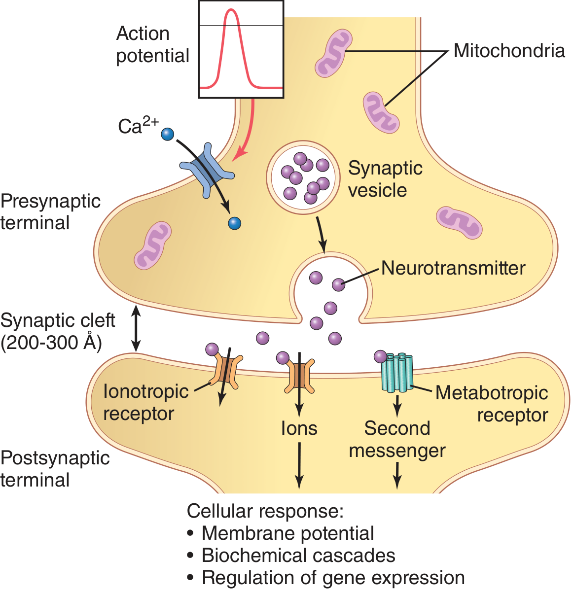
Figure: Chemical synapse - Guyton and Hall Textbook of Medical Physiology
2. Electrical Synapse
The two cells are coupled directly by gap junction channels through a much narrower intercellular gap of only 20-40 Å. Transmission is bidirectional and nearly instantaneous. These synapses are useful for synchronizing large groups of interconnected neurons - for example, hormone-secreting neurons in the hypothalamus use them to coordinate burst firing.
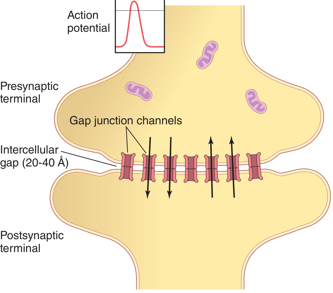
Figure: Electrical synapse - Guyton and Hall Textbook of Medical Physiology
Anatomy of a Chemical Synapse
- Presynaptic terminal (synaptic knob): The axon terminal of the sending neuron. Contains mitochondria and synaptic vesicles packed with neurotransmitter. A single motor neuron soma and its dendrites may receive 10,000 to 200,000 synaptic knobs from other neurons.
- Synaptic cleft: The narrow gap (200-300 Å) between the two membranes.
- Postsynaptic membrane: Usually on a dendrite or soma of the receiving neuron. Bears the receptors for the neurotransmitter.
How Chemical Synaptic Transmission Works
The sequence of events (per Katzung's Basic and Clinical Pharmacology):
- Action potential arrives at the presynaptic terminal.
- Voltage-gated Ca²⁺ channels open in the presynaptic membrane, allowing calcium to flow in.
- Rising intracellular Ca²⁺ triggers synaptic vesicle fusion with the presynaptic membrane.
- Neurotransmitter is released into the synaptic cleft by exocytosis.
- The neurotransmitter diffuses across the cleft and binds to receptors on the postsynaptic membrane.
- This produces either an EPSP (excitatory postsynaptic potential) - a brief depolarization - or an IPSP (inhibitory postsynaptic potential) - a hyperpolarization.
- When enough EPSPs summate (spatially or temporally), the postsynaptic neuron reaches threshold and fires its own action potential.
- Neurotransmitter action is terminated by reuptake into the presynaptic terminal (e.g., dopamine, serotonin, noradrenaline) or enzymatic degradation (e.g., acetylcholine by acetylcholinesterase).
Total delay from presynaptic action potential to postsynaptic response: ~0.5 ms (most of which is the time for Ca²⁺ channels to open and trigger release).
Postsynaptic Receptors: Two Categories
| Receptor Type | Mechanism | Speed | Example |
|---|---|---|---|
| Ionotropic | Neurotransmitter binding directly opens an ion channel | Fast (milliseconds) | AMPA, NMDA, GABA-A, nAChR |
| Metabotropic | Coupled to G-protein → second messenger cascade | Slow (seconds to minutes) | mGluR, GABA-B, muscarinic |
Excitation vs. Inhibition
- EPSP: Excitatory transmitter (e.g., glutamate) opens cation channels → membrane depolarizes toward threshold.
- IPSP: Inhibitory transmitter (e.g., GABA) opens Cl⁻ channels → membrane hyperpolarizes. The "shunting" effect (increased conductance) also makes it harder for EPSPs to reach threshold even without significant hyperpolarization.
- Presynaptic inhibition: Autoreceptors on the presynaptic terminal detect released transmitter and reduce further release. Transmitter "spillover" can also inhibit neighboring synapses.
The Electrical-to-Chemical-to-Electrical Transformation
As Neuroscience: Exploring the Brain puts it: "This electrical-to-chemical-to-electrical transformation of information makes possible many of the brain's computational abilities. Modification of this process is involved in memory and learning, and synaptic transmission dysfunction accounts for certain brain and muscle disorders. The synapse is also the site of action for many toxins and for many psychoactive and therapeutically useful drugs."
Clinical and Pharmacological Relevance
Virtually all CNS drugs work by modifying some step in synaptic transmission:
| Drug/Toxin | Site of Action |
|---|---|
| Reserpine | Blocks monoamine vesicle storage → depletes transmitter |
| Amphetamine | Induces catecholamine release from presynaptic terminal |
| Cocaine | Blocks reuptake of catecholamines → prolongs their effect |
| Tetanus toxin | Blocks neurotransmitter release |
| Anticholinesterases | Block acetylcholine degradation → prolong ACh action |
| Benzodiazepines / barbiturates | Potentiate GABA at GABA-A receptor |
Synaptic dysfunction underlies diseases including Alzheimer's disease (cholinergic synapse loss), Parkinson's disease (dopaminergic synapse degeneration), epilepsy (excessive excitation), depression (monoamine imbalance), and schizophrenia.
Sources: Guyton and Hall Textbook of Medical Physiology; Katzung's Basic and Clinical Pharmacology, 16th Ed.; Neuroscience: Exploring the Brain, 5th Ed.
This is a shared conversation. Sign in to Orris to start your own chat.