
Dkd deep search
"diabetic kidney disease" AND management
diabetic kidney disease DKD 2025 2026 guidelines treatment SGLT2 GLP1
"diabetic nephropathy" AND pathogenesis AND mechanisms
diabetic nephropathy kidney histology glomerulosclerosis
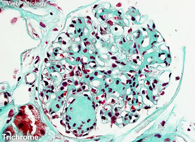
Masson's trichrome-stained renal cortex histology imaged at light microscopy reveals nodular glomerulosclerosis characteristic of diabetic nephropathy. The glomerulus shows circumscribed Kimmelstiel-Wilson nodules-eosinophilic mesangial nodules that expand the tuft and encroach on capillary loops. Diffuse mesangial sclerosis is evident as widespread mesangial matrix expansion, with increased basophilic mesangial tissue and accentuated blue-green collagen deposition in the extracellular matrix on Masson stain. The stain differentiates collagen (blue/green) from cytoplasm and nuclei (red/purple), highlighting the fibrotic nodules and mesangial expansion. Within the mesangium, there is progressive accumulation of matrix with variable cellularity; some capillary lumina appear narrowed due to nodular compression. The surrounding tubulointerstitial tissue may show mild chronic changes, consistent with chronic diabetic nephropathy. The overall pattern is typical of long-standing diabetes mellitus with nephropathy and correlates with progressive proteinuria, reduced glomerular filtration rate, and eventual renal insufficiency. Clinically, this finding supports diagnosis of diabetic kidney disease (diabetic nephropathy) and is essential for grading disease stage and guiding management. Differential diagnoses include nodular glomerulosclerosis due to amyloidosis or hypertensive nephrosclerosis, but the classic KW nodules and mesangial expansion favor diabetes. This image is valuable for medical education, pathology training, and research into diabetic renal pathology.
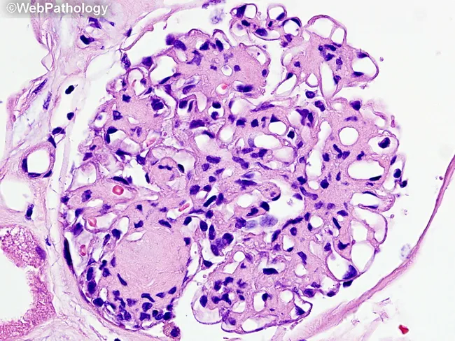
This is a renal biopsy histology slide examined under brightfield light microscopy. The primary subject is diabetic nephropathy with nodular glomerulosclerosis (Kimmelstiel-Wilson nodules) and diffuse mesangial sclerosis within the glomerular tuft of a renal cortex specimen. The glomerulus shows nodular mesangial expansion forming rounded, eosinophilic nodules that disrupt capillary loops, together with diffuse increased mesangial matrix. Capillary lumina are variably compressed, and arterioles frequently exhibit hyaline thickening consistent with diabetic microangiopathy. The cellular morphology is characterized by expanded mesangial cells with increased extracellular matrix and relatively sparse inflammatory infiltrate. The basement membrane appears thickened in cross-sectional elements. In this image, Masson’s trichrome is not visible; a subsequent stain would color collagen in blue, highlighting fibrotic remodeling. Clinically, these histologic features correlate with progressive proteinuria and reduced glomerular filtration rate in long-standing diabetes. Diagnostic significance rests on the combination of nodular sclerosis (Kimmelstiel-Wilson nodules) and diffuse mesangial sclerosis as hallmarks of diabetic nephropathy. Differential considerations include focal segmental glomerulosclerosis and hypertensive nephrosclerosis, but the nodular pattern is characteristic. This image is valuable for pathology education, nephrology training, and research into diabetic kidney disease progression and glycemic control effects. Representative for teaching clinics, this image supports correlating histology with clinical nephrology.
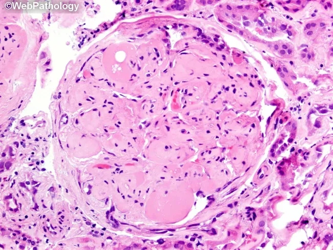
Imaging modality: brightfield histopathology. This slide shows a renal glomerulus from a biopsy specimen of diabetic nephropathy. The mesangial matrix is markedly expanded, producing round to nodular consolidations within the tuft. Several Kimmelstiel‑Wilson nodules are evident as acellular, hyaline nodules that distend the mesangial stalks and encroach on capillary loops. The glomerular basement membranes appear thickened on corresponding PAS‑positive and silver‑stained sections (referenced in adjacent images), consistent with nodular diabetic glomerulosclerosis. The surrounding cortex exhibits mild chronic interstitial changes and arteriolar hyalinosis in keeping with chronic diabetic kidney disease. The glomerulus displays relatively extensive mesangial deposition with focally patent capillary lumina, creating a characteristic nodular pattern. Overall, the morphology is diagnostic for diabetic microangiopathy with progressive nodular sclerosis. Clinically, these findings correlate with long‑standing hyperglycemia, proteinuria, and reduced GFR. Pathological significance: KW nodules indicate advanced disease and higher risk of progression to end‑stage renal disease. Differential considerations include non‑diabetic nodular glomerulosclerosis and other glomerulopathies, but the classic KW nodules and mesangial expansion strongly support diabetic nephropathy. This image is valuable for education, pathology review, and correlating histology with clinical diabetes management. It highlights key features for learners: mesangial expansion, KW nodules, PAS positivity, and altered capillary luminal flow in practice.
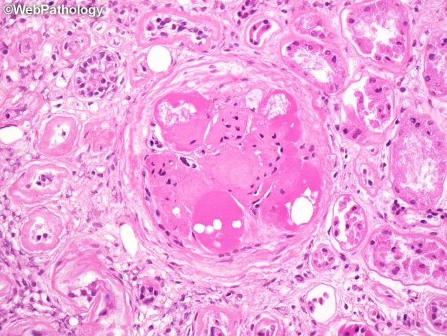
This is a renal biopsy histology image captured under bright-field light microscopy after Hematoxylin and Eosin staining. The focal structure is a glomerulus in the renal cortex showing near-complete global glomerulosclerosis with a markedly hyalinized tuft. Several hyaline caps outline the sclerotic core, appearing as pink, glassy deposits along capillary loops. Within these hyaline rims, lipid droplets and lipid-laden macrophages are evident, reflecting lipid accumulation associated with chronic diabetic microangiopathy. The surrounding tubulointerstitium displays mild interstitial fibrosis and preserved tubules in the periglomerular region. Collectively, the pattern is typical of advanced diabetic nephropathy with extensive glomerulosclerosis and hyaline arteriolosclerosis. The image highlights pathognomonic features such as noncellular sclerosis and lipid-laden inflammatory cells, which have prognostic implications for progressive renal insufficiency. Clinically, these findings correlate with long-standing diabetes mellitus, hypertension, proteinuria, and reduced GFR, indicating high risk for progression to end-stage kidney disease if glycemic control is poor. This specimen serves educational purposes in renal pathology, nephrology, and medical training to illustrate diabetic glomerulosclerosis, hyaline change, and lipid-related macrophage infiltration; it supports differential diagnosis with hypertensive nephrosclerosis and focal segmental glomerulosclerosis.
Diabetic Kidney Disease (DKD) - Deep Search
Sources: Goldman-Cecil Medicine, Brenner & Rector's The Kidney, Comprehensive Clinical Nephrology (7th Ed), NKF Primer, Goodman & Gilman's Pharmacology + PubMed 2025 literature + current guidelines
1. Overview & Epidemiology
2. Pathophysiology
2a. Hemodynamic Mechanism (Hyperfiltration)
2b. Hormones, Cytokines, and Signaling Pathways
- Transforming growth factor-β (TGF-β) - drives mesangial expansion and fibrosis
- Connective tissue growth factor (CTGF)
- Angiotensin II - the most clinically targetable mediator
- Vascular endothelial growth factor (VEGF) - increases glomerular permeability
- Endothelin and prostaglandins
- Nitric oxide dysregulation
2c. Genetics & Epigenetics
3. Stages of DKD - Natural History
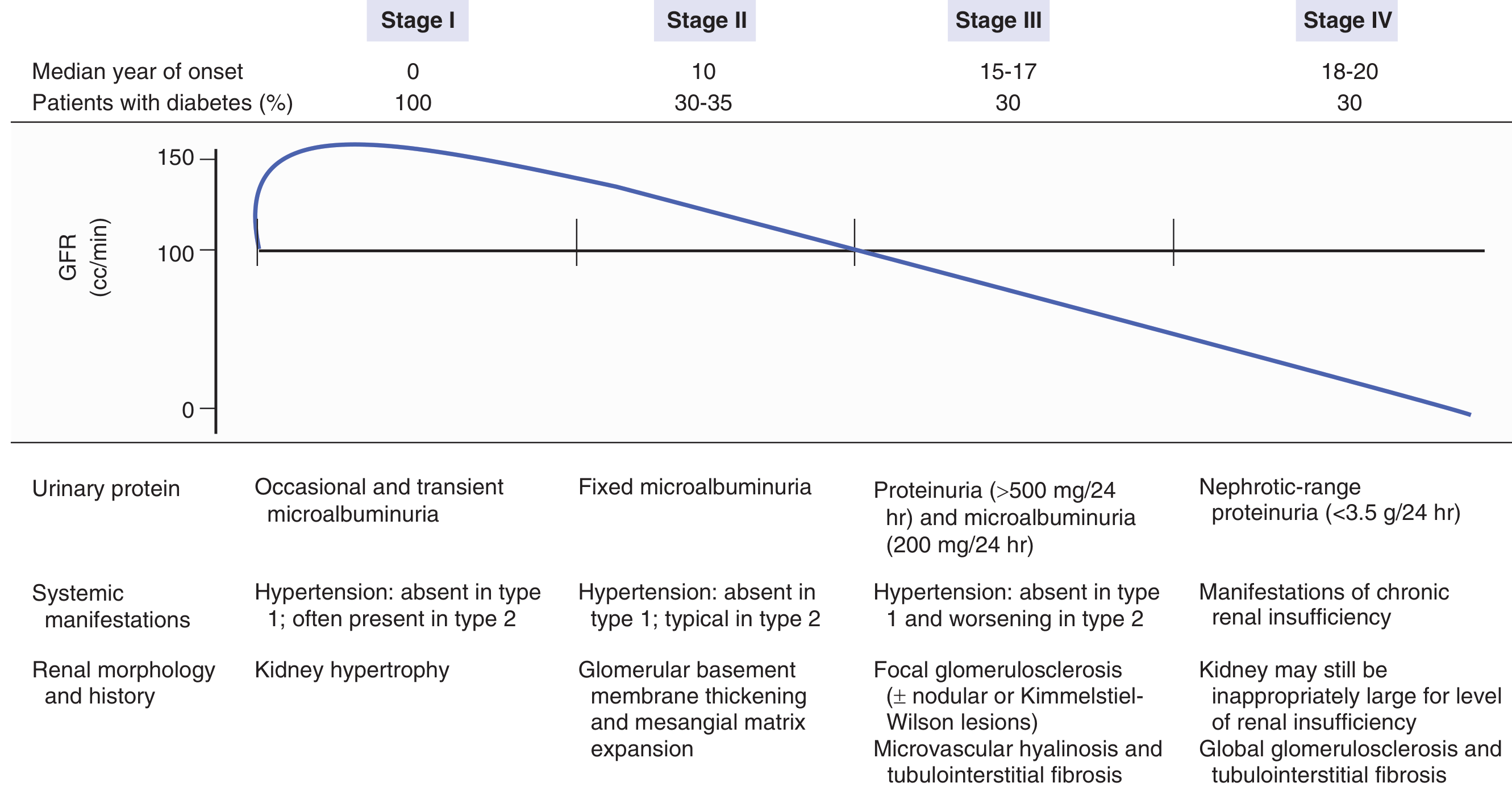
| Stage | Onset (years) | % of DM Patients | Key Features |
|---|---|---|---|
| I | 0 | 100% | Hyperfiltration (GFR +50%), kidney hypertrophy, occasional transient microalbuminuria |
| II | ~10 | 30-35% | Fixed microalbuminuria (30-300 mg/24h), GBM thickening, mesangial matrix expansion, HTN absent in T1D but typical in T2D |
| III | 15-17 | ~30% | Proteinuria >500 mg/24h + microalbuminuria, overt HTN, focal glomerulosclerosis ± Kimmelstiel-Wilson nodules |
| IV | 18-20 | ~30% | Nephrotic-range proteinuria (>3.5 g/24h), relentless GFR decline, progression to ESRD |
- Goldman-Cecil Medicine, pp. 1275-1276
4. Pathology
Renal Biopsy Findings
- Diffuse mesangial sclerosis - widespread mesangial matrix expansion (most common)
- Nodular (Kimmelstiel-Wilson) glomerulosclerosis - acellular, eosinophilic nodules in the mesangium, considered pathognomonic but present in only ~25% of cases. Kimmelstiel-Wilson nodules form when mesangial cells undergo mesangiolysis, and the overlying capillaries form microaneurysms.
- Glomerular basement membrane thickening
- Afferent AND efferent arteriolar hyalinosis - distinguishes DKD from hypertensive nephrosclerosis (isolated afferent hyalinosis)
- Tubulointerstitial fibrosis - correlates most closely with functional GFR decline
- "Capsular drop" and "fibrin cap" lesions - additional pathognomonic features
- Goldman-Cecil Medicine, p. 1276; Comprehensive Clinical Nephrology 7e, p. 432
5. Diagnosis
- Proteinuria within an appropriate time frame
- Retinopathy (present in 90-95% of T1DM-related DKD; 60-65% of T2DM-related)
- Absence of other causes of nephrotic syndrome or renal insufficiency
- Type 1 DM: begin microalbuminuria screening 5 years after diagnosis, then annually
- Type 2 DM: begin at time of diagnosis, then annually
6. Management - Stage-by-Stage Summary
| Stage | Interventions |
|---|---|
| I | Tight glycemic control (HbA1c ≤7%); consider ACE inhibitor or ARB for BP |
| II | Tight glycemic control; BP control; ACE inhibitor or ARB; SGLT2 inhibitor; Finerenone; smoking cessation; weight reduction; exercise; annual eye exam |
| III | BP control; ACE inhibitor or ARB; SGLT2 inhibitor; finerenone; protein restriction; smoking cessation |
| IV | BP control; ACE inhibitor or ARB; consider dialysis/transplant planning |
- Goldman-Cecil Medicine, pp. 3998-4010
7. Treatment Deep Dive
7a. Glycemic Control
- Target HbA1c ≤7% in most patients, with individualization to 7-8% in elderly or those at high hypoglycemia risk
- Tight control reduces microvascular complications in both T1DM and T2DM
- With declining GFR, medication adjustments are essential:
- Sulfonylureas: contraindicated when GFR <45 mL/min (stage 3b+) due to hypoglycemia risk
- Metformin: avoid when serum creatinine >1.7 mg/dL (or eGFR <30); can be used cautiously down to eGFR 30
- Insulin: requirements may decrease as GFR declines (reduced renal insulin clearance)
7b. RAAS Blockade (ACE Inhibitors / ARBs)
- First-line renoprotective therapy in DKD, particularly in patients with microalbuminuria or proteinuria
- ACE inhibitors and ARBs reduce intraglomerular pressure, decrease proteinuria, and slow GFR decline
- Do NOT combine ACE inhibitor + ARB (dual RAAS blockade) - increases hyperkalemia and AKI risk without added benefit (ONTARGET trial)
- Monitor potassium and creatinine closely after initiation
7c. SGLT2 Inhibitors - The New Cornerstone
| Drug | Trial | Key Result |
|---|---|---|
| Canagliflozin 100/300 mg | CREDENCE | Reduced composite of renal failure, CV death; 30% RRR for renal primary endpoint |
| Dapagliflozin 10 mg | DAPA-CKD | Reduced sustained ≥50% eGFR decline, ESKD, or renal/CV death |
| Empagliflozin 10/25 mg | EMPA-KIDNEY | Reduced CKD progression or CV death; benefit seen down to eGFR ~20 |
- Not approved for T1DM (risk of euglycaemic DKA)
- Withhold during acute illness, surgery, or prolonged fasting ("sick day rules")
- Risk of genital mycotic infections, Fournier's gangrene (rare)
- GLP-1 RA + SGLT2i combination may offer additive cardiorenal benefit
7d. GLP-1 Receptor Agonists
- Reduce albuminuria and slow GFR decline
- Primary evidence is cardiovascular outcome trials (LEADER, SUSTAIN-6, REWIND)
- The FLOW trial (semaglutide 1 mg weekly) specifically showed a 24% reduction in renal events in T2DM + CKD
- Recommended as adjunctive therapy, particularly in patients with obesity or high CV risk
- An international 2026 Delphi consensus (Handelsman et al., Adv Ther 2026) positions GLP-1 RAs + SGLT2i as complementary rather than redundant therapies in DKD + overweight/obesity
7e. Finerenone (Non-Steroidal MRA) - FDA Approved 2021
- Equally distributed in myocardial and kidney tissue (more balanced action)
- Binds different amino acids in MR ligand domain → different cardiac gene expression pattern
- Reduced cardiac fibrosis and inflammation vs. eplerenone in animal studies
- Low incidence of hyperkalemia in trials
7f. Blood Pressure Targets
- Target: <130/80 mmHg (ADA 2025, AHA/ACC); up to <140/90 mmHg acceptable in older or high-risk patients
- HOT trial subgroup: 51% reduction in major CV events with diastolic target ≤80 vs. ≤90 mmHg in diabetics
- ACE inhibitors or ARBs preferred as antihypertensive agents in DKD
- ACCORD trial showed no added renal benefit from BP <120 mmHg vs. <140 mmHg, but CV benefit persisted
8. Combination Therapy: The 2025-2026 Paradigm
Foundation:  ACE inhibitor or ARB
Add:         SGLT2 inhibitor (renal + CV protection)
Add:         Finerenone (anti-fibrotic, anti-inflammatory, MR blockade)
Consider:    GLP-1 RA (especially with obesity / high CV risk)
Optimise:    HbA1c, BP, lipids, weight, smoking cessation
9. Other Renal Complications in Diabetes
- Type IV RTA (hyporeninemic, hypoaldosteronemic) - metabolic acidosis + hyperkalemia in mild-moderate CKD; worsened by ACE inhibitors, ARBs, NSAIDs, heparin, K-sparing diuretics
- Urinary tract infections - increased bacterial and fungal infections including pyelonephritis, intrarenal abscess, perinephric abscess
- Renal artery stenosis - more common in T2DM; suspect if BP refractory or creatinine rises sharply after RAAS initiation
- Papillary necrosis with ureteral obstruction
- Contrast-induced AKI - risk is higher; use minimum contrast, pre-hydration
- Obstructive uropathy from autonomic bladder dysfunction
- Prerenal azotemia/ATN from gastroparesis-induced volume depletion
10. Recent Evidence Updates (2025-2026)
| PMID | Study | Key Finding |
|---|---|---|
| 41087076 | Saudi CPG for DKD 2025 | Multi-drug layered approach endorsed; SGLT2i + finerenone as standard add-ons to RAAS |
| 40847599 | Systematic review: pharmacotherapy in DKD | SGLT2i most validated; finerenone, GLP-1 RA recommended as adjuncts |
| 41272492 | Finerenone meta-analysis (Asian vs. non-Asian) | Comparable efficacy and safety across ethnicities |
| 40751239 | Incidence of DKD in Ethiopia (meta-analysis) | High prevalence of DKD in T2DM in sub-Saharan Africa; underdiagnosis common |
| NEJM 2025;393:1755 | Finerenone + empagliflozin in CKD/T2DM | Combination superior to either agent alone for composite cardiorenal outcome |
Key Teaching Points
- DKD is the leading cause of ESKD worldwide and results from the interplay of hemodynamic (intraglomerular hypertension), metabolic (AGEs, oxidative stress), and genetic/epigenetic factors.
- The four stages progress over 15-20 years; 30% of diabetics reach stage II, but progression is modifiable.
- The Kimmelstiel-Wilson nodule is pathognomonic but present in only ~25%; afferent + efferent arteriolar hyalinosis distinguishes DKD from hypertensive nephrosclerosis.
- Treatment is now a 4-drug combination: ACE inhibitor/ARB + SGLT2 inhibitor + finerenone ± GLP-1 RA.
- SGLT2 inhibitors provide renoprotection independent of glycemia, largely via tubuloglomerular feedback and intraglomerular pressure reduction.
- Finerenone (FDA 2021) targets the aldosterone-mediated inflammatory-fibrotic axis and complements rather than duplicates SGLT2 inhibitor mechanisms.
- Monitor for hyperkalemia with RAAS + finerenone; withhold SGLT2i during illness/fasting.