
House brattmans classification
House Brackmann classification facial nerve grading system
House Brackmann facial nerve grading scale classification
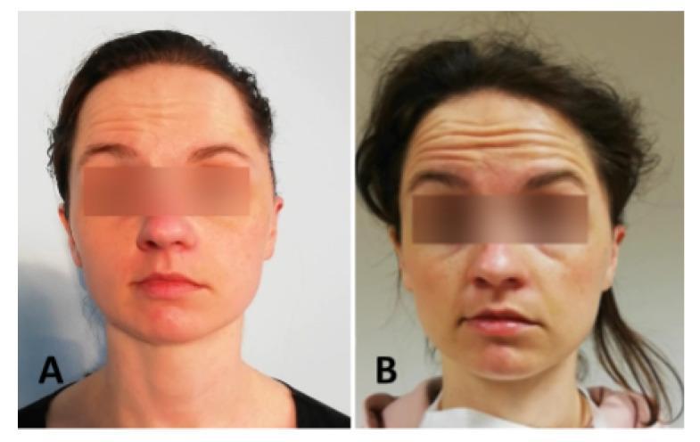
Clinical photograph comparing two stages of facial nerve dysfunction in a patient during an eyebrow elevation exercise. Image (A) represents a House-Brackmann (HB) grade VI classification, demonstrating total paralysis on the patient's left side with a complete absence of forehead wrinkling and eyebrow movement, resulting in marked asymmetry. Image (B) represents an HB grade III classification, showing moderate improvement in mimetic muscle function. In this stage, there is visible, though still asymmetric, forehead wrinkling and a noticeable arching/elevation of the eyebrows. The comparison illustrates the clinical assessment of the frontalis muscle and the cranial nerve VII (facial nerve) recovery process. The visual demonstrates key diagnostic indicators used in grading systems, such as the depth of forehead rhytids and the excursion distance of the eyebrows relative to the eyes.
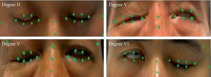
This set of clinical photographs illustrates various severities of facial paralysis, specifically focusing on the inability to close the eyes (lagophthalmos). The images are categorized by the House-Brackmann scale or similar facial nerve grading systems (FNGS 2.0). Degree II shows nearly complete eye closure with a minor slit remaining. Degree V (demonstrated in two panels) shows significant paresis with the eyelids remaining partially open, indicating severe dysfunction of the orbicularis oculi muscle. Degree VI shows total paralysis with a wide palpebral fissure and no voluntary movement. Green markers indicate the application of a facial landmark detection model (FP-FLDM) used to calculate the Eye Aspect Ratio (EAR). These markers trace the contours of the eyelids, medial and lateral canthi, and nasal bridge. The visual demonstrates the difficulty in automated landmark tracking in cases of severe facial asymmetry and pathological expressions. This material is relevant for neurology and ophthalmology education, focusing on cranial nerve VII assessment and the risk of corneal exposure.
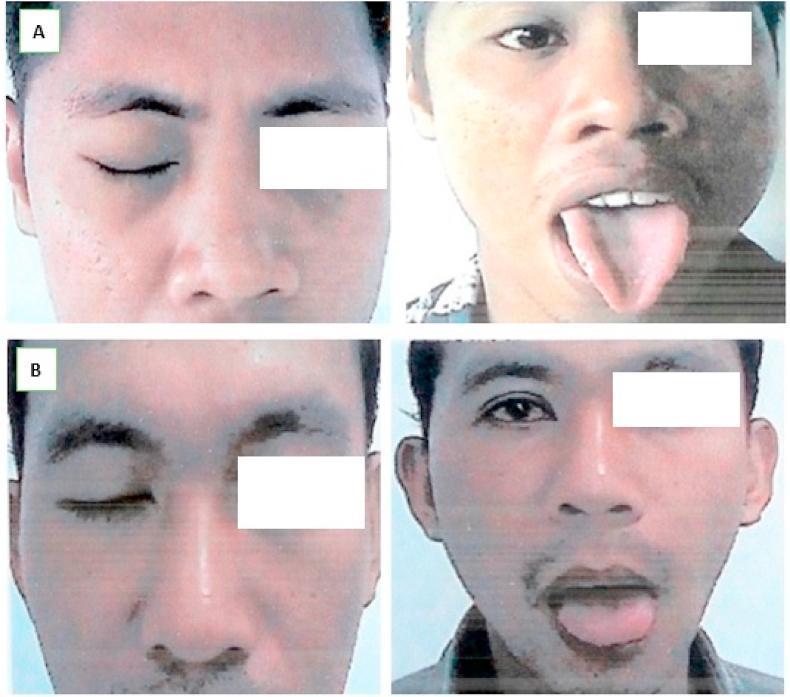
Clinical photograph showing two male patients (A and B) post-surgical reanimation for facial nerve palsy, evaluated using the House-Brackmann (HB) Scale. Each patient is shown in two views: one demonstrating eye closure and one demonstrating tongue protrusion. Patient A (top row) displays complete eye closure on the affected side and successful tongue protrusion with slight deviation, indicating progress after a split hypoglossal-facial anastomosis. Patient B (bottom row) likewise demonstrates functional eye closure and central tongue protrusion. The clinical objective shown is the assessment of motor function recovery in the facial and hypoglossal nerve distributions. Both cases illustrate HB Grade I results at 10 months post-operation, characterized by normal facial function in all areas. The images emphasize the success of nerve graft techniques in restoring facial symmetry and voluntary muscle control following skull base fractures.
House-Brackmann (HB) Grading System - Facial Nerve Classification
The 6-Grade Scale
| Grade | Description | Global Characteristics | At Rest | Forehead | Eye | Mouth |
|---|---|---|---|---|---|---|
| I | Normal | Normal facial function in all areas | Normal | Normal | Normal | Normal |
| II | Mild dysfunction | Slight weakness on close inspection; possibly very slight synkinesis | Normal symmetry and tone | Moderate to good function | Complete closure with minimum effort | Slight asymmetry |
| III | Moderate dysfunction | Obvious but not disfiguring difference; noticeable (not severe) synkinesis, contracture, or hemifacial spasm | Normal symmetry and tone | Slight to moderate movement | Complete closure with effort | Slightly weak with maximum effort |
| IV | Moderately severe dysfunction | Obvious weakness and/or disfiguring asymmetry | Normal symmetry and tone | None | Incomplete closure | Asymmetric with maximum effort |
| V | Severe dysfunction | Barely perceptible motion only | Asymmetry | None | Incomplete closure | Slight movement |
| VI | Total paralysis | No movement | - | - | - | - |
Key Clinical Points
- Grade I vs. VI are the least ambiguous (normal vs. complete paralysis). Grades II-V are most prone to inter-observer variability because multiple regional descriptors can overlap.
- The system assesses four regions: forehead, eye, nose, and mouth. Some experts recommend reporting each region individually for greater accuracy, especially in patients with differential facial function across regions.
- The grade primarily reflects eye function when a global score is used.
- The original HB system was designed for chronic facial nerve injury (includes criteria for synkinesis, asymmetry, and tone at rest), NOT for acute injury.
Important: FNGS 2.0 (Updated System)
- Excludes chronic injury criteria (synkinesis, resting asymmetry, tone)
- Is technically the correct scale to use for acute facial nerve injury (e.g., Bell's palsy, temporal bone trauma)
Limitations of the HB System
- Inter-observer agreement varies - disagreement among examiners can range from 13% to 44% when only a global score is used
- Fusion of static and dynamic criteria within a single grade contributes to variability
- A single global rating primarily reflects eye region function, potentially underrepresenting forehead or mouth deficits
- Not ideal for acute facial nerve injury (use FNGS 2.0 instead)
Clinical Images
- Cummings Otolaryngology Head and Neck Surgery, Table 171.1 (modified HB grading scale with unambiguous criteria per grade)
- Scott-Brown's Otorhinolaryngology Head & Neck Surgery Vol. 2, Table 112.2 (Facial palsy grading systems, p. 6452-6490)
Canal paresis in caloric test
caloric test canal paresis Jongkees formula vestibular
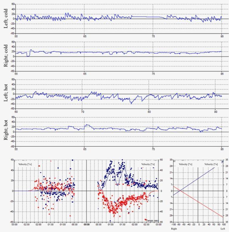
Diagnostic diagnostic traces and charts showing results of a caloric vestibular test, used to assess peripheral vestibular function. The top four panels display Electronystagmography (ENG) or Videonystagmography (VNG) recordings of slow phase velocity (SPV) in degrees per second (°/s) during bithermal caloric irrigation (30°C cold and 44°C hot) for both the left and right ears. The left ear panels demonstrate characteristic nystagmus responses with fluctuating SPV values. In contrast, the right ear panels ('Right; cold' and 'Right; hot') show flat traces with SPV peak values ≤ 5°/s, indicating a diagnosis of right vestibular areflexia. The bottom left quadrant contains a composite scatter plot of nystagmus velocity over time, with red and blue data points representing the fast and slow phases of nystagmus. The bottom right graph provides a summary caloric chart plotting peak velocities to quantify vestibular paresis or directional preponderance. This material is essential for neurology and otolaryngology students studying the diagnostic workup of vertigo, balance disorders, and unilateral vestibular loss.
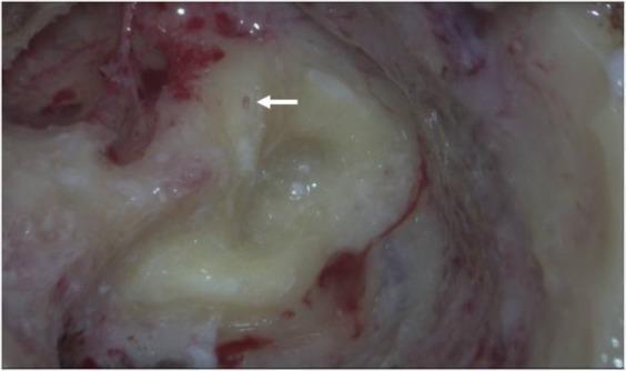
This clinical otologic photograph captures an intraoperative view of the left mastoid cavity during a revision labyrinthectomy in a patient with Meniere’s disease. The image focuses on the vestibular system, specifically the three semicircular canals previously treated with triple semicircular canal plugging (TSCP). The superior semicircular canal (SSC) and posterior semicircular canal (PSC) exhibit successful occlusion; the plugging fascia has undergone fibrotic and ossifying changes, resulting in the complete disappearance of the canal lumens. In contrast, the horizontal semicircular canal (HSC) demonstrates incomplete occlusion. A white arrow points to a small residual lumen within the HSC where endolymph fluid remains visible, indicating a failure of the initial plugging material to achieve full mechanical blockage. This visual evidence of incomplete occlusion highlights the anatomical basis for recurrent vestibular symptoms and provides clinical correlation for abnormal video head impulse test (v-HIT) or caloric test results in post-surgical patients.
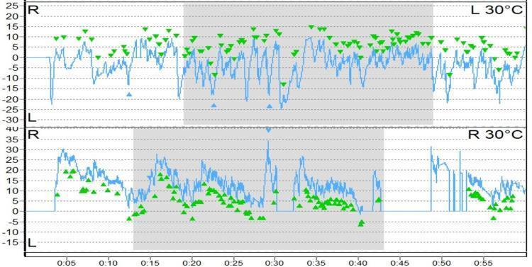
Diagnostic tracing showing Videonystagmography (VNG) results from a caloric reflex test, specifically illustrating the phenomenon of caloric inversion. The image contains two vertically stacked strip charts representing eye movement velocity (slow phase velocity) over time. The top graph is labeled 'L 30°C' (left ear cold irrigation) and the bottom graph 'R 30°C' (right ear cold irrigation). Each graph features a blue continuous line plot representing nystagmus and green arrowheads marking the peak velocity of the slow phase. A grey shaded box highlights the primary response window. Clinically, caloric inversion is characterized by nystagmus beating in a direction opposite to what is expected based on the temperature of the irrigation (COWS: Cold Opposite, Warm Same). In this tracing, the green arrowheads indicate a persistent vestibular response that deviates from standard physiological expectations. This finding is highly significant in neuro-otology as it often suggests central vestibular dysfunction or brainstem pathology, though technical artifacts such as tympanic membrane perforation must be ruled out.
Canal Paresis (Unilateral Weakness) in the Caloric Test
What is Canal Paresis?
Basis: The COWS Rule
| Irrigation | Expected Nystagmus Direction |
|---|---|
| Cold water | Opposite side (away from irrigated ear) |
| Warm water | Same side (toward irrigated ear) |
Mnemonic: COWS - Cold Opposite, Warm Same
Jongkees' Formula for Canal Paresis
Unilateral Weakness (Canal Paresis):
Directional Preponderance (DP):
- R = Right, L = Left
- W = Warm (44°C), C = Cold (30°C)
- Values = peak slow-phase velocity (°/sec)
Normal vs. Abnormal Cutoffs
| Parameter | Significant if... |
|---|---|
| Unilateral Weakness (Canal Paresis) | > 20% |
| Directional Preponderance | > 25% |
Five Main Abnormalities of the Caloric Response
| # | Finding | Implication |
|---|---|---|
| 1 | Bilateral absence of caloric nystagmus | Aminoglycoside ototoxicity, post-meningitis (see below) |
| 2 | Unilateral canal paresis | Vestibular neuritis, acoustic neuroma (schwannoma), labyrinthitis |
| 3 | Directional preponderance | Spontaneous nystagmus (non-specific; can be labyrinth to cortex) |
| 4 | Abnormal VOR suppression | Central lesion (failure to suppress nystagmus with fixation) |
| 5 | Caloric inversion | Central vestibular dysfunction / brainstem pathology |
Causes of Unilateral Canal Paresis
- Vestibular neuritis (most common peripheral cause)
- Vestibular schwannoma (acoustic neuroma)
- Labyrinthitis
- Meniere's disease
- Brainstem lesion at the VIII nerve root entry zone or vestibular nuclei (rare; CNS signs will usually be present)
Causes of Bilateral Caloric Hypofunction
| Cause | % |
|---|---|
| Neurological (cerebellar degeneration, post-meningitis, neuropathies) | 25% |
| Ototoxic (usually gentamicin) | 25% |
| Miscellaneous (autoimmune, Meniere's, head trauma) | 25% |
| Idiopathic | 25% |
Relationship Between Canal Paresis and Directional Preponderance
- Acutely: Complete left canal paresis dominates, with spontaneous right-beating nystagmus (right DP)
- Over time: Even if the canal paresis does not recover, the right ear's responses create asymmetry - warm irrigation (rightward nystagmus) stronger than cold irrigation (leftward) - leaving a right DP as the only residual finding
- DP in isolation is non-specific - it can result from lesions anywhere from the labyrinth to the cortex
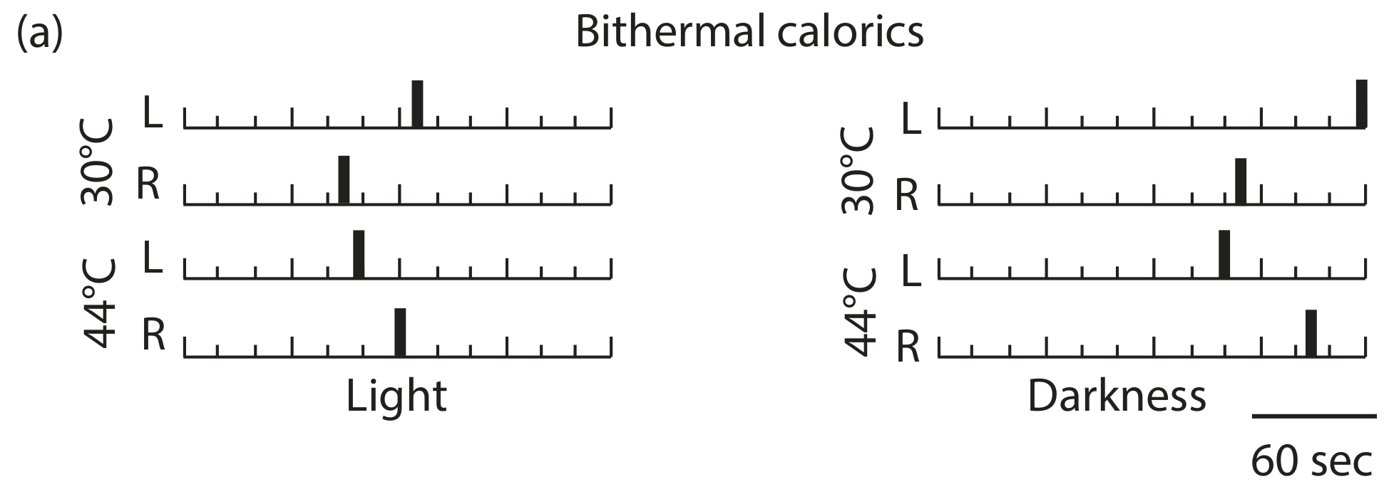
Clinical Caloric Bar Chart (Normal)
Important Technical Notes
- If minimal caloric responses are present, ice water (0°C) can be used to elicit any residual response
- Responses can be falsely reduced by cerumen impaction, tympanic membrane perforation, or poor technique
- Air irrigation can be used when the eardrum is perforated, but water gives more consistent responses
- For bilateral reduced calorics, supplement with rotational chair testing to define the extent of bilateral vestibular loss
- Shambaugh Surgery of the Ear, "Caloric Testing" (Jongkees formula, normative values)
- Scott-Brown's Otorhinolaryngology Head & Neck Surgery Vol. 2, "Caloric Tests" (five abnormalities, canal paresis causes)