
Lipoma pathology detailed
lipoma pathology
lipoma pathology histology variants molecular pathology MDM2 CDK4
lipoma histopathology mature adipocytes thin fibrous septa microscopy
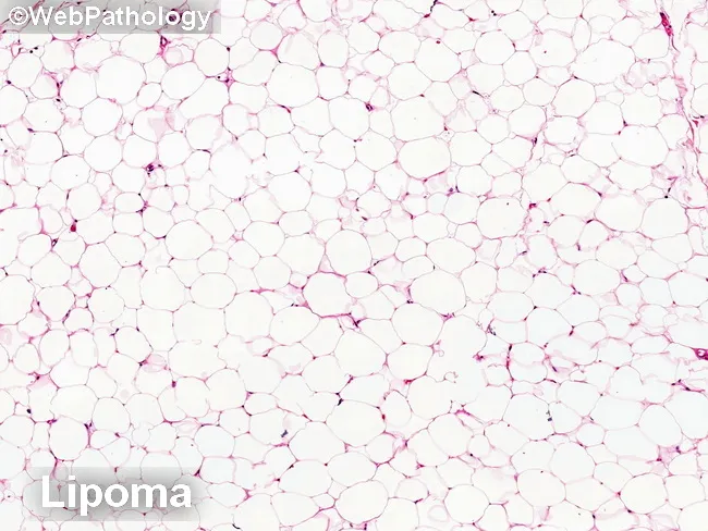
Imaging modality: Light microscopy of hematoxylin and eosin (H&E) stained formalin-fixed paraffin-embedded tissue section. Specimen: benign lipomatous lesion composed of mature adipocytes. Primary subject: adipose tissue within soft tissue (lipoma) with fibrous septa. Imaging perspective: histological cross-section through adipose lobules. The morphologic features: uniform round-to-oval adipocytes with small clear cytoplasm, peripherally placed nuclei, lacking significant cytologic atypia. Septa are relatively thin and occasionally show fibrous strands; no lipoblasts are evident. In this image, the background is comprised of many univacuolated adipocytes forming a lobulated architecture typical of lipoma. Compared to atypical lipomatous tumor/well-differentiated liposarcoma (ALT/WDL), this lesion shows minimal to no variation in adipocyte size, lack of hyperchromatic, atypical stromal cells in fibrous septa, and absence of necrosis or mitotic activity. Intramuscular lipoma variants may show atrophic skeletal muscle fibers adjacent to adipose tissue; in such cases, entrapped muscle fibers with hyperchromatic nuclei can mimic stromal atypia but lack lipoblasts and genetic alterations characteristic of WDL. The image corresponds to a benign lipomatous tumor and illustrates the core criteria used to distinguish lipoma from ALT/WDL in routine histopathology. Immunohistochemical or molecular markers, including MDM2 amplification status, can assist in differentiating ALT/WDL from benign lipomas in challenging cases. Accurate classification informs surgical margins and surveillance strategies.
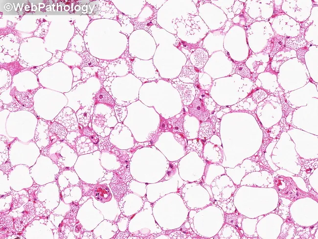
Imaging modality: Light microscopy of an H&E stained tissue section from a subcutaneous adipose lesion. Specimen: Excisional biopsy tissue of soft tissue mass. Location: Subcutaneous adipose tissue within the extremities or trunk, consistent with hibernoma lipoma-like variant. Visual features: The slide shows abundant univacuolated mature adipocytes with clear lipid droplets, minimal cytoplasm, and nuclei at the periphery, creating a pale, foamy appearance. The adipocytes are arranged in lobules and are separated by delicate fibrous septa. In this lipoma-like variant, the lesion is predominantly composed of mature white fat resembling a lipoma, with only scant brown fat differentiation or vacuolization. Vascular network: Thin capillary-rich stroma is present within septa, reflecting the vascular nature of brown fat components that may be variably represented. The overall histology lacks significant cellular atypia or lipoblasts. The sparse presence of multivacuolated cells, if present, and occasional mitochondria-rich cytoplasm may indicate residual brown fat differentiation, supporting a diagnosis within the spectrum of hibernoma rather than conventional lipoma. Clinical significance: Benign adipocytic tumor with lipoma-like histology; differentiating from liposarcoma is essential to avoid overtreatment; surgical excision is typically curative. Potential clinical use: diagnostic education, histopathology training, differential diagnosis evaluation, pathology atlases. Correlative imaging and clinical history enhance accuracy overall.
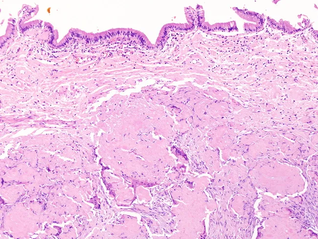
Comprehensive Description: This is a histopathology slide of a cutaneous skin biopsy examined with bright-field microscopy after hematoxylin and eosin staining. The specimen shows a perpendicular transection through epidermis, dermis, and subcutaneous fat. The epidermis exhibits marked acanthosis with elongation of rete ridges and mild hyperkeratosis, while the basal layer remains intact. The dermis contains variably dense fibrous stroma with sparse chronic inflammatory cells. Beneath the dermis, the subcutaneous tissue is dominated by mature adipocytes arranged in lobules separated by thin fibrous septa. No cytologic atypia, mitotic figures, or infiltrative tumor margins are evident. The overall architecture favors a benign adipose-rich process, such as a lipomatous lesion, with epidermal remodeling that may accompany adjacent subcutaneous fat expansion. There is no clear evidence of malignancy, inflammatory panniculitis, or granulomatous reaction in the field shown. Staining quality permits clear visualization of adipocytes (large clear cytoplasm), connective tissue septa, and epidermal layers. This image is useful for dermatopathology teaching, correlating histologic patterns of lipomatous subcutaneous lesions with overlying epidermal changes, and supporting differential diagnosis including lipoma, fibrolipoma, and, less likely, liposarcoma in cutaneous/subcutaneous tissue. It provides a reference for pattern recognition and morphology in skin biopsy evaluation. Clinically relevant across education and research.
https://www.pathologyoutlines.com/topic/softtissueadiposelip…
Lipoma: detailed pathology
1. Gross pathology
- Usually a well-circumscribed, soft, lobulated, yellow mass.
- Often has a thin fibrous capsule or pseudocapsule.
- The cut surface is homogeneous, greasy, and resembles normal adipose tissue.
- Conventional superficial lipomas are commonly small, but may grow large.
- Variants can differ:
- Fibrolipoma: firmer due to increased fibrous tissue.
- Angiolipoma: may be more nodular and sometimes painful.
- Intramuscular lipoma: can be poorly circumscribed because it infiltrates between skeletal-muscle fibers.
2. Microscopic pathology
- Lobules of mature univacuolated adipocytes.
- Uniform fat cells, generally similar in size and shape to normal white adipose tissue.
- Each adipocyte has clear cytoplasm because lipid dissolves during tissue processing and a small flattened peripheral nucleus.
- Lobules separated by delicate fibrous septa.
- A thin peripheral fibrous capsule may be present.
- Small capillaries are normal within septa.
- No significant variation in adipocyte size.
- No hyperchromatic or enlarged atypical stromal cells within fibrous septa.
- No true malignant lipoblasts.
- No necrosis.
- No significant mitotic activity.
- No infiltrative destructive growth, except that intramuscular lipoma may entrap muscle fibers.
3. Cytology
- Mature adipocytes.
- Sparse capillary fragments.
- No atypical stromal cells or lipoblasts.
4. Molecular pathology
- Rearrangements involving 12q13-15, commonly affecting HMGA2.
- Less commonly, rearrangements involving 6p21 with HMGA1.
- MDM2 amplification
- CDK4 amplification
5. Immunohistochemistry
- S100: highlights mature adipocytes, but is nonspecific.
- MDM2: negative in conventional lipoma.
- CDK4: negative in conventional lipoma.
- p16: generally negative or not diffusely expressed.
6. Histologic variants
| Variant | Main pathology features |
|---|---|
| Fibrolipoma | Mature fat with conspicuous dense fibrous connective tissue. |
| Angiolipoma | Mature adipose tissue plus numerous small capillary-type vessels, often with fibrin thrombi. Usually subcutaneous and may be painful. |
| Spindle-cell lipoma | Bland spindle cells, mature fat, thick “ropy” collagen, mast cells, and often myxoid stroma. Classically posterior neck, shoulder, and upper back of older men. CD34 is usually strongly positive; RB1 expression is often lost. |
| Pleomorphic lipoma | Closely related to spindle-cell lipoma; may have floret-like multinucleated giant cells but remains benign. |
| Intramuscular lipoma | Mature adipocytes extend between muscle bundles. May recur locally if incompletely excised, but lacks malignant atypia. |
| Chondroid lipoma | Rare deep lesion containing adipocytes, lipoblast-like cells, and cells in a chondromyxoid matrix. Can mimic myxoid liposarcoma. |
| Osteolipoma / chondrolipoma | Lipoma containing mature bone or cartilage metaplasia. |
| Hibernoma | Benign tumor of brown fat rather than conventional lipoma. Contains multivacuolated granular brown-fat cells and is more vascular. |
7. Differential diagnosis
A. Normal adipose tissue
- Lipoma is typically a discrete mass, often encapsulated and lobulated.
- Normal fat blends diffusely into adjacent subcutaneous tissue.
B. Atypical lipomatous tumor / well-differentiated liposarcoma
| Feature | Lipoma | ALT/WDL |
|---|---|---|
| Site | Usually superficial | Often deep soft tissue, especially extremity or retroperitoneum |
| Size | Usually smaller | Often >10 cm |
| Histology | Uniform mature adipocytes | Variable adipocyte size, fibrous septa containing atypical hyperchromatic stromal cells |
| Mitoses/necrosis | Absent | Usually low, but atypical stromal cells are diagnostic |
| MDM2/CDK4 | Not amplified | Usually amplified |
| Behavior | Benign | Locally recurrent, site-dependent risk; dedifferentiation risk is especially relevant in retroperitoneal disease |
C. Myxoid liposarcoma
- More myxoid matrix.
- Prominent delicate branching “chicken-wire” capillary network.
- Lipoblasts may be present.
- Molecularly associated with FUS::DDIT3 rearrangement, not MDM2 amplification.
D. Hibernoma
- Brown-fat differentiation, granular eosinophilic or multivacuolated cells.
- More vascular than ordinary lipoma.
E. Angiolipoma
- Numerous capillaries, often with fibrin microthrombi.
- Pain is more common clinically.
8. Pathology report example
Soft tissue, subcutaneous tissue, excision:
Lipoma, conventional type.
- Benign mature adipocytic neoplasm.
- No cytologic atypia, lipoblasts, necrosis, or increased mitotic activity identified.
- Completely excised, if margins can be assessed.
Soft tissue, deep thigh, excision:
Mature adipocytic neoplasm, favor lipoma. No significant cytologic atypia or mitotic activity identified. MDM2 immunostain is negative and MDM2 FISH shows no amplification, supporting lipoma over atypical lipomatous tumor/well-differentiated liposarcoma.
9. Prognosis
- Ordinary lipoma is benign and does not metastasize.
- Complete local excision is usually curative.
- Recurrence is uncommon, generally related to incomplete removal or an infiltrative intramuscular location.
- A lesion that is deep, rapidly enlarging, fixed, painful, recurrent, or large should be assessed carefully to exclude ALT/WDL or another sarcoma. The anatomical site, depth, and size materially affect the diagnostic and prognostic interpretation of lipogenic tumors (Fitzpatrick’s Dermatology, p. 2201).