
Epithelium general anatomy
types of epithelium classification diagram simple stratified
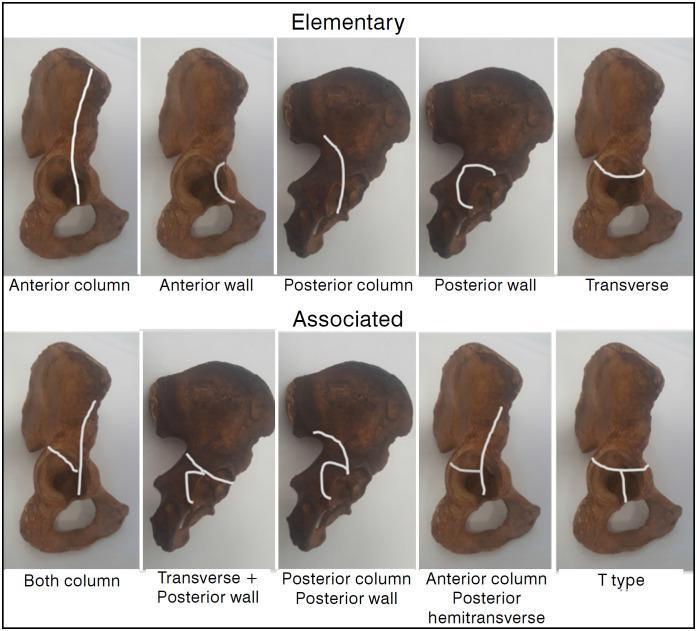
Educational diagram illustrating the Judet-Letournel classification of acetabular fractures using anatomical specimens of the hemipelvis. The visual is divided into two rows: 'Elementary' and 'Associated' fracture types. White lines superimposed on the specimens delineate specific fracture patterns. The 'Elementary' row features five simple patterns: Anterior column (vertical line through the pelvic brim), Anterior wall (localized articular segment), Posterior column (ischiadic notch through obturator foramen), Posterior wall (localized posterior rim), and Transverse (horizontal line bisecting the acetabulum). The 'Associated' row demonstrates complex configurations: Both column, Transverse plus posterior wall, Posterior column plus posterior wall, Anterior column with posterior hemitransverse (a vertical anterior column line joined by a horizontal posterior component), and T-type (a transverse fracture with an additional vertical stem extending through the obturator foramen). This orthopedic diagram serves as a clinical reference for orthopedic surgeons and medical students to understand pelvic trauma morphology and surgical planning.
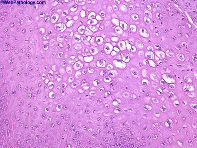
Photomicrograph of a genital mucosal biopsy stained with Hematoxylin and Eosin, viewed under brightfield light microscopy at low to moderate magnification (approx. 200x–400x). The section shows stratified squamous epithelium with condylomatous acanthosis and koilocytotic change. Koilocytes reveal enlarged, irregular nuclei with perinuclear halos and cytoplasmic clearing; binucleated cells are occasionally present. In addition, resinoid (raisinoid) nuclear changes with nuclear enlargement and hyperchromasia are evident in some keratinocytes, consistent with HPV-induced cytopathic effect. The epithelium displays mild to moderate dysplasia in foci, compatible with condyloma acuminatum harboring HPV-related dysplastic change. The underlying stroma is edematous without overt invasion. HPV types 16, 18, 31, and 33 are commonly associated with high-grade dysplasia in anogenital condylomas; detection by typing supports risk stratification for progression to high-grade intraepithelial neoplasia and carcinoma in susceptible mucosal sites. Note: This is a descriptive representation for imaging metadata; real-world interpretation requires clinical correlation and laboratory confirmation with HPV typing. The image exemplifies classic cytopathic effects of HPV infection in condylomatous epithelium. This description supports search queries for HPV-related anogenital lesions, koilocytosis, raisinoid nuclei, dysplasia, condyloma acuminatum, and high-risk HPV types (16/18/31/33); useful for educational, diagnostic, and research contexts. Clinically correlate with HPV typing and patient risk factors. Carefully.
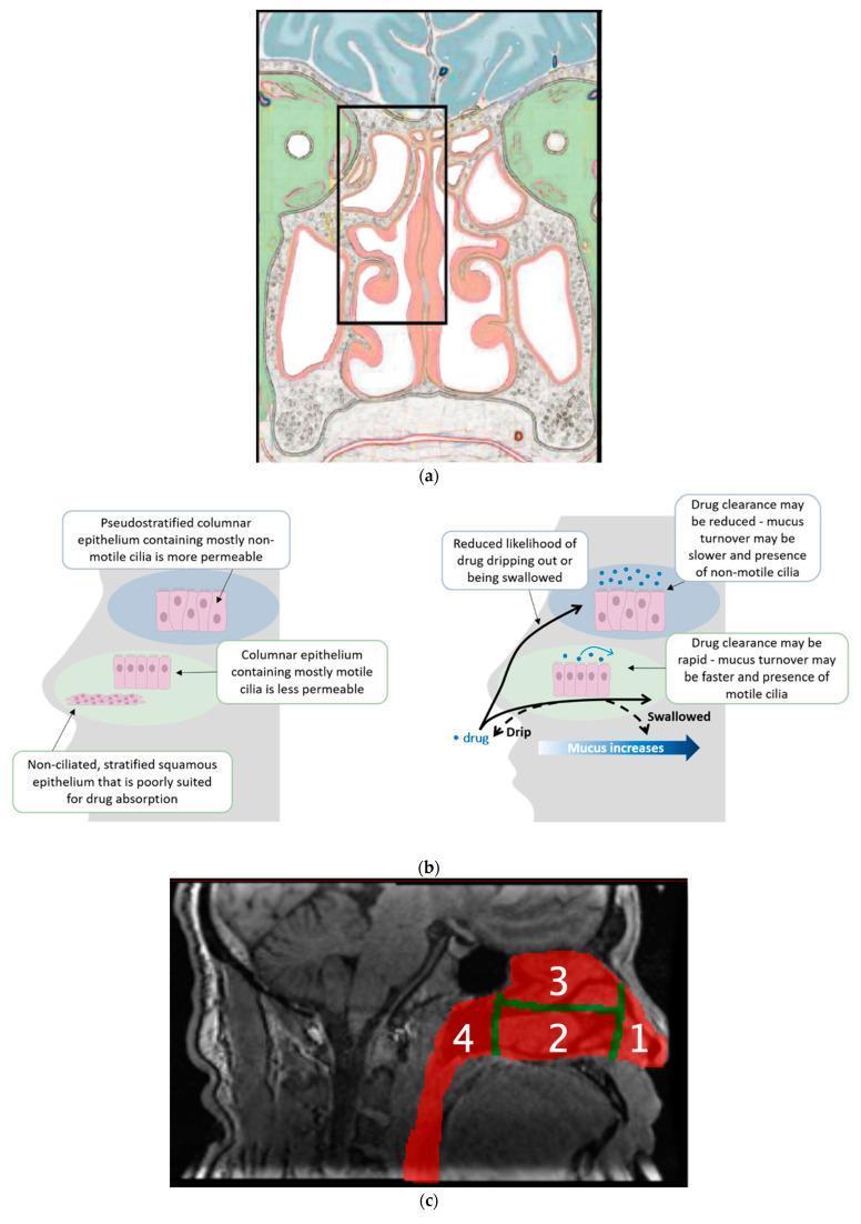
A multi-panel figure illustrating human nasal anatomy and drug delivery concepts. Panel (a) shows a coronal anatomical cross-section of the frontal head, focusing on the posterior nasal cavity. Key structures include the central nasal septum and lateral turbinates (conchae), with a black box highlighting the superior region enriched with olfactory epithelium. Panel (b) provides comparative histological diagrams and a sagittal head silhouette. The left diagram distinguishes three mucosal types: pseudostratified columnar epithelium (more permeable, non-motile cilia), columnar epithelium (less permeable, motile cilia), and non-ciliated stratified squamous epithelium. The right diagram depicts clearance mechanisms, showing how drugs delivered to the upper nasal space (UNS) avoid dripping or swallowing, unlike delivery to the lower nasal space where rapid mucus turnover and motile cilia facilitate clearance. Panel (c) is a sagittal MRI with a color-coded SPECT overlay dividing the nasal cavity into four functional regions for deposition analysis: (1) Vestibule, (2) Lower nasal space, (3) Upper nasal space (UNS), and (4) Nasopharynx. The figure demonstrates the anatomical and physiological basis for targeting the UNS in intranasal drug delivery.
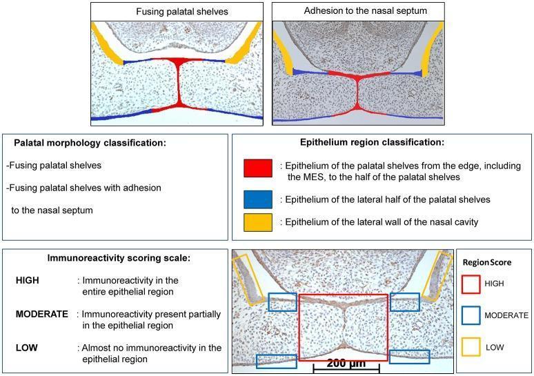
Educational diagram and histological composite illustrating palatal morphology classification and immunoreactivity scoring during palatogenesis. The upper panels show two histological stages: 'Fusing palatal shelves' and 'Fusing palatal shelves with adhesion to the nasal septum.' A color-coded epithelium region classification system is applied: RED indicates the epithelium from the midline epithelial seam (MES) to half the shelf width; BLUE identifies the lateral half of the palatal shelves; and YELLOW highlights the lateral wall of the nasal cavity as a control region. The lower section provides a semi-quantitative 'Immunoreactivity scoring scale' (HIGH, MODERATE, LOW) based on the distribution of staining (e.g., CXCL11, CXCR3, or HO-1) within these specific regions of interest. A representative immunostained section at E15 demonstrates high reactivity in the RED midline region, moderate in BLUE lateral regions, and low in the YELLOW nasal cavity control. This resource is designed for understanding embryonic craniofacial development and the biochemical signaling involved in secondary palate fusion.
simple squamous cuboidal columnar epithelium histology microscopy
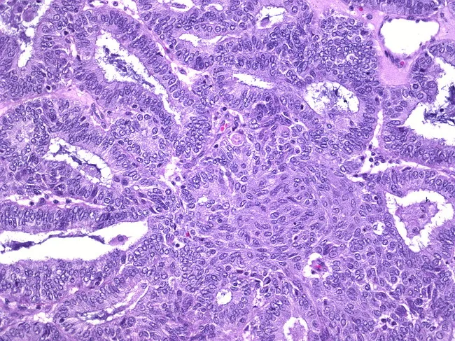
Imaging modality: Bright-field light microscopy of a hematoxylin and eosin stained ovarian tissue section. This histology shows an ovarian endometrioid carcinoma with focal squamous metaplasia (adenoacanthoma pattern). The architecture comprises back‑to‑back glandular structures and tubular glands lined by columnar to cuboidal epithelium with mild to moderate nuclear atypia and evident mitotic activity. In adjacent foci, nests and sheets of squamous cells display intercellular bridges and distinct squamous differentiation within the tumor, consistent with squamous metaplasia. The overall tumor demonstrates typical endometrioid morphology interlaced with solid and cribriform areas, within a fibromuscular ovarian stroma. The combination of glands with squamous differentiation characterizes the adenoacanthoma variant of ovarian endometrioid carcinoma; this feature has been reported in up to 50% of such tumors. Clinically, recognition of squamous metaplasia matters for accurate histologic grading, differential diagnosis against serous, mucinous, or teratomatous processes, and may influence prognosis and treatment planning. Immunohistochemical panels (e.g., CK7, PAX8, ER, PR) may corroborate endometrioid differentiation. This image is valuable for education, pathology practice, and research on divergent differentiation in ovarian epithelial neoplasms and the biological significance of squamous metaplasia within endometrioid carcinomas. This snapshot supports educational annotation, correlates histology with molecular profiling, and informs patient-specific management decisions in practice.
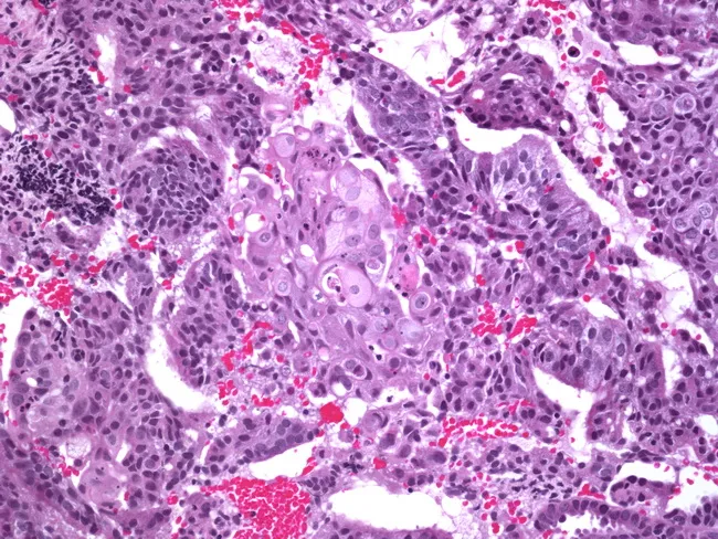
Imaging modality: Bright-field histology on a hematoxylin and eosin-stained formalin-fixed paraffin-embedded endometrial tissue section, viewed under light microscopy at high magnification (approximately 400x). The specimen shows endometrioid adenocarcinoma characterized by malignant, back-to-back glandular structures formed by columnar to cuboidal epithelium with oval, hyperchromatic nuclei and conspicuous nucleoli, and frequent mitotic figures. Intermixed within the glandular component are poorly formed squamous morules composed of small nests of squamoid cells with squamous differentiation, lacking substantial keratinization. The morules are surrounded by the glandular tumor and embedded in desmoplastic stroma with inflammatory infiltrate and occasional red blood cells in adjacent vasculature. The overall architecture demonstrates focal invasion into stromal tissue, consistent with carcinoma. The combination of gland-forming endometrioid morphology with focal squamous morules constitutes squamous differentiation within endometrial carcinoma. Clinically relevant implications include tumor heterogeneity, potential grading nuances, and differential considerations including adenosquamous carcinoma; immunohistochemical profiling may aid confirmation (e.g., p16, ER/PR status) though not shown. This histology supports a endometrial malignancy with mixed differentiation; appropriate surgical staging and adjuvant therapy decisions should consider invasion depth and lymphovascular invasion. This description aligns with endometrioid carcinoma with squamous morphology and morular differentiation. Further molecular profiling may refine prognosis and therapy planning for personalized care.
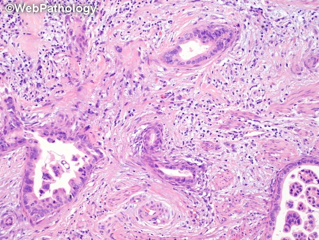
Histology: Light microscopy of a hematoxylin and eosin (H&E) stained paraffin section. Primary subject is a malignant gland-forming neoplasm with architectural glands showing round, oval, or irregular branching lumina. The neoplastic glands are lined by simple cuboidal to columnar epithelium and display features of glandular differentiation consistent with an adenocarcinoma spectrum. Invasive growth into the surrounding stroma is suggested by an irregular boundary and desmoplastic-like stromal response, though detailed invasion is not fully described in the image alone. The tumor microenvironment is notable for an inflammatory infiltrate comprising lymphocytes, plasma cells, neutrophils and eosinophils intermingled with malignant glands. Nuclear morphology of the epithelial component cannot be precisely assessed in this description, but applies typical malignant cytology. The pattern may be cribriform, tubuloacinar, or solid portions common to gland-forming carcinomas. Diagnostic significance lies in recognizing a malignant glandular neoplasm with accompanying inflammatory milieu, guiding differential diagnoses toward adenocarcinoma and its variants while excluding benign mimics. Immunohistochemistry and clinical correlation are essential to determine primary site and origin. Practical relevance includes educational illustration of glandular malignancy, pathology archives, and diagnostic consultation; aids in tumor grading, staging, and therapeutic decision-making where glandular cancers are suspected. Further study with additional stains would enhance specificity.
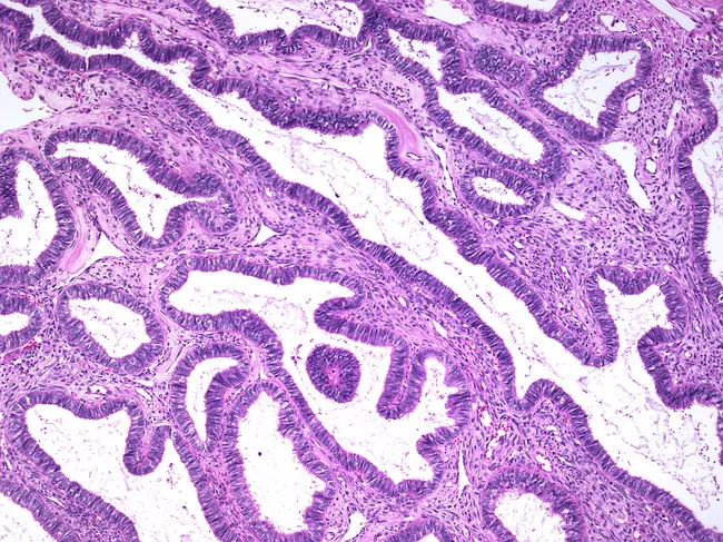
Imaging modality and technique: Light microscopy of formalin-fixed, paraffin-embedded ovarian tissue stained with Hematoxylin and Eosin (H&E). Primary subject: ovarian endometrioid cystadenoma, a benign epithelial neoplasm. Histology shows tubulocystic glands formed in back-to-back patterns and lined by cuboidal to columnar epithelium. The epithelium mimics normal endometrium, with glands exhibiting pseudostratified appearance but lacking cytologic atypia. Nuclei are uniform, with inconspicuous nucleoli and minimal pleomorphism; mitotic figures are rare. The stroma is typically fibrous with little desmoplasia; invasion of surrounding ovarian stroma is not evident. Some specimens may display squamous morules embedded within the epithelium, but without cytologic malignancy. Overall, the architecture favors a benign endometrioid phenotype rather than malignancy. Diagnostic significance: recognition of benign endometrioid lining helps distinguish endometrioid cystadenoma from ovarian endometrioid carcinoma, serous or mucinous cystadenomas with secondary endometrioid change, or mixed histology. Differential considerations include metastatic endometrium, mesonephric lesions, or other epithelial ovarian tumors. Clinical correlation includes patient age, imaging morphology of an adnexal mass, normal tumor markers, and definitive surgical pathology confirming benign behavior. Prognosis after complete excision is excellent; recommended follow-up per standard ovarian tumor protocols. Imaging-pathology concordance corroborates benign sequelae; avoid overtreatment; counsel patients on prognosis and routine gynecologic surveillance after surgical management completion.
pseudostratified transitional epithelium stratified squamous histology
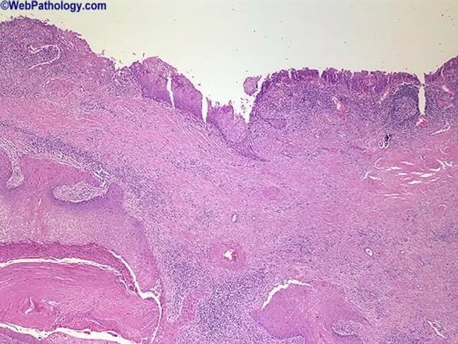
Renal pelvis histology obtained from a urinary tract lesion was imaged using light microscopy on an H&E-stained section. The specimen comprises renal pelvis mucosa lined by urothelium transformed by squamous metaplasia. The surface epithelium is thickened and organized into broad tongues of stratified squamous cells projecting into the underlying lamina propria. The metaplastic epithelium shows typical squamous differentiation with eosinophilic cytoplasm and intercellular bridges; underlying stroma displays mild chronic inflammatory infiltrate. In some areas the squamous epithelium invades the subepithelial tissue, consistent with early invasion rather than a purely reactive change. Normal urothelial architecture is disrupted; transitional epithelium is not intact over the superficial surface. The image highlights a pathologic sequence in which chronic irritation from calculi and infection fosters squamous metaplasia and raises the clinical suspicion for subsequent squamous cell carcinoma of the renal pelvis. Clinically, this pattern is associated with long-standing nephrolithiasis and potential for malignant transformation, particularly in kidneys with anomalous anatomy such as horseshoe kidney. The visualization emphasizes the need for thorough sampling to exclude invasive carcinoma and to guide management, which may include radical nephroureterectomy or targeted therapy depending on invasion depth and staging. This image thus serves educational and diagnostic utility in urologic oncology, nephrology, and surgical pathology.
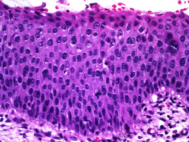
Histology image of mucosal epithelium showing features of squamous epithelial dysplasia on light microscopy. Prepared as a paraffin-embedded hematoxylin and eosin (H&E) stained section and viewed under brightfield microscopy at high magnification (~400x). The epithelium is stratified, with orderly basal cell layer but marked nuclear atypia throughout the lower and mid portions. Nuclear enlargement and hyperchromasia are evident, along with variation in nuclear size (anisokaryosis) and coarse, clumped chromatin. There is partial maturation at the superficial aspect, with surface layer showing more differentiated cells, suggesting some preserved maturation toward the lumen. Occasional mitotic figures are observed in the lower half of the epithelium, indicative of increased proliferative activity. Overall architecture reveals thickened epithelium (acanthosis) with preserved polarity in some cells but dysplastic changes in others. No invasion is evident in this field, though the mitotic activity and cytologic atypia raise concern for preinvasive squamous intraepithelial lesion. The imaging is relevant for diagnosis of epithelial dysplasia or squamous intraepithelial lesion and has clinical significance for risk stratification, biopsy correlation, and surveillance. Potential clinical applications include screening for precancerous lesions in mucosal surfaces, monitoring response to therapy, and educational demonstration of dysplastic cytology for medical students and residents.
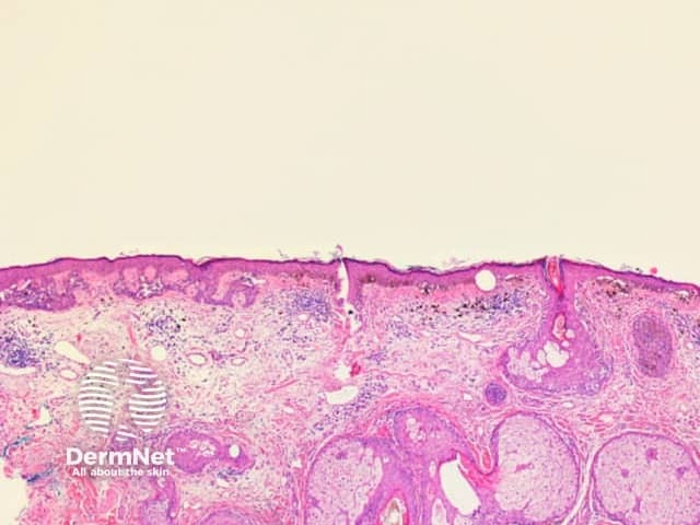
This is a light-microscopy histology image of human skin obtained from a skin biopsy. The section presents a cross-sectional view through the epidermis and dermis, highlighting classic skin architecture suitable for educational comparison. The epidermis shows stratified squamous epithelium with orderly keratinocyte maturation and a clearly visible keratin layer at the surface (stratum corneum). The epidermal-dermal junction appears intact. In the underlying dermis, dense collagenous connective tissue is intermingled with looser fibrous stroma, and adnexal elements such as hair follicles, sebaceous glands, and eccrine glands may be present depending on the field illuminated by the section. Hematoxylin provides purple nuclear detail, while eosin stains the cytoplasm and extracellular matrix pink, creating the familiar purple-pink contrast that delineates structures. No obvious atypia, dysplasia, or malignant invasion is discernible in this field, though assessment is limited to a single plane and context is clinical. The image serves as a reference for normal or near-normal skin histology and can assist in recognizing deviations arising from inflammatory dermatoses, hyperplasia, keratinization disorders, or benign adnexal lesions when correlated with patient history and additional levels. It is relevant for dermatology training, histopathology practice, and educational case discussions.
Epithelium - General Anatomy
Definition and General Properties
- Close cell apposition - cells are tightly packed, joined by specialized cell-to-cell junctions, with minimal intercellular space.
- Presence at a free surface - epithelium always lines a free surface exposed to the external environment, body cavities, or the lumen of tubes and organs.
- Avascular - no blood vessels cross the basal lamina. All nutrients reach epithelial cells by diffusion from underlying connective tissue or by absorption from the free surface.
- Rests on a basal lamina (basement membrane) - this is the structural attachment site anchoring epithelial cells to the underlying connective tissue.
- Polarity - cells have distinct apical (free surface), lateral, and basal domains.
- Cells are held together by specialized cell junctions: tight junctions, adherens junctions, desmosomes, and gap junctions.
Functions of Epithelium
- Protection - mechanical, chemical, microbial barrier (e.g., skin epidermis)
- Absorption - intake of nutrients (e.g., intestinal epithelium)
- Secretion - production of mucus, enzymes, hormones (e.g., glands)
- Sensation - special sensory epithelia (e.g., olfactory, taste)
- Exchange - gas and fluid exchange across thin layers (e.g., alveoli, capillaries)
- Lubrication - mesothelium secretes serous fluid to reduce friction between organs
Classification of Epithelium
By Number of Layers
| Term | Meaning |
|---|---|
| Simple | Single layer of cells; all cells contact the basement membrane |
| Stratified | Multiple layers; only basal cells contact the basement membrane |
| Pseudostratified | Appears stratified but is actually simple - all cells contact the basement membrane, but nuclei sit at different heights, creating a false layered appearance |
By Cell Shape
| Shape | Appearance |
|---|---|
| Squamous | Flat, scale-like cells; nucleus is flattened |
| Cuboidal | Roughly as wide as tall; nucleus is round and central |
| Columnar | Taller than wide; nucleus is elongated and basal |
Types of Covering (Lining) Epithelium
| Type | Typical Locations | Main Function |
|---|---|---|
| Simple squamous | Vascular system (endothelium), body cavities (mesothelium), Bowman's capsule (kidney), alveoli | Exchange, barrier, lubrication |
| Simple cuboidal | Small gland ducts, ovarian surface, kidney tubules, thyroid follicles | Absorption, secretion |
| Simple columnar | Small intestine, colon, stomach, gallbladder | Absorption and secretion |
| Pseudostratified columnar | Trachea and bronchial tree, ductus deferens, efferent ductules of epididymis | Secretion, conduit |
| Stratified squamous (nonkeratinized) | Oral cavity, esophagus, vagina, cornea | Protection, withstand abrasion |
| Stratified squamous (keratinized) | Skin epidermis | Protection, barrier to water loss |
| Stratified cuboidal | Large sweat gland ducts, ovarian follicle wall | Conduit |
| Stratified columnar | Larynx, parts of male urethra | Protection, secretion |
| Transitional (urothelium) | Renal calyces, ureter, urinary bladder, proximal urethra | Distensibility - allows stretch |
Special named epithelia: Endothelium = simple squamous lining of the cardiovascular system. Mesothelium = simple squamous lining of pericardial, pleural, and peritoneal cavities (secretes lubricating serous fluid).
Histological Examples
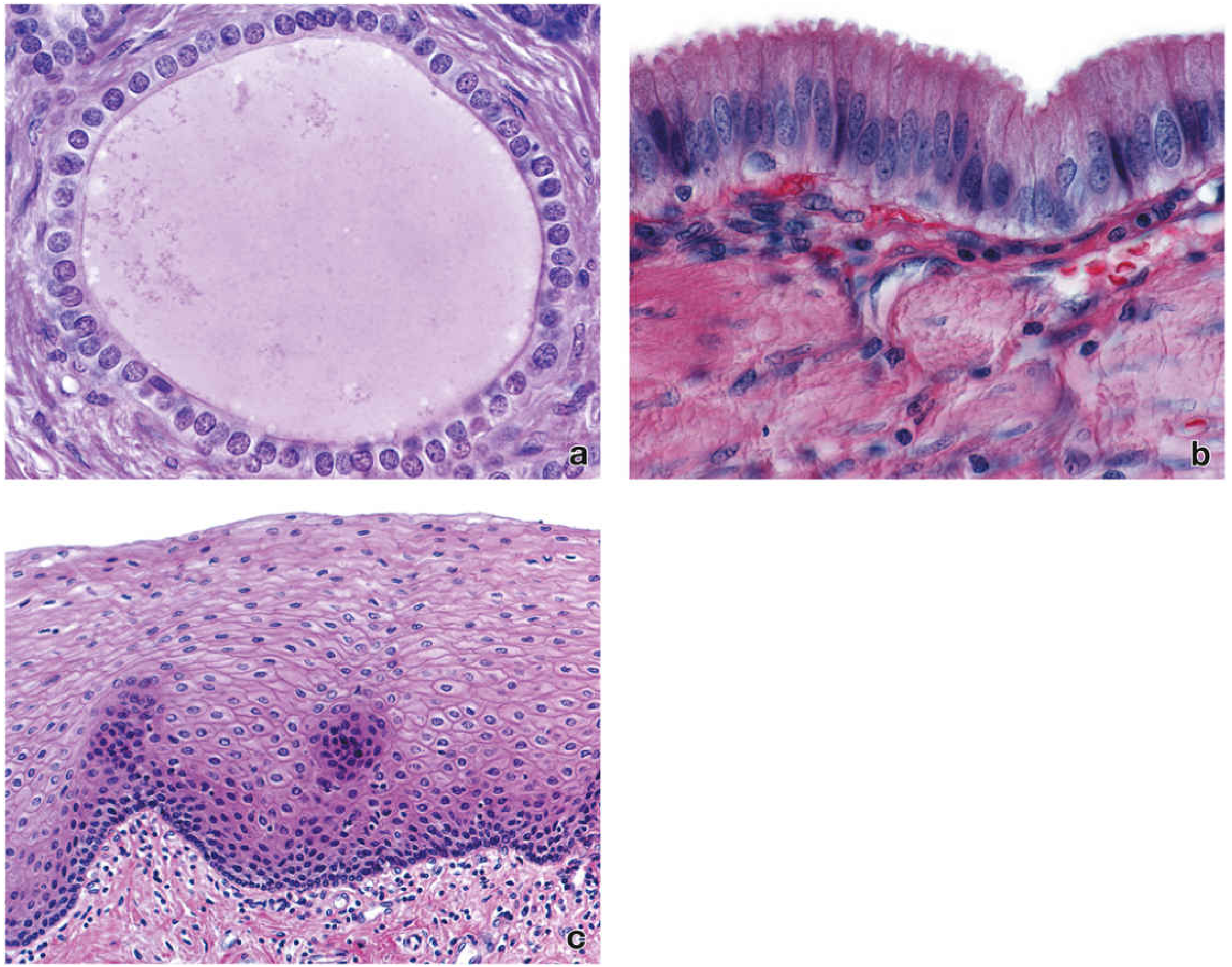
Surface Specializations of the Apical Domain
- Microvilli - finger-like projections that greatly increase surface area for absorption; packed densely they form a "brush border" (intestine) or "striated border"
- Stereocilia - long, non-motile microvilli-like projections (epididymis, inner ear hair cells)
- Cilia - motile projections that move mucus or fluids (respiratory tract, uterine tube)
- Keratinization - in stratified squamous epithelium exposed to the external environment, surface cells accumulate keratin and die, forming a tough waterproof layer (epidermis)
Glandular Epithelium
| Category | Description | Examples |
|---|---|---|
| Exocrine glands | Release secretions onto a surface (skin, mucosa) via ducts, or directly | Salivary glands, sweat glands, pancreas (exocrine portion) |
| Endocrine glands | Release secretions (hormones) into the bloodstream or lymphatics - no ducts | Thyroid, adrenal glands, pituitary |
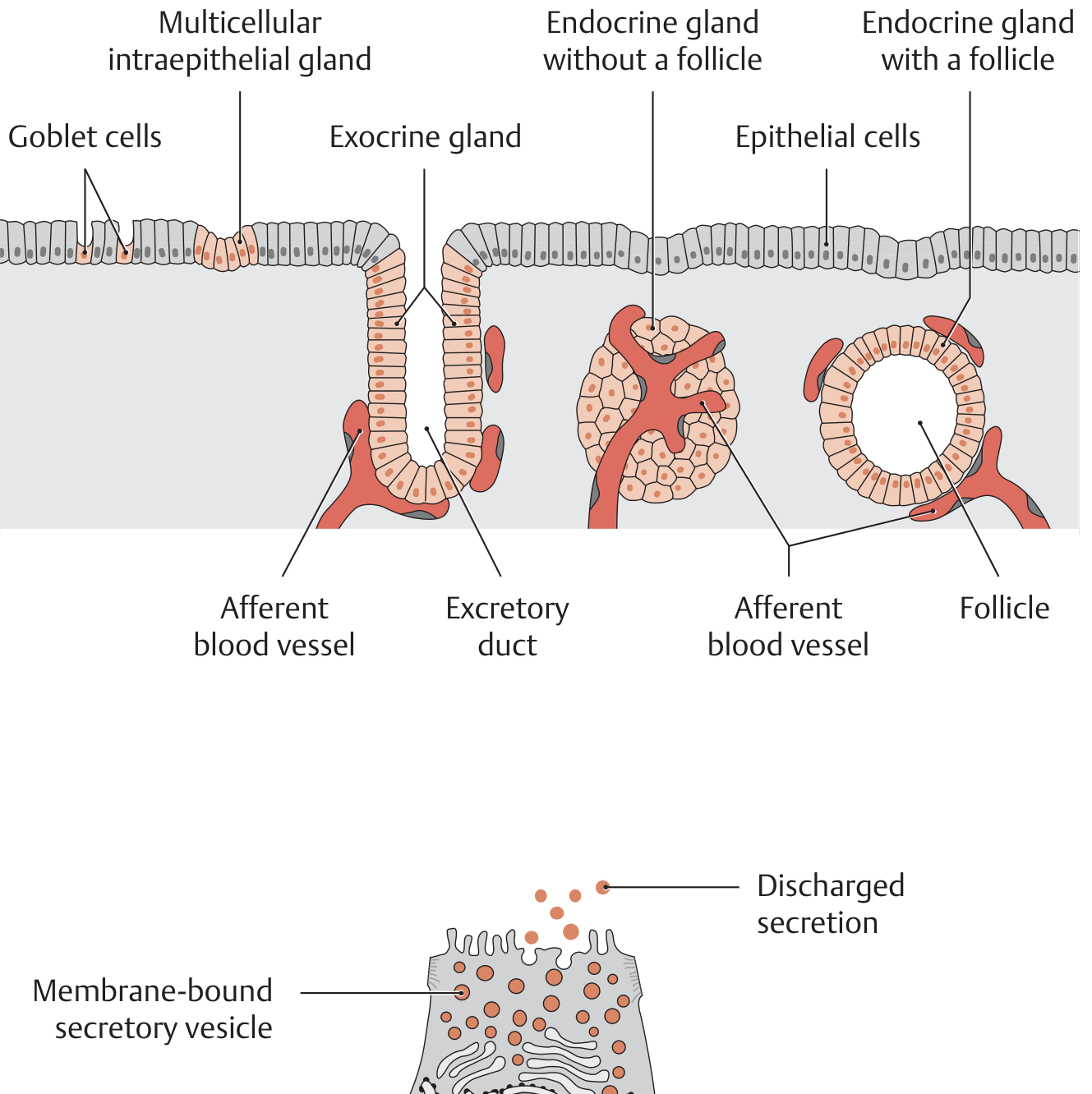
Secretory Mechanisms of Exocrine Glands
- Merocrine (eccrine/exocytosis) - secretory vesicles fuse with the apical membrane and discharge contents with no loss of cytoplasm. Most common (e.g., pancreatic acini, salivary glands).
- Apocrine - membrane-bound vesicles bud off the apical cell surface, releasing lipid-rich secretion enclosed in membrane (e.g., mammary glands, apocrine sweat glands).
- Holocrine - entire glandular cell disintegrates to become the secretion; basal cells continuously regenerate (e.g., sebaceous glands).
Morphological Classification of Exocrine Glands
- By duct structure: Simple (unbranched duct) vs. Compound (branched duct)
- By secretory unit shape: Tubular (tube-shaped), Acinar/alveolar (flask-shaped), Tubuloalveolar (mixed)
- By secretion type: Serous (watery, protein-rich), Mucous (thick, glycoprotein-rich), Mixed (seromucous)
Pseudostratified and Transitional Epithelium - Key Points
- Pseudostratified columnar epithelium: Looks multilayered because nuclei are at different heights, but every cell touches the basement membrane. Typically ciliated (respiratory tract). Classic example: trachea, where it moves mucus-trapped particles via mucociliary clearance.
- Transitional epithelium (urothelium): Lines the entire lower urinary tract (minor calyces to proximal urethra). Has the unique property of being able to stretch and accommodate large volume changes - cells flatten and "slide" over one another when the bladder fills, and round up when it empties. Surface "umbrella cells" are large, dome-shaped, and binucleate.
Summary Table
| Property | Epithelium |
|---|---|
| Vascularity | Avascular |
| Basement membrane | Always present |
| Cell polarity | Apical / lateral / basal domains |
| Cell junctions | Tight junctions, desmosomes, gap junctions, hemidesmosomes |
| Innervation | Yes (sensory nerve endings) |
| Renewal capacity | High (rapid mitosis in basal/stem cells) |
| Origin | Ectoderm, endoderm, or mesoderm |