
Fate of thrombosis
Thrombosis can resolve through several pathways: resolution (lysis by plasmin), organization (fibroblast ingrowth, collagen deposition), recanalization (new channels form through the organized thrombus), and propagation or embolization. The outcome depends on thrombus size, location, and the balance between coagulation and fibrinolysis.
Searching sources...
Reading File
Now let me search the medical library and images in parallel.
Finding Sources
Searching Images
fate of thrombus organization recanalization
The primary source is Robbins. Let me read that section along with the Basic Pathology section in parallel.
Reading File
Reading File
I now have excellent content from Robbins Pathologic Basis of Disease (the most authoritative source). Let me also fetch the key diagram showing the organized thrombus.
Loading Image
Loading Image
I now have all the information needed for a thorough, well-illustrated answer.
Fate of Thrombosis
Based on Robbins, Cotran & Kumar - Pathologic Basis of Disease, if a patient survives the initial thrombotic event, thrombi undergo one or more of four possible fates in the ensuing days to weeks:
1. Propagation
The thrombus enlarges by accumulating additional platelets and fibrin. This progressive growth can extend the occlusion and worsen ischemia distally. It is most significant in venous thrombosis (e.g., deep vein thrombosis extending proximally toward the iliofemoral system).
2. Embolization
The thrombus or a fragment dislodges and is carried by the bloodstream to distant sites. This is the most dangerous outcome:
- Venous thrombi (especially DVT) embolize to the pulmonary circulation (pulmonary embolism)
- Arterial/cardiac thrombi embolize to the brain, kidneys, spleen, or limbs, causing infarcts
3. Dissolution (Fibrinolysis)
Fibrinolytic enzymes, principally plasmin (derived from plasminogen via t-PA), digest the fibrin mesh and cause rapid shrinkage and total disappearance of the thrombus. Key points:
- This is most effective with fresh (recent) thrombi before extensive cross-linking of fibrin occurs
- Older thrombi have extensive fibrin cross-linking and are relatively resistant to lysis
- This is the rationale for why thrombolytic therapy (e.g., t-PA in acute MI or stroke) must be given within the first few hours of the thrombotic event
4. Organization and Recanalization
Older thrombi that are not lysed undergo organization: ingrowth of:
- Endothelial cells
- Smooth muscle cells
- Fibroblasts
This produces a mass of granulation tissue/connective tissue. Subsequently, recanalization occurs - capillary channels form through the organized thrombus, re-establishing continuity of the original lumen (though usually only partially).
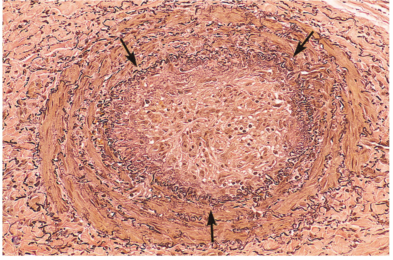
Fig. 4.14 - Organized thrombus in a thrombosed artery (Robbins PBD). Arrows mark the internal elastic lamina; the lumen is completely filled with organized thrombus.
With continued remodeling and contraction of mesenchymal elements, the thrombus may be incorporated into the vessel wall, eventually leaving only a fibrous lump as a remnant.
5. Septic Infection (Special Fate)
Occasionally, centers of thrombi undergo enzymatic digestion due to release of lysosomal enzymes from trapped leukocytes and platelets. In the setting of bacteremia, the thrombus becomes infected, forming an inflammatory mass that:
- Erodes and weakens the vessel wall
- May result in a mycotic aneurysm if unchecked
Mural Thrombi (Macroscopic Appearance)
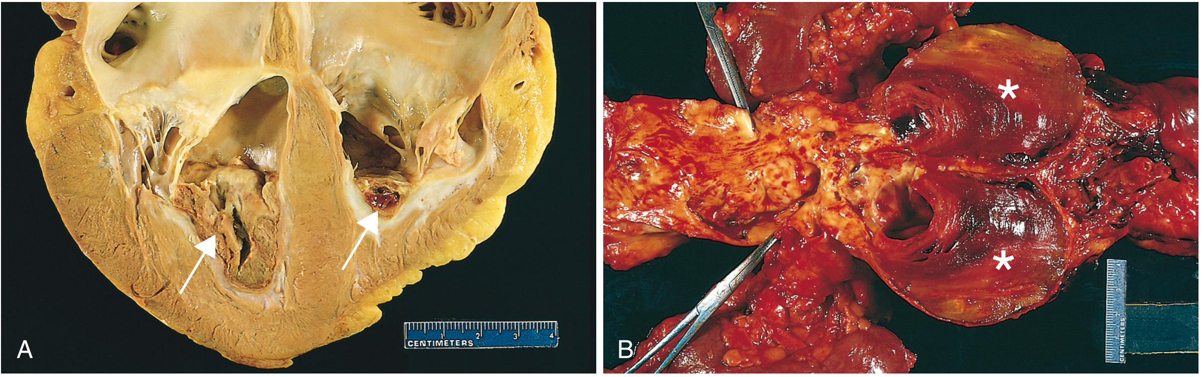
Fig. 4.13 - Mural thrombi in the heart and aorta (Robbins PBD).
Summary Table
| Fate | Mechanism | Clinical Significance |
|---|---|---|
| Propagation | Additional platelet/fibrin deposition | Progressive vascular occlusion |
| Embolization | Fragment detachment + transport | PE, stroke, peripheral infarct |
| Dissolution | Plasmin-mediated fibrinolysis | Complete resolution; t-PA window |
| Organization + Recanalization | Fibroblast/EC/SMC ingrowth; new channels | Partial restoration of flow; fibrous scar |
| Septic (mycotic) transformation | Bacterial seeding + enzymatic lysis | Vessel wall erosion; mycotic aneurysm |
- Robbins, Cotran & Kumar Pathologic Basis of Disease, pp. 129 (Fate of the Thrombus section)
- Robbins & Kumar Basic Pathology, p. 2495 (Virchow's triad and thrombosis overview)
This is a shared conversation. Sign in to Orris to start your own chat.