
Formation of trilaminar germ disc embryology
Reading File
The trilaminar germ disc forms during the 3rd week of embryogenesis through the process of gastrulation. Epiblast cells migrate through the primitive streak to form mesoderm and endoderm, leaving ectoderm on the surface - creating three primary germ layers.
Searching sources...
Finding Sources
Finding Sources
Finding Sources
Reading File
Finding Sources
Reading File
I now have the core content from Langman's. Let me also get the section on fate of germ layers and any images.
Reading File
Finding Sources
Reading File
Loading Image
Finding Sources
Reading File
Reading File
Finding Sources
Loading Image
Reading File
I now have comprehensive content from both Langman's and The Developing Human. Let me fetch one more key diagram showing gastrulation/primitive streak process.
Loading Image
I now have all the material needed. Let me compile a thorough, well-structured answer.
Formation of the Trilaminar Germ Disc
The trilaminar germ disc forms during the 3rd week of embryonic development through a process called gastrulation - the most significant event of the third week and the beginning of morphogenesis. The bilaminar disc (epiblast + hypoblast) is converted into a trilaminar disc (ectoderm + mesoderm + endoderm).
(Langman's Medical Embryology; The Developing Human, Clinically Oriented Embryology)
Background: The Bilaminar Disc (End of Week 2)
Before gastrulation begins, the embryonic disc has just two layers:
- Epiblast - the dorsal layer, facing the amniotic cavity (columnar cells)
- Hypoblast - the ventral layer, facing the yolk sac (cuboidal cells)
At this stage, the primitive streak forms as a faint groove in the caudal region of the epiblast - marking the very beginning of week 3.
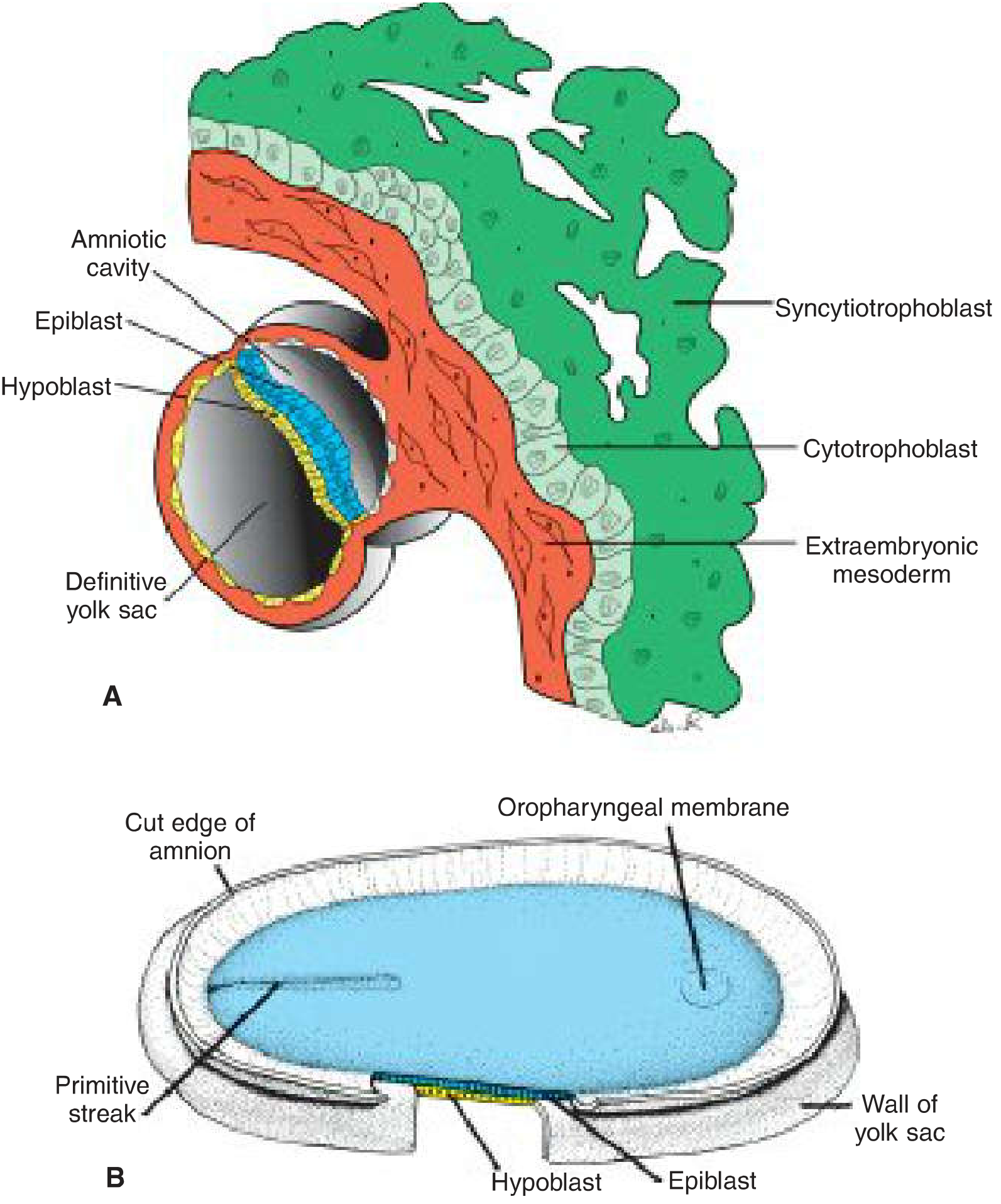
Step 1: Formation of the Primitive Streak (Day 15-16)
- The primitive streak appears as a narrow thickened groove on the dorsal epiblast surface, caudally in the midline
- It elongates cranially and its cranial end expands into the primitive node (Hensen's node), a slightly elevated region surrounding the primitive pit
- The streak establishes the embryo's:
- Craniocaudal axis
- Dorsal/ventral surfaces
- Left/right sides
Molecular control: FGF8, synthesized by streak cells, drives cell movement by down-regulating E-cadherin (which normally binds epiblast cells together). FGF8 also controls mesoderm specification by regulating BRACHYURY (T) expression. Additional signaling molecules include BMPs, Shh, Tbx16, Tgifs, and Wnts.
Step 2: Gastrulation - Formation of Mesoderm and Endoderm
Epiblast cells migrate toward and into the primitive streak in a process called invagination:
- Cells move to the streak and become flask-shaped
- They detach from the epiblast and slip beneath it
- Two fates then follow:
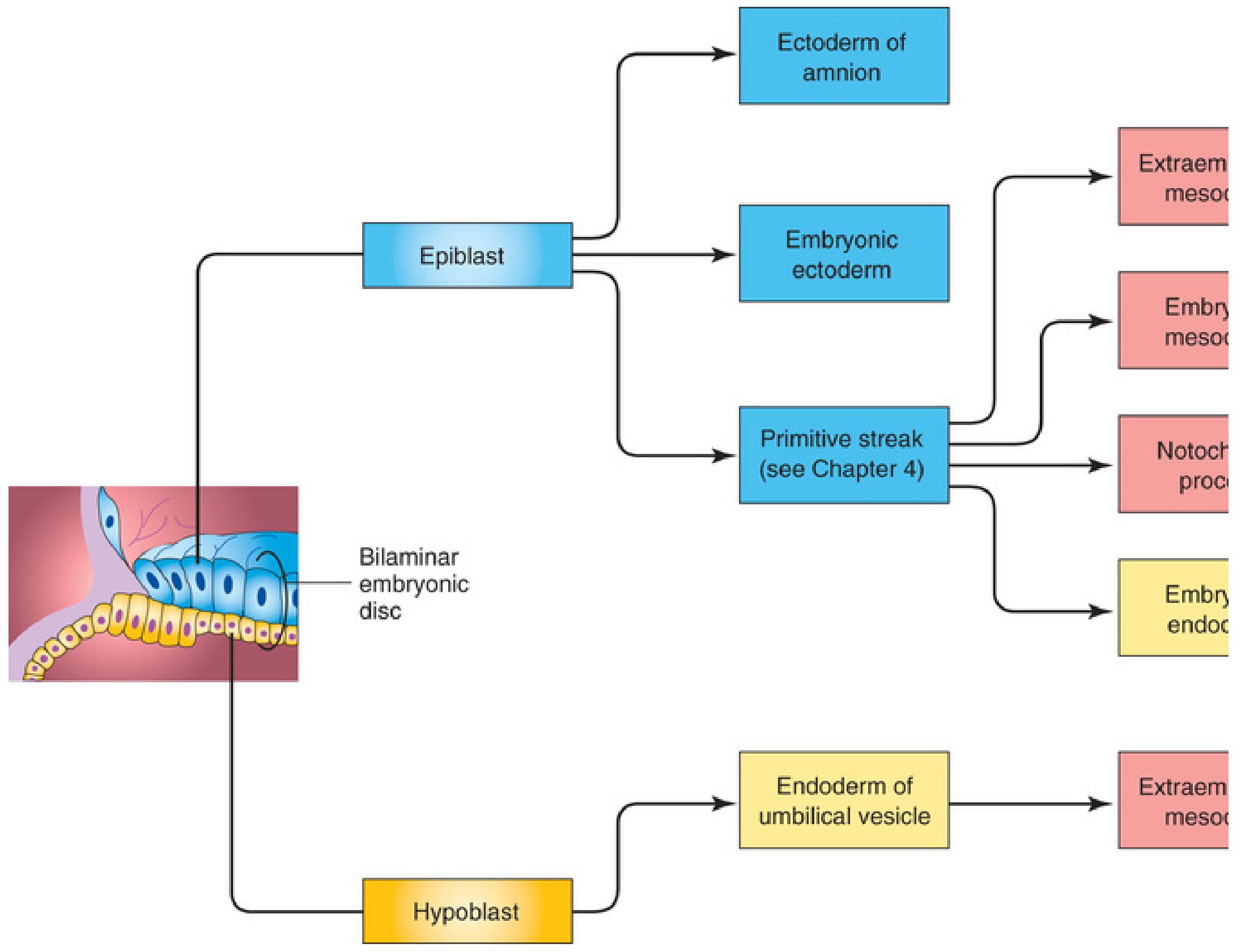
- Some cells displace the hypoblast cranially and laterally → become embryonic endoderm
- Others come to lie between the epiblast and the new endoderm → become intraembryonic mesoderm
- Cells remaining in the epiblast (those that did not invaginate) become embryonic ectoderm
"The epiblast, through the process of gastrulation, is the source of all of the germ layers." - Langman's Medical Embryology
Step 3: Spread of Intraembryonic Mesoderm
As more cells invaginate, mesoderm spreads:
- Laterally and cranially between ectoderm and endoderm
- Beyond the disc margin to connect with extraembryonic mesoderm covering the yolk sac and amnion
- Cranially on each side of the prechordal plate (toward the future forebrain)
Two membrane regions remain where ectoderm and endoderm are directly fused with no intervening mesoderm:
| Region | Location | Future structure |
|---|---|---|
| Oropharyngeal membrane | Cranial end | Future oral cavity |
| Cloacal membrane | Caudal end | Future anal opening |
Step 4: Formation of the Notochord
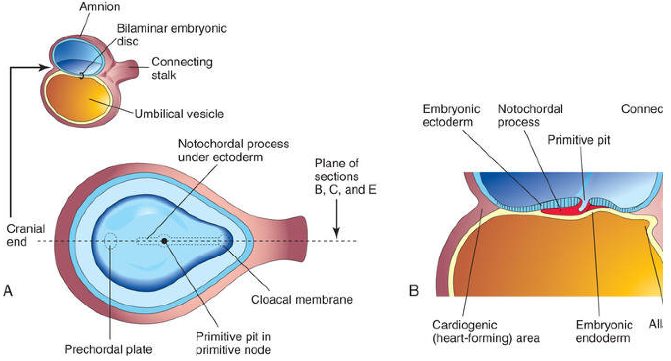
Simultaneously, prenotochordal cells invaginate through the primitive node/pit and migrate cranially in the midline:
- They intercalate into the hypoblast → form the notochordal plate
- As hypoblast is replaced by endoderm, notochordal plate detaches and forms the definitive notochord (a solid midline cord)
- The notochord extends from the prechordal plate (cranially) to the primitive pit (caudally)
The notochord is a key signaling center that:
- Induces neural tube formation (neurulation)
- Induces axial skeleton (vertebral column) formation
- Gives rise to the nucleus pulposus of intervertebral discs in adults
Result: The Trilaminar Germ Disc (End of Week 3)
By end of week 3, the embryo is a flat, ovoid trilaminar disc with:
| Germ Layer | Origin | Major Derivatives |
|---|---|---|
| Ectoderm | Remaining epiblast cells | Epidermis, CNS & PNS, eyes, inner ear, neural crest cells, head connective tissue |
| Mesoderm | Invaginated epiblast cells (between layers) | Skeletal muscle, blood cells, blood vessel lining, smooth muscle, serous linings, kidney, gonads, connective tissue (trunk), most cardiovascular system |
| Endoderm | Epiblast cells displacing hypoblast | Lining of GI and respiratory tracts, liver, pancreas, glandular organs |
Additional Events of Week 3
- Allantois forms as a diverticulum from the yolk sac into the connecting stalk (~day 16)
- Blood islands appear in the yolk sac wall and allantois → primitive vasculogenesis
- Primordial cardiovascular system forms; heart begins to beat on day 21-22
- Neural plate induced by the notochord (start of neurulation)
- Somites begin to form from paraxial mesoderm
Clinically Important Points
Primitive streak remnants → can persist and give rise to sacrococcygeal teratoma (most common tumor in neonates; ~1:35,000; 80% female; contains tissues from all 3 germ layers; usually benign)
Twinning: Monozygotic twins arise from splitting of the early embryo; if splitting occurs after primitive streak formation (day 13+), conjoined twins result.
Gastrulation = most critical period: Teratogens during week 3 cause major structural defects affecting multiple organ systems because all three germ layers are being established.
Left-Right axis: Cilia on the ventral surface of the primitive node generate fluid flow that establishes the L-R axis. Defects cause situs inversus or heterotaxy (situs ambiguus) - the latter carrying a 90% risk of complex congenital heart defects.
Sources: Langman's Medical Embryology (Chapter 5: Third Week of Development); The Developing Human: Clinically Oriented Embryology (Chapter 4: Third Week); Histology: A Text and Atlas with Correlated Cell and Molecular Biology
This is a shared conversation. Sign in to Orris to start your own chat.