
Brachial artery
Reading File
The brachial artery is the main arterial trunk of the upper arm, running from the lower border of teres major to the cubital fossa where it bifurcates into radial and ulnar arteries. Let me pull detailed textbook content now.
Searching sources...
Finding Sources
Finding Sources
Good results. Now let me read the most relevant sections in parallel.
Reading File
Reading File
Reading File
Reading File
Good. Let me now read the imaging anatomy section and the clinic notes, plus fetch the anatomy diagram images.
Reading File
Reading File
Reading File
Loading Image
Now let me fetch the main anatomy diagrams from Gray's and the Imaging Atlas:
Loading Image
Loading Image
Loading Image
Excellent images retrieved. I now have comprehensive content from multiple authoritative sources. Let me compile the full answer.
Brachial Artery
Overview
The brachial artery is the major arterial supply to the upper arm. It is the direct continuation of the axillary artery at the inferior border (lower margin) of the teres major muscle, and it terminates in the cubital fossa by dividing into the radial and ulnar arteries at the level of the neck of the radius. - Gray's Anatomy for Students
Course
Gray's Anatomy for Students provides a clear account:
- Proximal arm: The artery lies on the medial side of the arm, in the medial bicipital groove, in close relationship with the ulnar nerve and median nerve.
- As it descends, the median nerve initially lies lateral to the artery, then crosses superficially (anterior to the artery) from lateral to medial, eventually lying medial to the artery distally.
- Distal arm: The artery moves laterally, assuming a position approximately midway between the medial and lateral epicondyles of the humerus.
- Cubital fossa: The artery crosses anterior to the elbow joint, lying immediately medial to the tendon of the biceps brachii. Both structures are covered superficially by the bicipital aponeurosis (lacertus fibrosus).
- The pulse of the brachial artery is palpable along the entire ulnar border of the biceps brachii muscle.
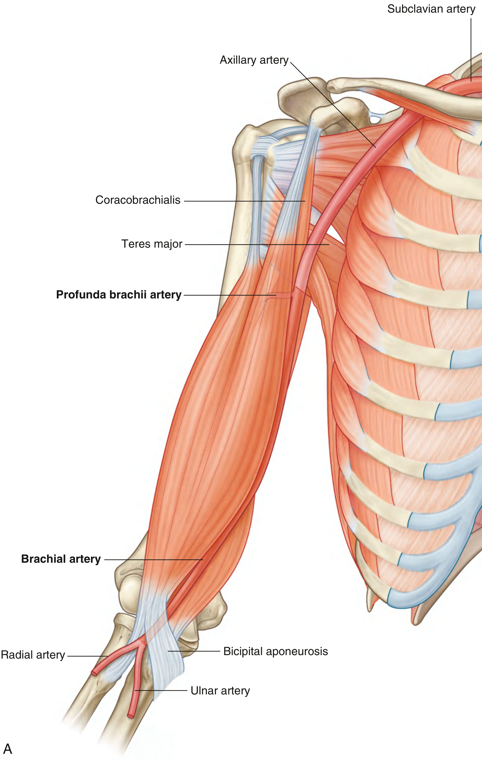
Fig. 7.66A - Brachial artery in context. (Gray's Anatomy for Students)
Branches
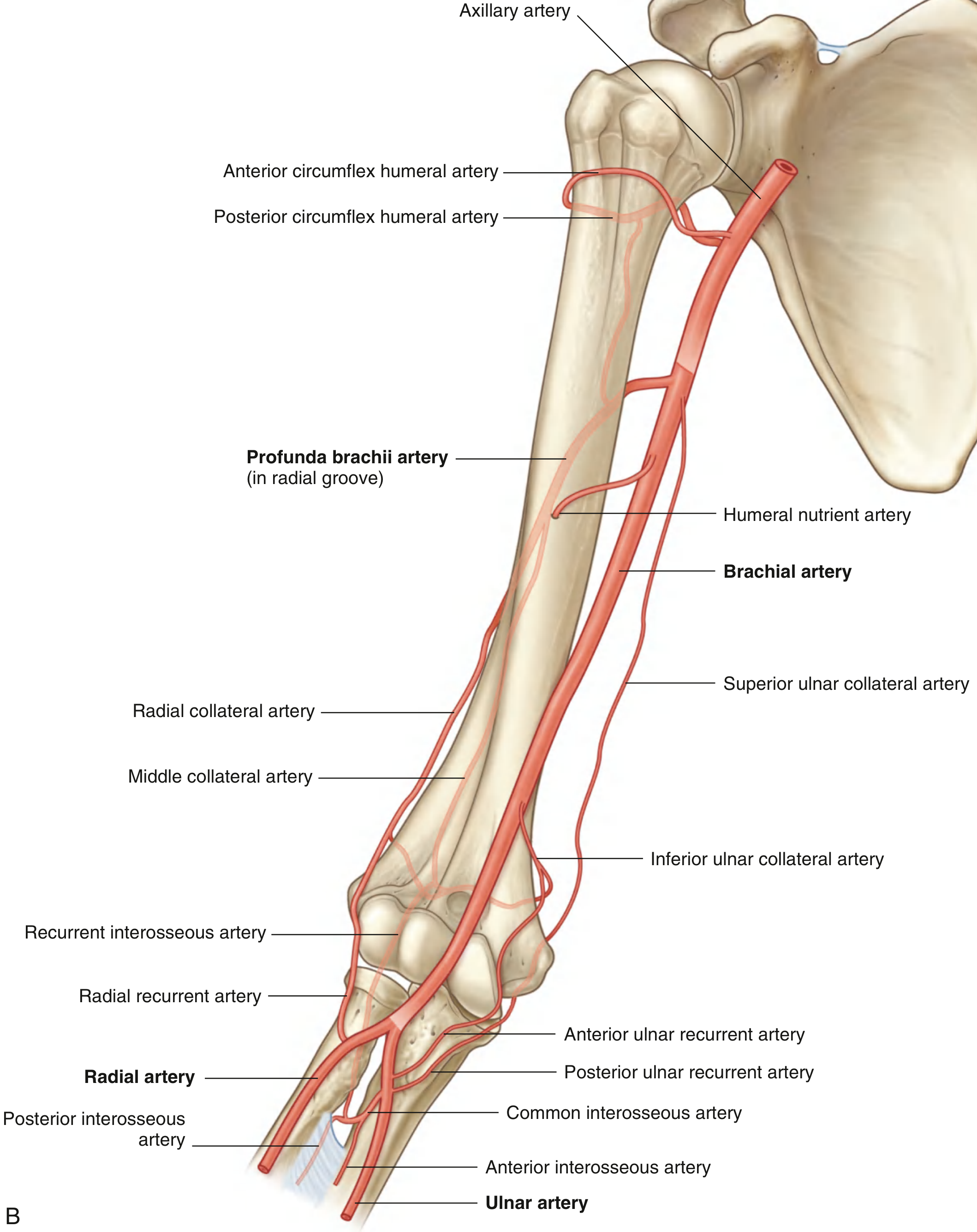
Fig. 7.66B - Branches of the brachial artery. (Gray's Anatomy for Students)
The branches of the brachial artery, in order, are: - THIEME General Anatomy and Musculoskeletal System / Fischer's Mastery of Surgery
| Branch | Notes |
|---|---|
| Profunda brachii (deep brachial artery) | Largest branch; arises near teres major level in 55-70% of cases. Passes with the radial nerve through the triangular interval and along the radial groove of the humerus. Terminates as the radial collateral and middle collateral arteries |
| Humeral nutrient arteries | Enter foramen on anteromedial humeral shaft |
| Superior ulnar collateral artery | Runs alongside the ulnar nerve; anastomoses with the posterior ulnar recurrent and inferior ulnar collateral arteries around the elbow |
| Inferior ulnar collateral artery | Contributes to the elbow arterial anastomosis |
| Muscular branches | To muscles of the anterior compartment |
| Radial artery (terminal) | Lateral terminal branch |
| Ulnar artery (terminal) | Medial terminal branch (usually larger) |
The profunda brachii is the single most important branch - it courses with the radial nerve in the radial groove on the posterior surface of the humerus, deep to the lateral head of triceps brachii. - Gray's Anatomy for Students, p. 876
Angiographic Anatomy
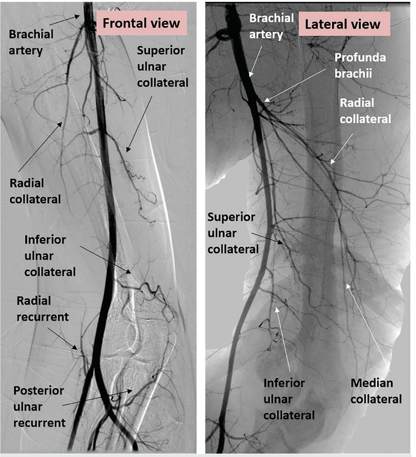
Frontal and lateral angiograms showing the brachial artery and its branches including profunda brachii, superior and inferior ulnar collaterals, radial collateral, and anastomoses at the elbow. (Imaging Anatomy, Vol. 3)
Relations
- Medially (proximal): Ulnar nerve and basilic vein
- Laterally: Median nerve (proximally), then biceps brachii and coracobrachialis
- Anteriorly: Skin, fascia, and (at the cubital fossa) the bicipital aponeurosis
- Posteriorly: Triceps brachii (proximally), then brachialis muscle
- Veins: Two brachial venae comitantes run alongside the artery (medial and lateral sides)
Elbow Anastomosis (Collateral Circulation)
An important anastomotic network surrounds the elbow joint, formed by:
- Profunda brachii → radial collateral + middle collateral
- Superior ulnar collateral + inferior ulnar collateral (from brachial artery)
- Radial recurrent artery (from radial artery)
- Anterior and posterior ulnar recurrent arteries (from ulnar artery)
- Recurrent interosseous artery
Clinical significance: The brachial artery can be ligated distal to the origin of the profunda brachii with relatively low risk because the elbow anastomotic network can establish adequate collateral circulation. - THIEME General Anatomy
Variations
The brachial artery shows several important anatomical variants: - Imaging Anatomy Vol. 3
-
Superficial brachial artery (4-12%) - the most common variant. Courses superficial to the median nerve (instead of deep to it). This variant is more vulnerable to injury due to its superficial position.
-
High division ("high bifurcation", ~20-25%) - the brachial artery divides into two branches (superficial + deep) while still in the arm:
- In ~10%: high-origin radial artery
- In ~3%: high-origin ulnar artery
- In ~7%: superficial brachial artery divides into radial + ulnar, while the main trunk becomes the common interosseous artery
-
Duplicated brachial artery (rare) - the axillary artery bifurcates into superficial and deep branches, both continuing as brachial arteries flanking the median nerve.
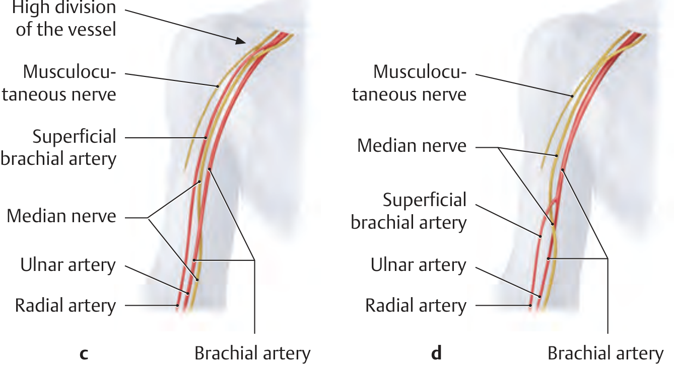
Variants of brachial artery division (c, d). In the "high division" pattern (~25%), the vessel divides into a superficial brachial artery and a brachial artery. The median nerve roots may be entrapped between them. (THIEME General Anatomy)
Clinical Relevance
Blood Pressure Measurement
The brachial artery is the standard site for non-invasive blood pressure measurement. The sphygmomanometer cuff compresses the brachial artery against the humerus in the mid-arm. The clinician auscultates Korotkoff sounds over the brachial artery in the cubital fossa. Normal range: 90-120 / 60-80 mm Hg. - Gray's Anatomy for Students
Arterial Pulse and Access
- The brachial pulse is palpable along the entire medial border of the biceps brachii.
- The cubital fossa location is used for arterial blood gas sampling and blood pressure auscultation.
- Unlike the radial artery at the wrist, the brachial artery lacks a comparable dual collateral supply at the same level, so arterial puncture here carries a somewhat higher risk of ischemia. - Roberts and Hedges' Clinical Procedures in Emergency Medicine
Brachial Artery Injuries
Injury is common with supracondylar fractures of the humerus (especially in children), elbow dislocations, and penetrating trauma. The artery's proximity to the median nerve means neurovascular bundle injuries are common together. The covered stent technique and open repair are described for penetrating injuries. - Fischer's Mastery of Surgery
Nerve Entrapment
Because of variable arterial anatomy, the median nerve or its roots may be entrapped between a superficial brachial artery and the main brachial/axillary artery, causing median nerve entrapment syndromes. - Imaging Anatomy Vol. 3
This is a shared conversation. Sign in to Orris to start your own chat.