
What is TRAP in osteoclastic activity
TRAP (Tartrate-Resistant Acid Phosphatase) is an enzyme expressed by osteoclasts and is one of the classic markers of osteoclastic bone resorption activity.
Searching sources...
Reading File
Finding Sources
Finding Sources
Reading File
Reading File
Here is a thorough answer based on authoritative textbook sources:
TRAP (Tartrate-Resistant Acid Phosphatase) in Osteoclastic Activity
What is TRAP?
TRAP is a 35-kDa iron-containing lysosomal enzyme (acid phosphatase isoform 5b) that is characteristically expressed by osteoclasts. Its name reflects the fact that, unlike most acid phosphatases, its activity is not inhibited by tartrate - a property that makes it biochemically distinct from other tissue phosphatases.
- Histology: A Text and Atlas with Correlated Cell and Molecular Biology, p. 599
- Firestein & Kelley's Textbook of Rheumatology, p. (block 1)
Role in Osteoclast Biology
| Aspect | Detail |
|---|---|
| Expression timing | Acquired during osteoclast differentiation and maturation, as preosteoclasts fuse into multinucleated giant cells |
| Cellular location | Found within the numerous lysosomes of the osteoclast |
| Secretion site | Released into the resorption lacuna (Howship lacuna) alongside cathepsin K and HCl |
| Function in resorption | Part of the enzymatic cocktail that degrades the organic bone matrix after acid demineralisation |
The osteoclast creates an extracellular lysosomal compartment beneath its ruffled border by:
- Secreting HCl via proton pumps (V-ATPase) to dissolve hydroxyapatite mineral
- Releasing matrix-degrading enzymes - TRAP, cathepsin K, and other cathepsins - to degrade collagen and non-collagenous proteins
TRAP as a Clinical Marker
TRAP is the most widely used biochemical marker of osteoclast activity and differentiation because:
- It is osteoclast-specific (also expressed in dendritic cells and activated macrophages, but the bone isoform - 5b - is relatively specific)
- Serum TRAP-5b levels reflect the number and activity of osteoclasts, making it a useful marker for:
- Osteoporosis monitoring
- Paget's disease of bone (markedly elevated)
- Bone metastases (elevated)
- Osteopetrosis - where TRAP is paradoxically elevated, being one of the few significant lab findings alongside elevated creatine kinase brain isoenzyme (Harrison's, block 46)
- Rheumatoid arthritis and inflammatory joint disease (osteoclast-mediated erosions)
TRAP in Osteoclast Differentiation (Histochemical Use)
In histology, TRAP staining (histochemical stain) is the gold-standard method to identify and count osteoclasts in bone sections:
- TRAP-positive cells stain red/pink on bone sections
- Used in research and pathology to quantify osteoclast number and distribution
- Particularly useful in studying osteopetrosis, Paget's disease, and inflammatory arthritis
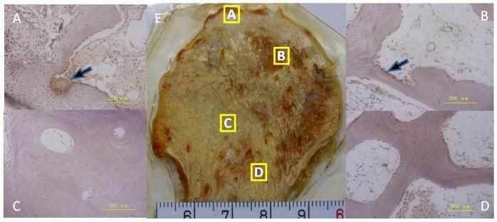
The image above shows TRAP-positive osteoclasts (black arrows) clustered along trabeculae in bone necrosis - regions of active resorption stain positively while healthy/sclerotic zones show markedly reduced TRAP activity.
TRAP and the RANK-RANKL Pathway
TRAP expression is downstream of the key osteoclastogenic signalling cascade:
M-CSF + RANKL (from osteoblasts/osteocytes) → RANK activation on precursors → differentiation → fusion into multinucleated osteoclast → TRAP expression + ruffled border formation → active bone resorption
Osteoprotegerin (OPG), produced by osteoblasts, acts as a decoy receptor for RANKL and inhibits this entire process, including TRAP expression.
Summary
TRAP is both a functional enzyme in osteoclast-mediated bone matrix degradation and a clinical/histochemical marker of osteoclastic bone resorption. Elevated serum TRAP-5b indicates increased osteoclast number or activity and is used in the diagnosis and monitoring of metabolic bone diseases.

437ffcd0-5b8f-4140-ad2b-7f26b1f8874d.jpeg
What is prl here
In this sleep physiology chart, PRL = Prolactin.
Searching sources...
Finding Sources
Reading File
PRL = Prolactin
In the context of this sleep chart, PRL stands for Prolactin - a pituitary hormone whose secretion is closely tied to the sleep cycle.
What the chart is showing
The image is a table comparing REM vs. NREM sleep for hormone levels. The arrows (↑ = increase, ↓ = decrease) show how four hormones change in each sleep stage:
| Hormone | REM | NREM |
|---|---|---|
| ACh (Acetylcholine) | ↑ | ↓ |
| 5-HT, NE (Serotonin, Norepinephrine) | ↓ | ↑ |
| GH (Growth Hormone) | ↓ | ↑ |
| PRL (Prolactin) | ↓ | ↑ |
Why Prolactin rises during NREM sleep
- Prolactin secretion rises 30-90 minutes after sleep onset, predominantly during NREM (slow-wave) sleep
- This is part of the broader sleep-associated neuroendocrine surge that also includes GH - both peak in the first third of the night during deep NREM sleep
- During REM sleep, prolactin secretion is relatively suppressed compared to NREM
This pattern is clinically relevant because:
- Sleep deprivation can suppress nocturnal prolactin release
- Hyperprolactinaemia can disrupt normal sleep architecture
- The NREM-linked prolactin surge is thought to have immunomodulatory and restorative roles during sleep
(Bradley and Daroff's Neurology in Clinical Practice)

5ea590ac-96d1-4b27-aed4-317a9109b337.jpeg
What is scn
In this slide, SCN = Suprachiasmatic Nucleus.
Searching sources...
Finding Sources
Reading File
SCN = Suprachiasmatic Nucleus
The slide shows this pathway:
Photosensitive SCN → NE → Pineal Gland → ↑ Melatonin → Sleep
Here is what each step means:
What is the SCN?
The Suprachiasmatic Nucleus (SCN) is a tiny paired nucleus located in the anterior hypothalamus, sitting just above (supra) the optic chiasm. It functions as the body's master circadian clock - a biological pacemaker that fires with a 24-hour rhythm and controls virtually all cyclical body functions, including the sleep-wake cycle.
How the pathway works (step by step)
| Step | What happens |
|---|---|
| Light hits the retina | Specialized retinal ganglion cells (containing melanopsin) detect light and send signals via the retinohypothalamic tract to the SCN |
| SCN is photosensitive | The SCN neurons are entrained (synchronized) by the light-dark cycle - light suppresses their melatonin-promoting output |
| SCN → NE | At night (darkness), SCN sends signals through a polysynaptic pathway to the superior cervical ganglion, which releases Norepinephrine (NE) onto the pineal gland |
| NE → Pineal Gland | NE stimulates pinealocytes in the pineal gland via β-adrenergic receptors |
| ↑ Melatonin | The pineal gland synthesizes and releases melatonin (from serotonin) - levels peak between 3-5 AM |
| Melatonin → Sleep | Melatonin acts on MT1/MT2 receptors in the SCN itself (feedback) and other brain areas to suppress the wake signal and promote sleep onset |
Key clinical points
- Light exposure at night suppresses SCN-driven melatonin release - this is why screen light disrupts sleep
- Melatonin supplements and drugs like ramelteon (MT1/MT2 agonist) exploit this pathway to treat insomnia and jet lag
- Blind individuals (no light input to SCN) can develop non-24-hour sleep-wake disorder because the SCN cannot be entrained to the environment
- The SCN drives circadian rhythms but does NOT generate the 90-minute REM/NREM cycling - that is controlled separately by brainstem REM-ON (ACh) and REM-OFF (5-HT, NE) neurons
(Guyton & Hall Medical Physiology; Stahl's Essential Psychopharmacology)
This is a shared conversation. Sign in to Orris to start your own chat.