Ma ak vetenarian hun . Ak male cat mara clinic ma aya hai neutering ka lya . Mujay neutering in male cats . Tamam tar knowledge chahyea vet book sa ak b point miss na ho or 100 authentic ho . Us ko asan English ma likh ka bajyen sequence ka sath . Jis ko parka ma ak acha vet ban saku or mujay 100 neutering ke Samaj a sakay . Ap jitna ho sakay explain karo vet books sa or apni knowledge b use kar sakti but I need 100 accuracy with 0 prcn error . Or last ma ya b explain karna k surgery sa phla Kya cheezian dakhni hai surgery ka bad Kya Dana hai kin cheezu ka khyal rakhna hai surgery or us ka types or matlab mujay ziada sa ziada knowledge chahyea .
male cat castration neutering surgical procedure technique
feline pre-anesthetic assessment preoperative evaluation cat surgery
🐱 COMPLETE GUIDE TO MALE CAT NEUTERING (CASTRATION / ORCHIECTOMY)
For the Practicing Veterinarian — Zero Points Missed
SECTION 1: BASIC TERMINOLOGY & ANATOMY
What is Neutering?
Relevant Anatomy
| Structure | Details |
|---|---|
| Testes | Paired oval organs in the scrotum. Produce testosterone + sperm |
| Scrotum | Skin pouch housing the testes. In cats it is well-furred and small |
| Epididymis | Coiled tube on the dorsal surface of each testis — stores sperm |
| Spermatic cord | Contains: ductus deferens + testicular artery + testicular vein + lymphatics + nerves |
| Tunica vaginalis | Peritoneal covering around the testis (important in closed vs open technique) |
| Cremaster muscle | Part of spermatic cord — can retract testis upward (important intraop) |
| Inguinal ring | Opening through which cord passes — relevant if testis is retained |
In cats, the testes are close to the inguinal ring compared to dogs. The scrotum is pendulous and sits just below the anus.
SECTION 2: INDICATIONS FOR NEUTERING
- ✅ Population control (most common)
- ✅ Behavioral: eliminate urine spraying (marking), roaming, aggression toward other cats
- ✅ Medical: testicular neoplasia, orchitis, epididymitis, scrotal trauma, cryptorchidism
- ✅ Hormonal diseases: testosterone-related conditions
- ✅ Owner request (routine elective)
SECTION 3: IDEAL AGE FOR NEUTERING
| Approach | Age |
|---|---|
| Traditional | 5–6 months (before sexual maturity) |
| Early / Pediatric neutering | 8–16 weeks (safe, accepted by AVMA & AAFP) |
| Post-pubertal | Any adult age — still safe and beneficial |
Cats reach puberty around 5–6 months. Neutering before this prevents urine spraying in ~90% of cases.
SECTION 4: PRE-SURGICAL ASSESSMENT ✅ (CRITICAL)
4.1 History Taking
- Age, breed, weight
- Vaccination status (especially if staying overnight)
- Last meal/water (fasting status)
- Any previous illness, surgery, anesthetic events
- Current medications (especially NSAIDs, steroids, anticoagulants)
- Is the cat indoor/outdoor? (affects post-op care)
- Any known bleeding disorders in the cat or littermates?
4.2 Physical Examination — Head to Tail
- Body weight, Body Condition Score (BCS 1–9)
- Temperature, Pulse, Respiration (TPR)
- Hydration status
- Mucous membrane color and CRT (capillary refill time <2 sec = normal)
- Auscultate for murmurs — cats can have occult HCM (hypertrophic cardiomyopathy)
- If murmur detected → consider echo before anesthesia
- Heart rate: normal cat = 140–220 bpm
- Auscultate lungs — rule out fluid, wheeze, crackles
- Respiratory rate: normal = 20–30 breaths/min
- Any masses, pain, organomegaly?
- Are both testes palpable in the scrotum?
- Size, consistency (firm and smooth = normal)
- Any signs of orchitis (swelling, heat, pain)?
- Any scrotal wounds or infection?
- Check for inguinal hernia
⚠️ If one or both testes are NOT in the scrotum → Cryptorchid cat → Requires a different, more complex surgery (see Section 7)
4.3 Pre-Anesthetic Bloodwork
| Test | Why |
|---|---|
| PCV / Total Protein | Baseline anemia, hydration |
| BUN / Creatinine | Renal function — affects drug metabolism |
| ALT / ALP | Liver function |
| Blood glucose | Hypoglycemia risk especially in young/small cats |
| Electrolytes (Na, K, Cl) | Important if cat is sick |
For young, healthy cats (<2 years) with no abnormalities on PE → minimum database acceptable (PCV + TP). Full panel preferred in cats >5 years or if any abnormality found.
4.4 ASA Physical Status Classification
| Class | Description |
|---|---|
| ASA I | Normal healthy cat — routine neuter |
| ASA II | Mild systemic disease (e.g., mild URI) |
| ASA III | Moderate disease |
| ASA IV | Severe, life-threatening disease |
| ASA V | Moribund |
Most routine neuters = ASA I or II
4.5 Fasting Protocol
- Adult cats: withhold food 8–12 hours before surgery
- Kittens (<8 weeks): max 2–4 hours (hypoglycemia risk)
- Water: can be available until 2–4 hours before surgery
4.6 Owner Consent
- Explain procedure, risks, alternatives
- Written consent form signed
- Discuss post-op instructions before surgery day
SECTION 5: ANESTHESIA PROTOCOL
5.1 Pre-Medication (IM injection, 15–30 min before induction)
- Dexmedetomidine 5–20 mcg/kg IM
- Butorphanol 0.2–0.4 mg/kg IM
- Ketamine 5–10 mg/kg IM
- Medetomidine or Dexmedetomidine 20–40 mcg/kg IM
- ± Butorphanol 0.2 mg/kg IM
- 4–6 mg/kg IM — useful for fractious cats
- Caution: prolonged recovery, not reversible
5.2 Induction
| Agent | Dose | Route |
|---|---|---|
| Propofol | 4–6 mg/kg IV (to effect) | IV — smooth induction |
| Alfaxalone | 2–3 mg/kg IV | IV — excellent in cats |
| Ketamine + Diazepam/Midazolam | 5 mg/kg + 0.25 mg/kg IV | IV |
| Mask induction | Isoflurane via mask | No IV access — less ideal |
5.3 Maintenance
- Isoflurane (most common) in oxygen via mask or endotracheal tube
- ETT intubation preferred (airway protection, ability to give O₂, IPPV if needed)
- Cat larynx is sensitive — use lidocaine spray before intubation
- ETT size: usually 3.0–4.5 mm for adult cats
5.4 Monitoring During Anesthesia
| Parameter | Normal Range |
|---|---|
| Heart rate | 120–200 bpm |
| SpO₂ | >95% (aim >98%) |
| Respiratory rate | 10–20 breaths/min |
| ETCO₂ | 35–45 mmHg |
| Temperature | 37.5–39°C — cats lose heat fast, use warm water blanket |
| Blood pressure | MAP >65 mmHg |
⚠️ Hypothermia is the most common anesthetic complication in cats — always use warm pads, wrap limbs, warm fluids.
5.5 Local Anesthesia (optional but excellent)
- Intratesticular block: Lidocaine 0.5–1 mg/kg per testis
- Reduces inhalant requirement, better analgesia
- Use before making the incision for best effect
SECTION 6: SURGICAL TECHNIQUE — STEP BY STEP
6.1 Patient Positioning
- Dorsal recumbency (on back) with hindlimbs pulled forward
- OR perineal position (cat in sternal with tail elevated)
- Most surgeons prefer dorsal recumbency for easier access
6.2 Surgical Site Preparation
- Clip fur over the scrotum (use a #40 blade — very gentle, scrotal skin is thin and fragile)
- Aseptic scrub: Chlorhexidine 2% scrub × 3 times, then chlorhexidine solution
- Drape the area (sterile field)
6.3 Two Main Techniques
TECHNIQUE 1: OPEN CASTRATION (Most Common in Cats)
- Stabilize the testis between thumb and forefinger — push it to the tip of the scrotum
- Single scrotal incision (most common) OR two separate incisions — one over each testis
- Incise the skin over the testis with a scalpel (#15 blade) — longitudinal incision
- Incise the tunica vaginalis — open it completely (this is what makes it "open")
- The testis pops out through the incision
- Strip the tunica vaginalis away from the spermatic cord using gauze
- Ligate the spermatic cord:
- In open technique → separate the ductus deferens from the vascular cord
- Tie the vascular cord around the ductus deferens (auto-ligation / self-tie technique)
- OR use hemostatic forceps + ligature (absorbable suture e.g., Vicryl 2-0 or 3-0)
- Transect the cord distal to the ligature
- Return the cord stump into the scrotum
- Repeat for the second testis through the same or second incision
- Scrotal incisions are NOT sutured in cats (left open to drain) — heals by second intention
TECHNIQUE 2: CLOSED CASTRATION
- Same positioning and prep
- Incise the scrotal skin
- DO NOT incise the tunica vaginalis — keep it intact
- Reflect the testis and spermatic cord out together (tunica intact around them)
- Ligate the entire cord + tunica together as one unit
- Transect distal to the ligature
- Incisions left open
- Very large/heavy testes
- Suspicion of enlarged inguinal ring (reduces risk of herniation)
- Older cats with greater fat in the cord
6.4 Ligation Methods
| Method | Details |
|---|---|
| Autoligation (self-tie) | Cord tied on itself — fast, no suture needed, excellent for routine open castration in young cats |
| Suture ligation | Absorbable suture (Vicryl 3-0 or PDS 3-0) — more secure, use in older/larger cats |
| Hemostatic clips | Quick, reliable alternative |
| Electrocautery / vessel sealing | Acceptable but risk of thermal injury if used too close to body wall |
6.5 Key Intraoperative Points
- ✅ Always verify both testes are removed — count them before closing
- ✅ Ensure no hemorrhage before releasing the cord stump
- ✅ If the cord retracts into the inguinal canal → gently apply pressure, don't pull forcefully
- ✅ Scrotal incisions in cats should not be sutured — open healing prevents seroma/infection
- ✅ The epididymis must come out with the testis (ensure it's not left behind)
- ✅ Use delicate tissue handling — cat scrotal skin is very thin
SECTION 7: CRYPTORCHID CATS — SPECIAL CASE
Definition
| Type | Location |
|---|---|
| Abdominal cryptorchid | Testis inside the abdomen |
| Inguinal cryptorchid | Testis in the inguinal canal or just under the skin near the inguinal ring |
| Unilateral | One testis in scrotum, one retained |
| Bilateral | Both testes retained |
Why MUST it be removed?
- Retained testes have 10× higher risk of neoplasia (especially Sertoli cell tumor, seminoma)
- Testosterone is still produced → behavioral issues remain
- NEVER castrate only the scrotal testis and leave the retained one
Surgery:
- Inguinal cryptorchid: Small inguinal incision → locate testis, dissect, ligate, remove
- Abdominal cryptorchid: Exploratory laparotomy or laparoscopy → locate testis along the path from kidney to inguinal ring → ligate and remove
SECTION 8: POST-OPERATIVE CARE — COMPLETE GUIDE
8.1 Immediate Recovery (First 1–2 Hours)
- Place in a warm, quiet recovery cage — away from noise
- Monitor every 15 minutes: RR, HR, temperature, mucous membrane color, CRT
- Keep sternal or lateral — never leave in dorsal recumbency unattended
- Extubate when swallowing reflex returns
- Monitor for hyperthermia (post-ketamine) or hypothermia (most common)
- Oxygen support if SpO₂ <95%
- Do NOT offer food or water until fully awake and swallowing normally (~2–4 hrs post-op)
8.2 Pain Management (CRITICAL — cats hide pain!)
| Drug | Dose | Route | Timing |
|---|---|---|---|
| Buprenorphine | 0.01–0.02 mg/kg | IV/IM/SQ or oral transmucosal (OTM) | Every 6–8 hrs for 24–48 hrs |
| Meloxicam (NSAID) | 0.1–0.2 mg/kg first dose, then 0.05 mg/kg | SQ or oral | Once daily for 3–5 days |
| Butorphanol | 0.2–0.4 mg/kg | IM/SQ | Every 4–6 hrs short-term |
| Robenacoxib | 1 mg/kg | Oral | Once daily |
⚠️ Never give Acetaminophen (Paracetamol) to cats — it is FATAL ⚠️ NSAIDs only after verifying normal kidney function and adequate hydration
8.3 Wound Care Instructions for Owner
- Do NOT clean the incision (open wound heals best undisturbed)
- Check the scrotal area twice daily for:
- Swelling (mild swelling is normal for 1–2 days)
- Discharge (small amount of serosanguinous = normal, pus = not normal)
- Excessive licking
- If cat is licking the wound → use Elizabethan collar (E-collar) — critical
- Keep indoors for minimum 5–7 days
- No bathing for 10 days
- Bedding should be clean and dry (avoid cat litter for 3–5 days — can stick to wound; use paper-based litter instead)
8.4 Activity Restriction
- Restrict jumping and running for 5–7 days
- Confine to one room if necessary
- No rough play with other animals
8.5 Feeding Post-Op
- Small meal the evening of surgery (if fully alert)
- Normal feeding the next day
- Inform owner: neutered cats have lower caloric needs (~20–30% reduction in metabolism) → risk of obesity post-neuter → switch to neutered/indoor cat food
8.6 Follow-Up
- Recheck at 24–48 hours if any concerns
- Suture removal: Not required (scrotal incisions left open)
- If sutured skin incision (cryptorchid surgery) → suture removal at 10–14 days
SECTION 9: COMPLICATIONS — KNOW THEM ALL
Intraoperative Complications
| Complication | Cause | Management |
|---|---|---|
| Hemorrhage | Slipped ligature, torn vessel | Apply pressure, re-ligate, check cord before releasing |
| Tearing of cord | Excessive traction | Careful dissection, retrieve retracted cord, ligate |
| Evisceration | Inguinal hernia undetected, tunica opened excessively | Emergency — push bowel back, close inguinal ring, convert to closed technique |
| Anesthetic reaction | Drug sensitivity | Stop anesthesia, support airway, treat hypotension |
Post-Operative Complications
| Complication | Signs | Management |
|---|---|---|
| Hemorrhage / Hematoma | Swollen, discolored scrotum, pale MM, tachycardia | Minor: monitor. Major: re-explore, re-ligate |
| Scrotal swelling (seroma) | Fluid swelling, no pain | Usually resolves; warm compress if persistent |
| Infection / Abscess | Pain, heat, pus, fever | Antibiotics, drainage if abscess forms |
| Excessive licking / self-trauma | Wound inflammation | E-collar, topical antiseptic |
| Dehiscence (wound opening) | Wound edges separated | Usually not sutured so less relevant; if cryptorchid incision → re-suture |
| Urinary issues | Inability to urinate | Check for urethral spasm or pre-existing obstruction |
| Hypothermia | Low body temp in recovery | Active warming, warm IV fluids |
| Retained testicle (missed) | Cat still sprays after neuter | Check testosterone level → if elevated → missed testis → re-explore |
| Post-neuter weight gain | Reduced metabolism | Dietary management, owner education |
SECTION 10: SPECIFIC CONSIDERATIONS
Pediatric (Early) Neutering — Special Notes
- Very safe at 8–16 weeks if weight >1 kg
- Use pediatric anesthetic protocols — lower drug doses
- Hypothermia risk is very high — aggressive warming
- Recovery is usually faster than adults
- Penile spine atrophy begins within weeks of castration (normal — no clinical significance)
- Urethral diameter is NOT significantly affected by early neuter (old belief is disproven)
Behavioral Changes Post-Neutering
| Behavior | Effect |
|---|---|
| Urine spraying (marking) | Eliminated in ~85–90% of cats |
| Inter-male aggression | Significantly reduced |
| Roaming | Significantly reduced |
| Sexual vocalization | Eliminated |
| Affection toward owner | Often increases |
Note: Behavioral changes may take 4–8 weeks as testosterone clears from the body
When to DELAY Surgery
- Active infection anywhere in the body
- Fever (temp >39.5°C)
- Severe dehydration or hypovolemia
- Uncontrolled systemic disease
- Recent anesthetic complications needing investigation
- Active upper respiratory infection (risk of respiratory depression under anesthesia)
SECTION 11: QUICK REFERENCE SUMMARY TABLE
| Phase | Key Actions |
|---|---|
| Pre-op | Full PE, check both testes, bloodwork, fasting, consent, IV catheter, premedication |
| Anesthesia | Premedicate → induce → maintain → monitor TPR + SpO₂ + ETCO₂ + temp |
| Intraop | Clip, scrub, drape, open/closed technique, confirm both testes removed, hemostasis |
| Recovery | Warm, quiet, monitor, extubate at swallowing reflex, pain management |
| Post-op | Analgesia 3–5 days, E-collar, restrict activity, paper litter, recheck if needed |
| Owner ed. | Diet change, behavioral expectations, warning signs to watch |
SECTION 12: WARNING SIGNS — CALL THE VET IMMEDIATELY
- 🔴 Not urinating within 12–24 hours post-op
- 🔴 Excessive bleeding from the scrotal area
- 🔴 Pale or white gums
- 🔴 Breathing difficulty
- 🔴 Temperature <37°C or >40°C
- 🔴 Not eating for >24 hours after surgery
- 🔴 Dragging the hindquarters or weakness
- 🔴 Large, hot, painful scrotal swelling
- 🔴 Pus or foul smell from the wound
FINAL CHECKLIST — BEFORE YOU BEGIN SURGERY
- Both testes confirmed in scrotum (if not → cryptorchid protocol)
- ASA status assessed
- Pre-op bloodwork reviewed
- Fasting confirmed (adult: 8–12 hrs)
- Owner consent signed
- IV catheter placed
- Pre-medication given, 15–30 min wait
- Induction smooth, ETT placed and secured
- Monitoring equipment connected and working
- Local block administered (intratesticular lidocaine)
- Surgical site clipped and aseptically prepared
- Sterile instruments and drape ready
- Recovery area prepared and warm
Same ab dogs ka b batayen . Same usi traha jasa upar cats ka bataya . Mujay tamam tar knowledge chahyea. With out missing any point . Last ma Kuch important question Jo pusha jtay hai cat or dogs neutering ka bra wo b likh Dana topic ka last ma baqi sare cheezian cover ho ak b miss na ho tamam tar vet books plus your knowledge
male dog castration orchiectomy surgical technique procedure
canine preoperative assessment anesthesia protocol neutering
🐶 COMPLETE GUIDE TO MALE DOG NEUTERING (CASTRATION / ORCHIECTOMY)
For the Practicing Veterinarian — Every Point Covered
SECTION 1: BASIC TERMINOLOGY & ANATOMY
What is Neutering in Dogs?
Complete Reproductive Anatomy of the Male Dog
| Structure | Description & Clinical Relevance |
|---|---|
| Testes | Paired oval organs. Produce testosterone (Leydig cells) + sperm (Sertoli cells). Left testis often slightly lower |
| Scrotum | Two-chambered skin pouch between inguinal region and perineum. Well-developed in dogs unlike cats |
| Epididymis | Coiled tubular structure on the dorsolateral surface of each testis. Head (caput), Body (corpus), Tail (cauda). Must be removed with testis |
| Ductus deferens (Vas deferens) | Transports sperm from epididymis to urethra. Part of the spermatic cord |
| Spermatic cord | Contains: testicular artery + testicular vein (pampiniform plexus) + ductus deferens + lymphatics + autonomic nerves + cremaster muscle |
| Tunica vaginalis | Double-layered peritoneal covering around the testis and cord. Parietal layer (outer) + Visceral layer (inner, adheres to testis) |
| Tunica albuginea | Dense fibrous capsule directly surrounding testicular parenchyma |
| Cremaster muscle | Can retract testis — relaxes under anesthesia, making surgery easier |
| Prepuce & Penis | Not involved in castration but examine for abnormalities |
| Inguinal canal & ring | Path testes descend through during development. Important in cryptorchidism and to prevent herniation |
| Prostate gland | Accessory sex gland — surrounds the urethra. Regresses significantly after castration — very important clinically |
Key Anatomical Differences: Dogs vs Cats
| Feature | Dog | Cat |
|---|---|---|
| Scrotum location | Between inguinal area and perineum | Just below the anus |
| Scrotum size | Large, well-developed | Small, compact |
| Scrotal incision closure | Usually sutured | Usually left open |
| Prescrotal approach | Common in dogs | Not used in cats |
| Testes size | Large (breed-dependent) | Small |
SECTION 2: INDICATIONS FOR NEUTERING
Elective Indications
- ✅ Population control (most common reason worldwide)
- ✅ Behavioral: Inter-male aggression, roaming, mounting, urine marking
- ✅ Owner request
Medical / Therapeutic Indications
- ✅ Benign Prostatic Hyperplasia (BPH) — most important medical indication in dogs
- ✅ Perineal hernia — testosterone contributes to pelvic diaphragm weakening
- ✅ Perianal (circumanal/hepatoid) gland adenoma — testosterone-dependent tumor
- ✅ Prostatic cysts, prostatic abscess (combined with other treatment)
- ✅ Testicular neoplasia — Sertoli cell tumor, Leydig cell tumor, Seminoma
- ✅ Orchitis / Epididymitis (severe/chronic)
- ✅ Scrotal trauma, avulsion, neoplasia
- ✅ Cryptorchidism — retained testis has 10x neoplasia risk
- ✅ Testosterone-driven aggression (adjunct to behavioral modification)
- ✅ Hormonal alopecia (Sertoli cell tumor-associated feminization syndrome)
SECTION 3: OPTIMAL AGE FOR NEUTERING
| Approach | Age | Notes |
|---|---|---|
| Traditional elective | 6–12 months | Before or just after sexual maturity |
| Early / Pediatric | 8–16 weeks (>2 kg) | AVMA approved; safe with proper protocol |
| Large/Giant breeds | Many specialists recommend 12–18 months | Musculoskeletal development concerns (see controversies below) |
| Adult / Any age | Safe at any age | Benefits still achieved for medical indications |
⚠️ Breed-Specific Timing Controversy
- Joint disorders (CCL rupture, hip dysplasia)
- Certain cancers (osteosarcoma, mast cell tumor, hemangiosarcoma)
SECTION 4: PRE-SURGICAL ASSESSMENT ✅
4.1 Complete History
- Signalment: Age, breed, weight
- Purpose of the dog (working, pet, breeding)
- Vaccination and deworming status
- Fasting status (very important)
- Previous surgeries, anesthetic history
- Current medications (steroids, NSAIDs, anticoagulants, heartworm prevention)
- Bleeding tendency (bruising easily, prolonged bleeding from cuts)
- Known hereditary diseases in breed (e.g., von Willebrand disease in Dobermanns, Rottweilers)
- Any current illnesses, vomiting, diarrhea, coughing
4.2 Complete Physical Examination
- Body weight and BCS (1–9 scale)
- Temperature: Normal = 38–39.2°C
- Heart rate: Normal = 60–140 bpm (breed-dependent; athletic dogs can be 40–50 bpm)
- Respiratory rate: Normal = 10–30 breaths/min
- Mucous membrane color (pink = normal), CRT (<2 seconds)
- Hydration status
- Auscultate for murmurs (grade and characterize)
- Check femoral pulse quality and synchrony
- Arrhythmias — if detected → ECG before surgery
- Large/giant breeds → consider cardiac screening
- Lung auscultation — bilateral air entry
- Brachycephalic breeds (Bulldog, Pug, Boxer): full upper airway assessment — may need pre-op airway management
- Palpate for masses, organomegaly, pain
- Palpate prostate via rectal exam if indicated (older dogs)
- Gait, weight-bearing — for recovery planning
- Are both testes present and fully descended into the scrotum?
- Compare size and symmetry (asymmetry → neoplasia possible)
- Consistency: firm and smooth = normal; hard = neoplasia; soft = atrophy
- Epididymis palpable separately (dorsolateral of testis)
- Check for swelling, heat, pain (orchitis/epididymitis)
- Inspect scrotal skin: wounds, dermatitis, trauma
- Assess prepuce, penis, inguinal lymph nodes
- Check for inguinal hernia
4.3 Pre-Anesthetic Diagnostics
| Test | Indication |
|---|---|
| PCV + Total Protein | Minimum database for young healthy dogs |
| Complete Blood Count (CBC) | Anemia, infection, thrombocytopenia |
| Biochemistry panel | Renal (BUN, Creatinine), Hepatic (ALT, ALP, GGT), Glucose, Albumin, Electrolytes |
| Coagulation screen (PT/APTT) | Breeds at risk of von Willebrand disease, or if bleeding history |
| Buccal mucosal bleeding time (BMBT) | Platelet function assessment — especially Dobermann, Rottweiler |
| ECG | If arrhythmia or cardiac murmur detected |
| Thoracic radiographs | If cardiac/respiratory abnormality or >7 years |
| Abdominal ultrasound | If mass, cryptorchidism, prostatic disease suspected |
| Urinalysis | Older dogs, or prostatic disease |
- <2 years, healthy: PCV + TP minimum (full panel preferred)
- 2–6 years: CBC + basic biochem
-
6 years: Full panel + urinalysis + thoracic radiographs
- Any age with abnormality: Full panel
4.4 ASA Classification
| Class | Status |
|---|---|
| ASA I | Normal healthy dog — routine elective neuter |
| ASA II | Mild disease (e.g., mild obesity, controlled epilepsy) |
| ASA III | Moderate systemic disease (compensated cardiac, mild renal) |
| ASA IV | Severe systemic disease (uncompensated, life-threatening) |
| ASA V | Moribund |
Elective neuter should only proceed in ASA I–II ideally. ASA III requires careful protocol modification.
4.5 Fasting Protocol
| Category | Food | Water |
|---|---|---|
| Adults (>6 months) | 8–12 hours | Free until 2–4 hrs before |
| Puppies (<12 weeks) | 2–4 hours maximum | Free until 1–2 hrs before |
| Puppies (12 wks – 6 months) | 4–6 hours | Free until 2 hrs before |
| Diabetic dogs | Individualized — feed half ration morning of surgery | Monitor glucose closely |
4.6 Coagulation Breed Warnings
| Breed | Risk |
|---|---|
| Dobermann Pinscher | Von Willebrand Disease Type I (very common — ~70%) |
| Rottweiler | vWD |
| German Shepherd | vWD |
| Shetland Sheepdog | vWD |
| Golden Retriever | Hemophilia A possible |
| Greyhound | Prolonged thiopental recovery, increased bleeding tendency |
Always perform BMBT or vWF antigen test in at-risk breeds before surgery.
4.7 Informed Consent
- Procedure explained (irreversible)
- Risks of anesthesia discussed
- Benefits listed (behavioral, medical)
- Post-op care instructions given
- Alternatives mentioned (chemical castration, vasectomy — not routine but owner should know)
- Written consent signed
SECTION 5: ANESTHESIA PROTOCOL FOR DOGS
5.1 Pre-Medication (IM, 15–30 minutes before induction)
- Acepromazine 0.02–0.05 mg/kg IM (max 3 mg total) + Butorphanol 0.2–0.4 mg/kg IM
- ⚠️ Avoid acepromazine in: brachycephalics, epileptics, hypovolemic, giant breeds
- Dexmedetomidine 5–20 mcg/kg IM + Butorphanol 0.2 mg/kg IM
- Or Dexmedetomidine + Methadone 0.3 mg/kg IM (excellent analgesia)
- Morphine 0.5 mg/kg IM + Acepromazine 0.02 mg/kg IM
- Good pre-emptive analgesia
- Dexmedetomidine 20 mcg/kg + Ketamine 5 mg/kg IM ("kitty magic" equivalent for dogs)
5.2 Induction
| Drug | Dose | Notes |
|---|---|---|
| Propofol | 4–6 mg/kg IV (to effect) | Drug of choice — smooth, titratable |
| Alfaxalone | 2–3 mg/kg IV | Excellent alternative |
| Ketamine + Diazepam | 5 mg/kg + 0.25 mg/kg IV (mix 1:1) | Good in compromised patients |
| Thiopental | 10–12 mg/kg IV | Less used now; avoid in Greyhounds |
Always pre-oxygenate (3–5 min O₂ by mask) before induction in brachycephalics, obese dogs, and respiratory cases
5.3 Airway Management
- Endotracheal intubation always preferred in dogs
- ETT size selection:
| Dog Weight | Approximate ETT Size |
|---|---|
| 2–5 kg | 5.0–6.0 mm |
| 5–10 kg | 6.0–8.0 mm |
| 10–20 kg | 8.0–10.0 mm |
| 20–40 kg | 10.0–14.0 mm |
| >40 kg | 14.0–18.0 mm |
- Inflate cuff until no air leak
- Confirm bilateral chest expansion and capnograph reading
- Brachycephalic breeds: use shorter, wider tube; have difficult airway kit ready
5.4 Maintenance
- Isoflurane (most common) at 1.5–2.5% in oxygen
- Sevoflurane — faster recovery, good alternative
- IV fluid support: Lactated Ringer's or Normal Saline 5–10 ml/kg/hr intraoperatively
- Fluid maintenance helps maintain blood pressure and renal perfusion
5.5 Anesthetic Monitoring
| Parameter | Target |
|---|---|
| Heart rate | 60–130 bpm |
| SpO₂ | >95% (aim >98%) |
| ETCO₂ | 35–45 mmHg |
| Blood pressure (MAP) | >65 mmHg (systolic >90 mmHg) |
| Respiratory rate | 8–20 breaths/min |
| Temperature | 37.5–39°C |
| Plane of anesthesia | No purposeful movement, stable jaw tone, centered eye |
⚠️ Hypothermia common in small dogs. Use: warm water blanket (Bair Hugger), bubble wrap for limbs, warm IV fluids, warm prep solutions.
5.6 Local Anesthesia — Highly Recommended
- Intratesticular block: Lidocaine (2%) 1–2 mg/kg per testis (max dose 4 mg/kg total lidocaine)
- Inject directly into testicular parenchyma, 2–3 minutes before incision
- Spermatic cord block: infiltrate around the cord at the level of the inguinal ring
- Benefits: reduces inhalant requirement, significantly better intraop and post-op analgesia, faster recovery
SECTION 6: SURGICAL APPROACHES — TWO MAIN APPROACHES IN DOGS
APPROACH 1: PRE-SCROTAL (PRESCROTAL) APPROACH ✅ Most Common
- Scrotal incision in dogs has higher complication rate (swelling, licking, hematoma)
- Prescrotal approach keeps incision away from the highly vascular, reactive scrotal skin
- Faster healing, less post-op swelling
- Position: Dorsal recumbency, hindlimbs pulled cranially
- Clip from umbilicus to scrotum (include prepuce and scrotum)
- Aseptic prep: Chlorhexidine scrub × 3, final chlorhexidine or dilute povidone-iodine
- Sterile drape
- Push one testis cranially into the prescrotal area using thumb and forefinger
- Incise skin on the midline just cranial to the scrotum over the testis (~2–4 cm depending on testis size)
- Incise subcutaneous tissue and fascia
- The testis (with tunica vaginalis) is pushed out through the incision
- Open or Closed technique applied (see Section 6.3)
- Ligate and transect spermatic cord
- Return cord stump
- Push the second testis cranially into the same incision (if reachable) or extend incision
- Close the incision in layers:
- Subcutaneous tissue: absorbable suture (Vicryl 2-0 or 3-0, simple continuous)
- Skin: non-absorbable (Nylon 3-0) interrupted, OR intradermal absorbable (Monocryl 3-0)
APPROACH 2: SCROTAL APPROACH (SCROTAL CASTRATION)
- Scrotal trauma, neoplasia, severe infection requiring scrotal ablation
- Perineal hernia surgery (to combine scrotal castration with herniorrhaphy)
- Older, large dogs where prescrotal approach gives insufficient exposure
- Prostatic surgery combined with castration
- Position: Perineal position (sternal with tail elevated) OR dorsal recumbency
- Clip and prep scrotal area
- Incision directly over each testis on the scrotum (one or two incisions)
- Technique same as open/closed after skin incision
- Scrotal skin may be left open OR sutured depending on condition
Scrotal Ablation (Scrotectomy)
- Performed when scrotal skin is diseased (neoplasia, trauma, severe dermatitis)
- Remove entire scrotal skin along with testes
- Closure: simple interrupted or walking sutures to reduce dead space
- Higher complication rate — more hemorrhage, swelling, healing time
SECTION 7: SURGICAL TECHNIQUE — OPEN vs CLOSED
TECHNIQUE 1: OPEN CASTRATION
- After the testis is exteriorized, incise the parietal tunica vaginalis longitudinally
- The testis and epididymis are pushed out through the slit
- Strip the tunica away from the spermatic cord using gauze (epididymal ligament may need to be broken down carefully)
- Separate the cord into:
- Vascular cord (testicular artery + vein + lymphatics)
- Ductus deferens (+ its small artery)
- Ligate each separately or together:
- For large dogs: separate ligation of vascular cord and ductus deferens (more secure)
- For small dogs: can be ligated together
- Use absorbable suture (Vicryl 2-0 or PDS 2-0): transfixation ligature preferred for vascular cord (pass needle through cord, then wrap around)
- Transect cord 0.5 cm distal to ligature
- Check stump for hemorrhage — hold with forceps 30 seconds
- Release and confirm dry field
- Return cord stump gently
TECHNIQUE 2: CLOSED CASTRATION
- After the testis is exteriorized, do NOT incise the tunica vaginalis
- Ligate the entire cord + tunica vaginalis together as one unit
- More secure against herniation
- Preferred in:
- Obese dogs (large inguinal ring)
- Older dogs with patulous inguinal ring
- Dogs with inguinal hernia risk
- Breeds prone to inguinal hernia (Pekingese, Poodle, Basset Hound)
7.1 Ligation Techniques
| Method | Details | Best For |
|---|---|---|
| Simple encircling ligature | Absorbable suture tied around cord | Small dogs, young cats |
| Transfixation ligature | Needle through cord, suture wraps twice | Large dogs — most secure |
| Double ligation | Two ties proximal, one distal — transect between | Large/giant breeds |
| Figure-of-8 suture | Interlocking pattern | Adds security |
| Hemostatic clips (LigaClips) | Fast, reliable | Any size |
| Vessel sealing device (LigaSure) | Thermal sealing | Excellent in large dogs |
| Autoligation (self-tie) | Cord tied on itself | NOT recommended in dogs — testes and cords too large |
SECTION 8: CRYPTORCHIDISM IN DOGS — COMPLETE COVERAGE
Definition
Prevalence
- 1.2–10% of male dogs
- Most common breed predispositions: Chihuahua, Poodle, Pomeranian, Yorkshire Terrier, Dachshund, Boxer, Shetland Sheepdog, Persian (cat)
Types of Cryptorchidism
| Type | Description |
|---|---|
| Unilateral | One testis in scrotum, one retained (right > left retained) |
| Bilateral | Both testes retained — dog is infertile but still produces testosterone |
| Abdominal | Retained testis inside abdominal cavity |
| Inguinal (subcutaneous) | Testis in inguinal canal or under inguinal skin |
| Prescrotal | Near external inguinal ring but not fully descended |
Why MUST Retained Testis Be Removed?
- 10x higher risk of neoplasia (Sertoli cell tumor, seminoma, Leydig cell tumor)
- Sertoli cell tumor → estrogen production → feminization syndrome: gynecomastia, symmetric alopecia, pendulous prepuce, bone marrow suppression (aplastic anemia in severe cases)
- Testosterone still produced → behavioral issues not resolved
- Legal/ethical obligation: do not castrate only the scrotal testis and leave the retained one
- Retained testis cannot be left as "just remove the scrotal one" — owner must be fully informed
Locating the Cryptorchid Testis
- Palpate inguinal area (most common location)
- Palpate along the inguinal canal
- If not palpable → ultrasound to locate (most reliable)
- Abdominal testis typically found between kidney and inguinal ring, near the caudal pole of the kidney
Surgical Approach Based on Location
| Location | Surgical Approach |
|---|---|
| Inguinal (palpable) | Small inguinal incision over the testis → exteriorize → standard ligation |
| Abdominal (not palpable) | Paramedian or midline celiotomy (laparotomy) → explore from kidney toward inguinal ring → find testis → ligate and remove |
| Laparoscopic approach | Minimally invasive → preferred in well-equipped practices |
Intraoperative Tip for Abdominal Cryptorchid
- Start exploration at the caudal pole of the ipsilateral kidney (testicular vessels originate from aorta near renal artery)
- Follow the ductus deferens from the urethra backward — it leads to the testis
- Testis may be small, pale, and soft (atrophied)
SECTION 9: VASECTOMY (ALTERNATIVE TO CASTRATION)
- Surgically cut and ligate the ductus deferens bilaterally — dog becomes sterile but NOT neutered
- Testosterone still produced — NO behavioral benefits
- Very rarely performed in practice
- Relevant to discuss with owners who want population control without behavioral/hormonal changes
SECTION 10: POST-OPERATIVE CARE — COMPLETE
10.1 Immediate Recovery (0–2 Hours)
- Place in warm, quiet recovery kennel
- Lateral recumbency until fully awake
- Monitor every 15 minutes: HR, RR, temp, MM color, CRT
- Extubate when active swallowing reflex returns (dogs can be extubated with cuff slightly inflated to clear secretions)
- Brachycephalic breeds: extubate late (keep ETT in until very awake — they need it longer)
- Active warming: warm water blankets, foil emergency blankets, warmed IV fluids
- Oxygen support if needed (SpO₂ <95%)
- Offer small amount of water once fully alert (2–4 hrs post-op)
- Small meal 4–6 hrs post-op or next morning
10.2 Pain Management Protocol
| Drug | Dose | Route | Frequency | Duration |
|---|---|---|---|---|
| Meloxicam (NSAID) | 0.2 mg/kg loading, then 0.1 mg/kg | SQ/oral | Once daily | 3–5 days |
| Carprofen | 4.4 mg/kg | SQ/oral | Once daily or 2.2 mg/kg BID | 3–5 days |
| Buprenorphine | 0.01–0.02 mg/kg | IV/IM/SQ | Every 6–8 hrs | 24–48 hrs |
| Methadone | 0.1–0.3 mg/kg | IM/SQ | Every 4–6 hrs | First 24 hrs |
| Tramadol | 2–5 mg/kg | Oral | Every 8–12 hrs | 3–5 days |
| Gabapentin | 5–10 mg/kg | Oral | Every 8–12 hrs | Optional for anxious/painful dogs |
⚠️ NSAIDs contraindicated if: renal disease, dehydration, GI ulcers, concurrent steroid use ⚠️ Always confirm pre-op renal function before NSAIDs
10.3 Wound Care
- Keep clean and dry
- No bathing for 10–14 days
- Check daily for: swelling, discharge, redness, dehiscence
- Small swelling around scrotal area for 2–3 days = normal (remaining scrotal skin may look swollen)
- E-collar (Elizabethan collar) — MANDATORY to prevent licking
- Suture removal at 10–14 days (if non-absorbable skin sutures used)
- If intradermal closure → no removal needed
- More reactive — expect more swelling
- Ice pack (covered with cloth) for first 24 hrs helps reduce swelling
- Strict E-collar
10.4 Activity Restriction
- Leash walks only for 10–14 days
- No running, jumping, swimming, or rough play
- Confine to house / small area
- No off-leash exercise until full wound healing confirmed
10.5 Scrotal Changes Post-Op
- The empty scrotum often looks swollen initially — owners frequently worry about this
- Explain: "The scrotum is empty, may look puffy for 1–2 weeks, then flattens and becomes less prominent"
- In adult/large dogs the scrotum persists as a skin flap
- In puppies neutered early → scrotum regresses significantly
10.6 Diet Post-Neuter
- Testosterone reduction → decreased metabolic rate by 20–30%
- Neutered dogs have significantly increased risk of obesity
- Recommend:
- Switch to neutered/light formula food within 4–8 weeks
- Reduce calorie intake by ~20%
- Regular exercise
- Monthly weight checks for first 6 months
10.7 Behavioral Changes Timeline
| Behavior | Expected Change | Timeframe |
|---|---|---|
| Inter-male aggression | Reduced in ~60–70% | Weeks to months |
| Mounting/humping | Reduced in ~70–80% | Weeks |
| Urine marking indoors | Reduced in ~50–60% | Weeks to months |
| Roaming | Significantly reduced | Weeks |
| Testosterone-driven dominance | Variable | Months |
Note: Learned behaviors (practiced for long time) may persist even after castration. Behavioral modification still needed.
SECTION 11: COMPLICATIONS — ALL OF THEM
Intraoperative Complications
| Complication | Cause | Management |
|---|---|---|
| Hemorrhage | Slipped ligature, vessel torn | Apply pressure, re-ligate; if internal → re-explore |
| Cord retraction into abdomen | Excessive traction, thin cord | Gentle pressure, extend incision, retrieve cord, re-ligate |
| Evisceration | Inguinal hernia undetected, tunica opened wide | Emergency — reduce bowel, close inguinal ring (polypropylene), use closed technique |
| Incomplete castration | Epididymis left behind, cord cut too distal | Ensure epididymis removed with testis |
| Bladder laceration (cryptorchid surgery) | Mistaking bladder for retained testis | Careful identification, close laceration in two layers |
| Ureter/vessel damage (cryptorchid) | Blind dissection | Systematic approach, identify testicular vessels first |
| Anesthetic death | Overdose, undiagnosed cardiac/respiratory disease | Full pre-op workup, careful monitoring |
Post-Operative Complications
| Complication | Signs | Management |
|---|---|---|
| Scrotal/prescrotal hematoma | Swelling, bruising, discomfort | Minor: cold compress, monitoring. Severe: re-explore, evacuate, re-ligate |
| Seroma | Fluctuant, non-painful fluid swelling | Usually resolves spontaneously; aspiration if large |
| Wound infection/abscess | Pain, heat, pus, fever, lethargy | Antibiotics (amoxicillin-clavulanate or cefalexin), drainage if abscess |
| Wound dehiscence | Suture failure, wound opening | Re-suture under sedation/anesthesia after debridement |
| Self-trauma | Licking, chewing wound | Strict E-collar, topical antibiotic if infected |
| Urinary issues | Stranguria, pollakiuria | Rule out pre-existing condition, urethral spasm (transient) |
| Scrotal dermatitis | Inflamed remaining scrotal skin | Topical hydrocortisone, keep dry |
| Post-op hemorrhage (internal) | Pale MM, tachycardia, abdominal pain, collapse | EMERGENCY — IV fluids, blood transfusion, re-explore immediately |
| Retained epididymis | Ongoing sperm granuloma, swelling | Surgical removal |
| Testosterone still present weeks later | Persistent marking/aggression | Normal — takes 4–8 weeks to clear; retest at 8 weeks if concerned |
| Incomplete castration | Testosterone test elevated months later | Re-explore for retained tissue or missed cryptorchid testis |
| Obesity | Weight gain 3–6 months post-op | Dietary management |
| Urinary incontinence | Uncommon in males (more common in females) | Rule out neurogenic cause |
| Hypothermia in recovery | Low temp | Active warming |
SECTION 12: SPECIAL CASES & CONSIDERATIONS
Brachycephalic Dogs (Bulldog, Pug, Boxer, French Bulldog, Shih Tzu)
- High anesthetic risk
- Pre-op: assess nares, palate, laryngeal saccules — may need concurrent BOAS correction
- Have difficult airway equipment ready (smaller ETT sizes, stylet, laryngoscope)
- Pre-oxygenate 5 minutes before induction
- Never leave unattended during recovery — maintain airway until fully alert
- Extubate late — keep ETT in as long as possible
- Position in sternal recovery
Giant Breeds (Great Dane, St. Bernard, Irish Wolfhound)
- Drug doses based on lean body weight, not total weight
- IV fluid volumes calculated carefully
- Double ligation on large vascular cord — always
- Monitor for GDV risk post-op (avoid stress eating)
- Longer surgery time → greater hypothermia risk
Obese Dogs
- Increase surgical difficulty — more fat in spermatic cord
- Longer recovery from anesthesia
- Higher respiratory depression risk
- Use closed technique to reduce herniation risk
- Reduce inhalant doses (fat is a reservoir for volatile anesthetics)
Old Dogs (>8 years)
- Full pre-op workup mandatory
- More likely to have: BPH, prostate cysts, perianal adenomas (good medical indications for castration)
- Higher anesthetic risk — reduce drug doses, IV fluid support more important
- Ensure excellent analgesia (older dogs feel pain acutely)
- Slower recovery
Dogs with Testicular Neoplasia
- Sertoli cell tumor: may have feminization syndrome (bone marrow suppression → check CBC)
- Seminoma: usually benign but can metastasize
- Leydig (interstitial) cell tumor: usually benign, small, soft, often incidental
- Send histopathology on removed testes — always inform owner
- Post-op monitoring for metastasis if malignant
Dogs with Perineal Hernia
- Castration is always performed concurrently with herniorrhaphy
- Testosterone causes pelvic diaphragm atrophy → hernia recurrence without castration
- Approach: perineal position for hernia repair, then reposition for castration or use scrotal approach in perineal position
SECTION 13: POST-OP INSTRUCTIONS — FULL OWNER EDUCATION
- ✅ Keep E-collar on at ALL times until suture removal
- ✅ Leash walk only for 10–14 days (no running, jumping)
- ✅ Check wound twice daily — mild swelling first few days is normal
- ✅ No bathing for 10–14 days
- ✅ Use paper-based or no litter around the wound area
- ✅ Give all medications as directed (finish the full course)
- ✅ Small meal tonight; normal feeding tomorrow
- ✅ The scrotum may look swollen/puffy for a few days — this is normal
- 🔴 Excessive swelling or hard swelling at surgical site
- 🔴 Active bleeding or blood-soaked bandage
- 🔴 Pale, white, or blue gums
- 🔴 Breathing difficulty or labored breathing
- 🔴 Not eating for >24 hours
- 🔴 Not urinating
- 🔴 Temperature >39.5°C or <37°C
- 🔴 Dog is extremely lethargic or cannot stand
- 🔴 Wound is open or sutures are missing
- 🔴 Pus or foul smell from the wound
- Reduce food portions by ~20% (neuter reduces metabolism)
- Schedule weight checks monthly for first 3 months
- Behavioral changes take 4–8 weeks to appear
SECTION 14: DOGS vs CATS — KEY SURGICAL DIFFERENCES AT A GLANCE
| Feature | Dog | Cat |
|---|---|---|
| Standard approach | Prescrotal (midline) | Scrotal |
| Incision closure | Yes — subcutaneous + skin | No — left open |
| Autoligation | NOT used | Commonly used in open technique |
| Scrotal ablation | Sometimes needed | Almost never |
| Anesthesia complexity | Higher (more body weight variation) | Simpler overall |
| Cryptorchid complexity | Higher (larger dogs, more fat) | Moderate |
| Post-op swelling | More common and pronounced | Usually minimal |
| E-collar | Essential | Essential |
| Litter change needed | No | Yes (paper litter) |
SECTION 15: IMPORTANT EXAM & VIVA QUESTIONS — CATS AND DOGS BOTH
🔵 ANATOMY QUESTIONS
Testicular artery, testicular vein (pampiniform plexus), ductus deferens (vas deferens), lymphatic vessels, autonomic nerve fibers, and cremaster muscle.
It is the peritoneal covering of the testis — parietal and visceral layers. In open castration it is incised; in closed castration it is kept intact. The closed technique protects against herniation through the inguinal ring.
The epididymis is the coiled tube on the dorsolateral surface of the testis where sperm mature. YES — it must always be removed with the testis. Leaving it behind causes sperm granuloma and ongoing inflammation.
Leydig (interstitial) cells → testosterone. Sertoli cells (seminiferous tubules) → support spermatogenesis. Spermatogonia → sperm.
🔵 SURGICAL TECHNIQUE QUESTIONS
Open: tunica vaginalis is incised, testis freed from it, cord structures ligated separately. Closed: tunica kept intact, entire cord + tunica ligated as one unit. Closed is safer when inguinal ring is large (reduces herniation risk).
Scrotal skin in dogs is highly vascular and reactive — scrotal approach leads to more hemorrhage, post-op swelling, seroma formation, and licking. The prescrotal midline approach gives excellent access while avoiding these complications.
Cat scrotal skin is thin, small, and heals excellently by second intention with lower complication rate. Dog scrotal skin is larger, more vascular, and sutured dead space reduces hematoma/seroma. The prescrotal incision in dogs goes through multiple tissue layers requiring layered closure.
A ligature where the suture needle passes through the center of the cord/vessel, and the suture is then tied around both sides — prevents slippage. Used in large dogs where the vascular cord is thick and a simple encircling ligature may slip.
Do not panic. Apply gentle pressure around the inguinal area. Extend the incision if needed. Gently retrieve the cord with tissue forceps or a right-angle forceps. Religate. If cord is retracted deep into abdomen → exploratory laparotomy may be needed.
🔵 CRYPTORCHIDISM QUESTIONS
Failure of one or both testes to fully descend into the scrotum. Diagnosed definitively at 6 months — testes should have descended by then.
10x higher risk of neoplasia (Sertoli cell tumor, seminoma). Also still produces testosterone so behavioral issues persist. Leaving only the scrotal testis removed while leaving the retained one is negligent practice.
Begin at the caudal pole of the ipsilateral kidney (testicular vessels arise near the renal artery). Follow the vessels/ductus deferens distally toward the inguinal ring. Testis is found along this path. Ultrasound pre-op is very helpful to confirm location.
Unilateral cryptorchid → the scrotal testis may be fertile (though sperm quality may be reduced). Bilateral cryptorchid → infertile (abdominal temperature destroys sperm), but testosterone still produced. Cryptorchidism has a heritable component — cryptorchid dogs should NOT be used for breeding.
🔵 ANESTHESIA & PHARMACOLOGY QUESTIONS
Provides pre-emptive local analgesia — reduces intraoperative nociception and inhalant requirements, and provides better post-op pain control. Injection into testicular parenchyma 2–3 minutes before incision. Very safe and highly effective.
Contraindicated in: epileptic dogs (lowers seizure threshold), hypovolemic/anemic dogs (vasodilation worsens hypotension), brachycephalic breeds (respiratory depression risk), and giant breeds (exaggerated hypotension).
Anticholinergic agent — reduces vagal tone, prevents bradycardia (especially with dexmedetomidine or opioids which can cause bradycardia), reduces airway secretions. Dose: 0.02–0.04 mg/kg SQ/IM.
Atipamezole (Antisedan) — given IM at 5x the dexmedetomidine dose (in mcg/kg). Reverses sedation, bradycardia, and vasoconstriction. Very useful in recovery to speed up return to normal.
Bradycardia, hypotension (MAP <60 mmHg), respiratory depression (RR <8), cyanotic mucous membranes, absent jaw tone, absent palpebral reflex, fixed dilated pupils. Immediate action: reduce/stop inhalant, assist ventilation, treat hypotension.
🔵 POST-OP & COMPLICATIONS QUESTIONS
Assess: Is it a hematoma (firm, painful, discolored) or seroma (soft, fluid, non-painful)? Small hematoma → monitor, cold compress. Large hematoma → re-explore, evacuate, identify bleeding vessel, re-ligate. Seroma → usually resolves; aspirate if large.
How much blood? Dripping or flowing? Is the dog pale, lethargic? Any swelling in the scrotal/prescrotal area? Is the dog on NSAIDs (could affect platelet function)? Small ooze from skin → clean, apply pressure, add suture if needed. Significant bleeding → come in immediately, may need re-exploration.
Yes, completely normal. Testosterone takes 4–8 weeks to fully clear from the body post-castration. Additionally, learned behaviors may persist longer. Reassure owner, review again at 6–8 weeks. If persistent at 3 months → check testosterone levels to rule out incomplete castration or retained tissue.
Measure serum testosterone level:
- Basal testosterone <0.1 ng/ml = successfully castrated
- Or perform hCG stimulation test: inject hCG, recheck testosterone 60 minutes later — if no rise → castrated. If rise → residual testicular tissue present
Absorbable: Polyglactin 910 (Vicryl) 2-0 or 3-0, or Polydioxanone (PDS) 2-0. These are safe, reliable, and self-absorbing. Non-absorbable sutures (silk) can cause granuloma reactions and are generally avoided for internal ligation.
Sertoli cell tumor is the most common testicular tumor in dogs, especially in cryptorchid testes. It produces estrogen leading to feminization syndrome: symmetric bilateral alopecia, hyperpigmentation, gynecomastia, pendulous prepuce, atrophy of the contralateral testis, libido loss, and in severe cases bone marrow suppression → non-regenerative anemia, thrombocytopenia. Treatment = castration (remove both testes + check for metastasis).
To assess the prostate gland. BPH (benign prostatic hyperplasia) is common in intact male dogs >5 years and resolves within weeks of castration. Prostatitis, prostatic cysts, and prostatic carcinoma should be differentiated. Prostatic carcinoma is NOT hormone-responsive and castration does not help — important to differentiate before surgery.
🔵 CAT-SPECIFIC VIVA QUESTIONS
Cat scrotal skin is thin, poorly vascularized, and heals rapidly by second intention. Suturing risks trapping infection and secretions. Open drainage = lower infection and complication rate.
Maximum 2–4 hours — young kittens have minimal glycogen stores and are highly susceptible to hypoglycemia. Monitor blood glucose intraoperatively.
The spermatic cord is tied on itself (like a granny knot using the cord itself) — no suture material needed. Appropriate for young, small cats with a thin, pliable cord. NOT recommended for adult large cats, or dogs where cord is thick.
- Testosterone has not fully cleared (takes 4–8 weeks) — wait and reassess at 3 months. 2) Incomplete castration or retained testicular tissue — check testosterone. 3) Learned behavior — may persist regardless. 4) Anxiety or stress-related marking — behavioral modification needed. 5) Missed cryptorchid testis.
Lidocaine 2% — maximum total dose in cats: 4 mg/kg (cats are very sensitive to lidocaine toxicity above this dose). Inject 0.5–1 mg/kg per testis slowly into the parenchyma.
FINAL SUMMARY CHECKLIST — DOGS
Before Surgery:
- Both testes confirmed scrotal (if not → cryptorchid protocol)
- Breed-specific risks assessed (vWD, brachycephalic, large breed timing)
- Age-appropriate bloodwork done and reviewed
- Fasting confirmed (adults: 8–12 hrs)
- ASA status assigned (I–II for elective)
- Owner consent signed
- IV catheter placed and fluids started
- Pre-medication given, 15–30 min wait
- Warm recovery area prepared
During Surgery:
- Induction smooth, ETT placed and cuff inflated
- Monitoring: SpO₂, ETCO₂, BP, temp, HR
- Active warming in place
- Intratesticular block given before incision
- Sterile prep and drape applied
- Both testes removed and confirmed
- Hemostasis confirmed before cord released
- Layered closure — subcutaneous + skin
After Surgery:
- Recovery in warm, quiet environment
- Pain management given
- E-collar applied before patient goes home
- Written post-op instructions given to owner
- Diet change counseling provided
- Follow-up appointment scheduled (10–14 days for suture removal)
Jasa dog ka banaya hai wasa he cat ka lya banao last ma question b add karyen . Or easy or more explaination I wanna become perfect and knowledgeable vet .
feline castration orchiectomy surgical technique open closed scrotal
cat anesthesia protocol premedication induction maintenance feline surgery
Ma ak vetenarian hun . Ak male cat mara clinic ma aya hai syaping ka lya . Mujay spaying in female cats ka tamam tar knowledge chahyea vet book sa ak b point miss na ho or 100 authentic ho . Us ko asan English ma likh ka bajyen sequence ka sath . Jis ko parka ma ak acha vet ban saku or mujay 100 prcn spaying ke Samaj a sakay . Ap jitna ho sakay explain karo vet books sa or apni knowledge b use kar sakti but I need 100 accuracy with 0 prcn error . Or last ma ya b explain karna k surgery sa phla Kya cheezian dakhni hai surgery ka bad Kya Dana hai kin cheezu ka khyal rakhna hai surgery or us ka types or matlab mujay ziada sa ziada knowledge chahyea .
female cat spaying ovariohysterectomy surgical technique procedure feline
feline ovariectomy ovariohysterectomy anatomy uterus ovaries cat surgery
🐱 COMPLETE GUIDE TO FEMALE CAT SPAYING (OVARIOHYSTERECTOMY / OVARIECTOMY)
For the Practicing Veterinarian — Every Single Point Covered
SECTION 1: BASIC TERMINOLOGY
What is Spaying?
Two Types of Spaying:
| Procedure | What is Removed | Standard? |
|---|---|---|
| Ovariohysterectomy (OHE) | Both ovaries + entire uterus (uterine body + horns) | Traditional gold standard in North America |
| Ovariectomy (OVE) | Both ovaries only — uterus left in place | Increasingly preferred in Europe, now accepted worldwide |
Important: Both are equally acceptable. Research (Van Goethem et al., 2006; Okkens et al., 1997) shows that ovariectomy alone is sufficient because without ovarian hormones (estrogen), the uterus becomes inactive and disease (pyometra) does not develop in a healthy uterus. However, if the uterus is already diseased → OHE must be performed.
SECTION 2: COMPLETE REPRODUCTIVE ANATOMY OF THE FEMALE CAT
2.1 Ovaries
- Small, oval, pink-yellow organs
- Located just caudal to each kidney (left ovary = caudal to left kidney, right ovary = caudal to right kidney)
- Suspended by the mesovarium (part of the broad ligament)
- Covered by a bursa (ovarian bursa) — a thin fat-filled membrane that envelops the ovary
- In cats the bursa is very prominent and contains fat — this is why the ovary is harder to identify visually; you must feel for it
- Right ovary is slightly more cranial and often harder to reach (closer to right kidney which sits more cranially in cats)
2.2 Uterine Horns (Cornua)
- Two long, thin tubes extending from the ovaries to the uterine body
- In a normal non-pregnant cat: pencil-thin (2–3 mm diameter)
- In estrus/pregnant cat: larger and more vascular
- Supported by the mesometrium (part of the broad ligament)
2.3 Uterine Body
- Short (1–2 cm) — much shorter than in dogs
- Lies dorsal to the bladder, ventral to the colon
- Divides cranially into the two horns (bifurcation)
- Ends caudally at the cervix
2.4 Cervix
- Connects uterine body to the vagina
- In OHE: the ligation is placed at the level of the uterine body (just cranial to the cervix) — the cervix itself is NOT removed
2.5 Broad Ligament
- Sheet of peritoneum that suspends the uterus and ovaries from the dorsal body wall
- Contains blood vessels, lymphatics, fat
- Has three parts:
- Mesovarium: suspends ovary
- Mesosalpinx: surrounds uterine tube (fallopian tube)
- Mesometrium: suspends uterine horn and body
2.6 Proper Ligament of the Ovary
- Short fibrous cord connecting the caudal pole of the ovary to the tip of the uterine horn
- Must be identified and incorporated in the ligation
2.7 Suspensory Ligament of the Ovary
- Attaches the cranial pole of the ovary to the body wall (near last rib)
- Must be stretched/broken to exteriorize the ovary during surgery
- In cats it is relatively thin — can be broken by gentle traction (unlike dogs where it is much tougher)
2.8 Ovarian Blood Supply
- Ovarian artery and ovarian vein — enter at the hilus of the ovary through the mesovarium
- Must be properly ligated before transecting
2.9 Uterine Blood Supply
- Uterine artery and uterine vein — branch from the vaginal artery (branch of internal iliac)
- Run in the mesometrium along the uterine body and horns
- Must be ligated during OHE when ligating the uterine body
2.10 Ureter — CRITICAL STRUCTURE
- Runs in or near the broad ligament (mesometrium)
- Located just medial and ventral to the uterine horn
- MOST COMMON SERIOUS COMPLICATION of spaying = accidentally ligating or cutting the ureter
- Always identify the ureter before placing any ligature near the uterine body
SECTION 3: PHYSIOLOGY — WHY SPAYING WORKS
Reproductive Cycle of the Cat
| Feature | Details |
|---|---|
| Seasonally polyestrous | Multiple estrus cycles during breeding season (spring–early fall in temperate climates; year-round in indoor cats with artificial light) |
| Induced ovulator | KEY FACT: Cats DO NOT spontaneously ovulate — ovulation is triggered by coitus (mating) or manual stimulation of the vagina |
| Estrus cycle length | 14–21 days (7–10 days in estrus if no mating, 8–10 days of anestrus between cycles) |
| Age of puberty | 4–12 months (average 5–6 months) |
| Gestation | 63–65 days |
What Happens After Spaying?
- Removal of ovaries → elimination of estrogen and progesterone
- No more estrus cycles, no more calling/rolling behavior
- No more pregnancy risk
- Progesterone-dependent conditions (pyometra, mammary tumors) prevented
- Metabolism slows → obesity risk increases
SECTION 4: INDICATIONS FOR SPAYING
Elective
- ✅ Population control (most common)
- ✅ Eliminate estrus behavior (calling, rolling, attracting males)
- ✅ Owner request
Medical / Therapeutic
- ✅ Pyometra (open or closed cervix) — EMERGENCY OHE
- ✅ Uterine neoplasia (rare in cats but occurs)
- ✅ Ovarian cysts or ovarian neoplasia
- ✅ Uterine torsion (rare but emergency)
- ✅ Uterine prolapse
- ✅ Fetal death / dystocia (when fetuses cannot be delivered)
- ✅ Chronic endometritis
- ✅ Mammary neoplasia prevention — spaying before first estrus reduces risk by 91% (Overley et al., 2005)
- ✅ Pseudopregnancy (uncommon in cats compared to dogs)
- ✅ Hormonal alopecia
SECTION 5: OPTIMAL AGE FOR SPAYING
| Approach | Age | Notes |
|---|---|---|
| Traditional | 5–6 months (before first estrus) | Most common worldwide |
| Early / Pediatric | 8–16 weeks (>1 kg body weight) | AVMA, AAFP endorsed — very safe |
| During estrus | Possible but higher risk | More vascular, friable tissue — increased hemorrhage risk |
| During pregnancy | Can be performed (pregnancy termination) | Discuss with owner; more complex |
| Adult / Any age | Safe | Medical indication-based |
Mammary Tumor Prevention — Critical Data:
| Timing of Spay | Mammary Tumor Risk Reduction |
|---|---|
| Before 1st estrus | 91% reduction |
| After 1st estrus | 86% reduction |
| After 2nd estrus | 11% reduction |
| After 2+ years | Minimal benefit for tumors |
This data is one of the strongest arguments for early spaying — share with every owner.
SECTION 6: PRE-SURGICAL ASSESSMENT ✅
6.1 Complete History
- Age, breed, weight
- Reproductive history: Has she ever been in heat? Is she currently in heat? Has she been bred? Any pregnancies/litters?
- Date of last estrus / last mating (to assess if pregnant)
- Vaccination and deworming status
- Fasting status
- Any medications (especially progesterone-based contraceptives — these increase pyometra risk!)
- Any previous illness or surgery
- Any abnormal vaginal discharge
- Any signs of illness: lethargy, polyuria/polydipsia, vomiting, weight loss
6.2 Complete Physical Examination
- Body weight and BCS (1–9)
- Temperature: Normal = 38–39.2°C
- Heart rate: Normal cat = 140–220 bpm
- Respiratory rate: Normal = 20–30 breaths/min
- Mucous membrane: pink, moist. CRT <2 seconds
- Hydration status
- Auscultate carefully — HCM (Hypertrophic Cardiomyopathy) is extremely common in cats
- Any murmurs → consider echocardiogram before anesthesia
- Maine Coon, Ragdoll, British Shorthair — highest HCM risk
- Bilateral lung auscultation
- Cats in respiratory distress → stabilize before ANY anesthesia
- Gently palpate for: uterine enlargement, masses, pain
- Enlarged uterus = pregnancy or pyometra
- Palpate mammary glands for nodules (mammary tumors possible even in young cats with hormone exposure)
- Check vulva for discharge:
- No discharge = normal
- Mucopurulent discharge = open pyometra (emergency)
- Bloody discharge = check for uterine or vaginal pathology
- Is the cat currently in estrus? (Signs: vocalization/calling, rolling on floor, lordosis posture, rubbing against things, raised hindquarters)
- Palpate mammary glands — 4 pairs in cats (8 glands total)
6.3 Is the Cat Pregnant?
- Confirm by: abdominal palpation (from day 18–20), ultrasound (from day 18), or radiographs (after day 42 — skeletal mineralization)
- If pregnant: discuss options with owner — continue pregnancy or spay (terminates pregnancy — OHE performed with fetuses inside uterus)
- If spaying a pregnant cat: more vascular, technically more demanding, more blood loss expected
6.4 Is the Cat in Estrus?
- Spaying during estrus is possible but has increased risks:
- Uterus and ovaries are more vascular → greater hemorrhage risk
- Tissues are more friable
- Many surgeons prefer to wait 2–3 weeks after estrus ends if possible
- If owner insists on spaying during estrus → proceed with extra care and proper hemostasis
6.5 Pre-Anesthetic Diagnostics
| Test | When |
|---|---|
| PCV + Total Protein | Minimum for young healthy cats |
| CBC | Any illness, suspected infection, pyometra |
| Biochemistry (BUN, Creatinine, ALT, Glucose) | Cats >3 years, or any illness |
| Full panel | Cats >6 years, or HCM suspected |
| Coagulation (PT/APTT) | If bleeding tendency or liver disease |
| Progesterone level | If you suspect diestrus or pseudo-pregnancy |
| Urinalysis | If PU/PD, suspected kidney disease |
| Thoracic radiographs | Murmur detected, respiratory signs, cat >7 years |
| Echocardiogram | Confirmed murmur, HCM-prone breed |
| Abdominal ultrasound | Suspected pregnancy, pyometra, uterine disease |
6.6 ASA Classification
| Class | Example |
|---|---|
| ASA I | Young healthy cat, routine elective spay |
| ASA II | Mild disease — early URI, mild anemia |
| ASA III | Moderate disease — compensated HCM, mild pyometra |
| ASA IV | Severe — open pyometra with sepsis, decompensated HCM |
| ASA V | Moribund — septic shock, uterine rupture |
Elective spay = ASA I–II only. Emergency (pyometra) can be ASA III–V — proceed with stabilization.
6.7 Fasting Protocol
| Category | Food Fast | Water |
|---|---|---|
| Adults (>6 months) | 8–12 hours | Until 2–4 hrs before |
| Kittens (<8 weeks) | 2–4 hours maximum | Until 1–2 hrs before |
| Kittens (8 wks – 6 months) | 4–6 hours | Until 2 hrs before |
| Pregnant cats | 6–8 hours | Until 2–4 hrs before |
6.8 Informed Owner Consent
- Explain: irreversible procedure — permanent sterilization
- Explain: two techniques (OHE vs OVE) and why you're choosing one
- Benefits: no more heat cycles, population control, pyometra prevention, mammary tumor risk reduction
- Risks: anesthesia risk, hemorrhage, infection, accidental ureter ligation, wound complications
- Post-op care requirements
- Written consent signed before any premedication given
SECTION 7: ANESTHESIA PROTOCOL — COMPLETE
7.1 Pre-Medication (IM, 15–30 minutes before induction)
- Dexmedetomidine 10–20 mcg/kg IM + Butorphanol 0.2–0.4 mg/kg IM
- Excellent sedation, analgesia, muscle relaxation
- Dexmedetomidine 10–20 mcg/kg IM + Buprenorphine 0.01–0.02 mg/kg IM
- Buprenorphine provides superior and longer-lasting analgesia (6–8 hrs)
- Ketamine 10–20 mg/kg IM + Dexmedetomidine 20–40 mcg/kg IM + Butorphanol 0.2 mg/kg IM
- This combination → full sedation in 5–10 minutes, allows IV catheter placement
- 4–6 mg/kg IM
- For fractious cats where other options fail
- Longer, rougher recovery — not ideal
⚠️ Acepromazine is less commonly used in cats now. If used: 0.02–0.05 mg/kg IM. Avoid in: cardiac disease, hypovolemia, very young kittens.
7.2 IV Catheter Placement
- After premedication takes effect (10–15 min), place cephalic vein catheter
- Flush with heparinized saline
- Allows: induction drug administration, IV fluids, emergency drug delivery
- Mandatory for all spay surgeries — spaying is more invasive than neutering (enter abdomen)
7.3 Induction
| Drug | Dose | Notes |
|---|---|---|
| Propofol | 2–6 mg/kg IV (to effect) | Drug of choice — smooth, titratable, rapid |
| Alfaxalone | 1–3 mg/kg IV | Excellent alternative, good in cardiac cats |
| Ketamine + Midazolam | 5 mg/kg + 0.25 mg/kg IV | If propofol not available |
| Alfaxalone IM | 2–3 mg/kg IM | If no IV access after premedication |
Always give slowly to effect — over 30–60 seconds. Never bolus the full dose at once.
7.4 Endotracheal Intubation — MANDATORY for Spaying
- Spaying = abdominal surgery (open abdomen) → ETT is essential for:
- Airway protection
- Oxygen delivery
- Inhalant maintenance
- Ability to ventilate if needed
- Cats have very sensitive larynges — laryngospasm is common
- Always spray the larynx with lidocaine (1–2 drops of 2% lidocaine) and wait 30–60 seconds before intubating
- Use gentle technique — don't force the tube
- ETT sizes: 2.5–4.5 mm internal diameter
- <2 kg cat → 2.5–3.0 mm
- 2–4 kg cat → 3.0–3.5 mm
-
4 kg cat → 3.5–4.5 mm
- Confirm placement: bilateral chest movement, capnograph reading, no gastric sounds on auscultation
7.5 Maintenance Anesthesia
- Isoflurane 1.5–2.5% in 100% oxygen — most commonly used
- Sevoflurane — faster induction/recovery, excellent in cats
- IV fluid rate: 5–10 ml/kg/hr (Lactated Ringer's Solution or 0.9% NaCl)
- Fluids maintain blood pressure, support kidneys, compensate for any blood loss
7.6 Intraoperative Monitoring
| Parameter | Normal Target |
|---|---|
| Heart rate | 120–200 bpm |
| SpO₂ (pulse oximetry) | >95% (aim >98%) |
| ETCO₂ (capnography) | 35–45 mmHg |
| Blood pressure (MAP) | >65 mmHg (systolic >90 mmHg) |
| Respiratory rate | 10–20 breaths/min |
| Body temperature | 37.5–39°C |
| Depth of anesthesia | No purposeful movement, present palpebral reflex (light plane), central-ventral eye position |
⚠️ Hypothermia — cats lose body heat RAPIDLY. Spaying takes longer than neutering → greater hypothermia risk. Always use: warm water circulating blanket (Bair Hugger ideal), foil wrap on limbs, warm prep solutions, warm IV fluids.
7.7 Pre-Emptive Local Anesthesia — Reduces Pain Dramatically
- Before the skin incision, infiltrate along the planned incision line
- Bupivacaine 0.25% (0.5 mg/kg max) or Lidocaine 2% (max 4 mg/kg)
- Wait 3–5 minutes before incising
- After exteriorizing the ovary, drip a few drops of bupivacaine 0.25% onto the ovarian pedicle before clamping
- Excellent, simple, very effective
- Inject bupivacaine into the broad ligament before tearing/ligating
- Reduces both intraop and post-op pain
SECTION 8: PATIENT PREPARATION FOR SURGERY
8.1 Positioning
- Dorsal recumbency (on the back)
- Forelimbs gently stretched cranially and tied
- Hindlimbs gently stretched caudally and tied
- The ventral abdomen must be accessible from xiphoid to pubis
8.2 Clipping
- Clip from xiphoid cartilage to pubis (entire ventral midline)
- Width: at least 3–4 cm either side of midline
- Use #40 blade — careful not to cut skin (especially in kittens with very thin skin)
- Clip against the hair direction first if fur is long, then with the grain for close clip
8.3 Aseptic Skin Preparation
- First: Remove loose hair with damp gauze
- Second: Apply chlorhexidine scrub (2–4%) or povidone-iodine scrub
- Scrub in concentric circles from center (incision site) outward — never back to center
- Repeat × 3 minimum (alternating scrub and spirit/saline rinse)
- Final prep: Chlorhexidine solution 0.5% spray or povidone-iodine solution
- Allow to dry
8.4 Sterile Draping
- Apply sterile drapes to isolate the surgical field
- At minimum: 4 towel clamps + drape sheet with fenestration over incision site
- Ideally: full sterile body drape
SECTION 9: SURGICAL TECHNIQUE — COMPLETE STEP BY STEP
9.1 Instrument Setup
- Scalpel handle + #15 blade (skin and linea incision)
- Rat-tooth tissue forceps
- Mayo scissors (blunt-blunt)
- Metzenbaum scissors (fine dissection)
- Mosquito hemostatic forceps (curved) × 4–6
- Straight hemostatic forceps × 2–3
- Spay hook (feline size — Snook hook or similar)
- Needle holders
- Absorbable suture: Vicryl (polyglactin 910) 2-0 and 3-0, or PDS (polydioxanone) 3-0
- Non-absorbable: Nylon 3-0 for skin (if not using intradermal)
- Sterile gauze, irrigation fluid (sterile saline)
9.2 APPROACH 1: VENTRAL MIDLINE APPROACH ✅ (Standard for OHE and OVE)
STEP 1: SKIN INCISION
- Identify the umbilicus (midpoint of ventral abdomen)
- In cats: incision is placed just caudal to the umbilicus on the ventral midline
- Length: 2–3 cm in a normal young cat (adjust to cat's size)
- Use scalpel (#15 blade) — single confident stroke through skin
- Incise along the linea alba (the white, avascular midline where the two rectus abdominis muscles meet)
💡 How to find the linea alba: Press the skin — you can feel the slightly firm white line running down the center. In young kittens and thin cats it is easily visible.
STEP 2: SUBCUTANEOUS TISSUE INCISION
- After the skin, you encounter yellow subcutaneous fat
- Separate with curved mosquito forceps or Metzenbaum scissors (blunt dissection)
- Identify the linea alba — it appears as a white, shiny, glistening fibrous line
- This is avascular — incision here causes minimal bleeding
STEP 3: ENTERING THE ABDOMINAL CAVITY (Celiotomy)
- Grasp the linea alba with rat-tooth forceps and tent it upward (lift away from underlying organs)
- Make a small stab incision with the scalpel
- Insert the tip of curved blunt scissors into the hole
- Extend the incision craniocaudally along the linea alba to match your skin incision
- Insert a finger to confirm you are in the peritoneal cavity (you will feel the abdominal organs)
- Protect underlying organs at all times — always tent the linea before cutting
⚠️ Bladder lies just below this incision — always tent the linea before stab incision to avoid puncturing the bladder.
STEP 4: LOCATING THE UTERINE HORN (Using the Spay Hook)
- Insert the spay hook (Snook hook) into the abdominal incision
- Angle it toward the lateral body wall (either left or right side)
- Sweep the hook along the inside of the body wall, hooking medially
- The uterine horn or broad ligament catches on the hook
- Gently withdraw the hook — the uterine horn comes up through the incision
- Confirm it is uterine horn: pink, tubular, smooth structure
- Follow the horn cranially toward the ovary
💡 If you can't find the uterus: Try angling the hook more dorsally. In obese cats, lots of fat can hide the uterus. Gently explore with your finger to locate the uterine horn. The bladder is ventral and midline — avoid it.
STEP 5: EXTERIORIZING THE FIRST OVARY
- Once you have the uterine horn in your hand, follow it cranially toward the ovary
- The ovary is at the cranial end of the horn, surrounded by the ovarian bursa (fat-filled membrane)
- Apply gentle traction on the uterine horn caudally
- The suspensory ligament of the ovary will become taut — this is normal
- In cats: gently stretch or break the suspensory ligament by applying steady upward traction (it is thin in cats — can be broken with gentle pressure)
- This allows the ovary to be fully exteriorized through the incision
- Confirm you have the complete ovary (the ovarian bursa + fat surrounding it)
⚠️ Do NOT cut the suspensory ligament with scissors — you risk cutting the ovarian artery/vein which run very close to it.
STEP 6: LIGATING THE OVARIAN PEDICLE (Proximal to Ovary)
-
Apply two hemostatic forceps across the ovarian pedicle (above the ovary, on the body-wall side)
- Forceps 1: more proximal (toward body wall) — this is where ligation will be placed
- Forceps 2: just proximal to the ovary as a safety clamp
-
Create a window in the mesovarium (thin tissue between the ovary and the suspensory ligament) for safe ligature placement
-
Ligate the pedicle:
- Pass Vicryl 2-0 or 3-0 suture around the pedicle
- Perform a transfixation ligature: pass the needle through the center of the pedicle, wrap around both sides, and tie — this prevents slippage
- In small young cats: a simple encircling ligature is usually sufficient
- Place ligature proximal to the forceps (between forcep and body wall)
-
Apply a second encircling ligature just distal to the first (two ligatures for security)
-
Transect the pedicle between the distal forcep and the distal ligature
-
Release the proximal clamp slowly — inspect for any hemorrhage for 30–60 seconds
- No bleeding → safe to proceed
- Bleeding → replace clamp immediately, re-ligate
-
Gently return the pedicle into the abdomen — do not lose sight of it until you confirm no bleeding
STEP 7: LIGATING THE PROPER LIGAMENT AND MOVING TO UTERINE HORN
- After the ovarian pedicle is ligated, you have the ovary + uterine horn exteriorized
- The proper ligament of the ovary (connects caudal ovary to uterine horn tip) should be ligated or incorporated in the ovarian pedicle ligation in OVE
- In OHE: continue holding the uterine horn and trace it caudally toward the uterine body
STEP 8: LIGATING THE SECOND OVARY
- Follow the uterine horn from the first ovary, across the uterine body bifurcation, to the opposite uterine horn
- Trace it cranially to the second ovary
- Repeat Steps 5–7 for the second ovary
- Both ovarian pedicles are now ligated
STEP 9: LIGATING THE UTERINE BODY (OHE only)
- Exteriorize the uterine body — it lies between the uterine horn bifurcation and the cervix
- Identify the ureters — they pass just medial and ventral to the uterine body — gently trace and confirm their position before ANY ligature placement here
- Identify the uterine blood vessels in the mesometrium (broad ligament) on both sides
- Create a window in the broad ligament on each side (between the uterine body and the ureters) to allow ligature passage
- First ligate the uterine body:
- Place two hemostatic forceps across the uterine body (below the uterine horn bifurcation, above the cervix)
- Pass Vicryl 2-0 suture around the uterine body between the cervix and the caudal clamp
- Transfixation ligature — most secure
- May also incorporate the broad ligament blood vessels in this ligation
- Transect the uterine body between the two clamps
- Inspect the cervical stump — no bleeding, no urine leakage
- Return the cervical stump gently into the pelvic canal
⚠️ NEVER accidentally ligate the ureter — if urine is not produced post-op → check for ureter entrapment as a priority
IN OVARIECTOMY (OVE) — STOPPING EARLIER
- After both ovarian pedicles are ligated and both ovaries are removed (Steps 5–8)
- The uterine horns and body are left in place
- Confirm no bleeding from either pedicle
- Close the abdomen
- The uterus, without hormonal stimulation, will atrophy and become inactive
STEP 10: CONFIRMING HEMOSTASIS
- Inspect both ovarian pedicle stumps — no bleeding
- Inspect the uterine body stump (if OHE) — no bleeding
- Inspect the broad ligament for any small vessel bleeding
- If any bleeding → replace clamp, re-ligate, or use electrocautery on small vessels
- Irrigate the abdomen with warm sterile saline if contamination or significant blood
- Count instruments and swabs — ensure nothing left inside (never close until confirmed)
STEP 11: ABDOMINAL CLOSURE (Three Layers)
- Most important layer — provides structural support, must hold
- Suture: Vicryl 2-0 or PDS 2-0
- Pattern: Simple continuous (most common) or simple interrupted
- Bites: 3–4 mm from edge, 3–4 mm apart
- Grab the linea alba on both sides — do not include underlying organs
- Tie securely — test by gently pulling
- First and last suture must be well-anchored with square knots
- Close dead space — prevents seroma
- Suture: Vicryl 3-0
- Pattern: Simple continuous or cruciate
- Eliminates tension on the skin
- Option A: Intradermal (subcuticular) closure — Monocryl 3-0 or Vicryl 3-0 — no suture removal needed, excellent cosmetic result, preferred in cats
- Option B: Simple interrupted skin sutures — Nylon 3-0 — require removal at 10–14 days
- Option C: Skin staples — quick, effective
💡 Intradermal closure is highly recommended in cats — reduces the chance of self-trauma to sutures, no removal needed, and cats tolerate it better.
9.3 APPROACH 2: FLANK APPROACH (LATERAL APPROACH)
- Trap-Neuter-Return (TNR) programs (field conditions)
- When ventral approach is contraindicated (severe ventral skin disease)
- Some continental European practices
- Left flank approach most common
- Position: Right lateral recumbency (for left flank approach)
- Clip: Left flank — triangle between last rib, lumbar vertebral transverse processes, ilium
- Incision: 2–3 cm through skin → external abdominal oblique → internal abdominal oblique → transversus abdominis → peritoneum
- Each muscle layer is split (blunt dissection) parallel to muscle fibers — no cutting of muscle fibers
- Enter peritoneum, locate left uterine horn, exteriorize left ovary, ligate, repeat for right side through the same flank incision (reach across)
- Closure: Each muscle layer separately with absorbable suture + skin
- Less visibility and access than ventral midline
- Harder to fully exteriorize both ovaries especially the right one
- More muscle layers to close
SECTION 10: CRYPTORCHIDISM EQUIVALENT IN FEMALES — OVARIAN REMNANT SYNDROME
What is it?
Causes:
- Accessory ovarian tissue (ectopic ovarian tissue) present at time of surgery — rare
- Incomplete removal of ovarian tissue during original spay — most common
- The ovarian bursa contains the ovary — if the bursa is not properly identified and the surgeon cuts through it, a portion of ovarian cortex can remain
Signs:
- Spayed cat still shows estrus behavior (calling, rolling, attracting males)
- Occurs weeks to years after spaying
- Vaginal cytology during estrus period shows cornified epithelial cells (as in intact estrus)
- Ultrasound may or may not identify the remnant
Diagnosis:
- Progesterone level post-mating stimulus or GnRH stimulation test
- Ultrasound — may visualize remnant cystic structure
- Definitive diagnosis at re-exploration
Treatment:
- Re-exploratory laparotomy — find and remove all remnant ovarian tissue
- Most common location: near the original ovarian pedicle site or along the mesovarium
- Can be very difficult to find if small
⚠️ Prevention is better than cure: always ensure complete ovary removal and visually and tactilely confirm complete ovary is in your hand before ligating.
SECTION 11: PYOMETRA — EMERGENCY SPAYING
What is Pyometra?
| Type | Description | Urgency |
|---|---|---|
| Open pyometra | Cervix is open — purulent discharge visible from vulva | Urgent but slightly more stable |
| Closed pyometra | Cervix is closed — pus accumulates inside — uterus distends | EMERGENCY — risk of uterine rupture and septic peritonitis |
Signs:
- Lethargy, anorexia, fever (sometimes hypothermia in severe cases)
- PU/PD (polyuria/polydipsia)
- Vomiting, abdominal distension
- Vaginal discharge (open) or none visible (closed)
- Palpable uterus (sometimes)
- Lab: leukocytosis (left shift), elevated BUN/Creatinine (renal compromise from endotoxins), hyperglobulinemia
Surgical Management (Emergency OHE):
- Stabilize first: IV fluids (correct dehydration and shock), antibiotics (amoxicillin-clavulanate + metronidazole, or ampicillin + enrofloxacin)
- Oxygen if needed
- Then emergency OHE:
- Handle the uterus VERY gently — fragile, pus-filled, risk of rupture
- Ligate the uterine body FIRST before manipulating too much (to prevent spillage)
- If uterus ruptures → copious abdominal lavage with warm sterile saline
- Post-op IV antibiotics for 5–7 days
- Close monitoring of renal function post-op
SECTION 12: SPAYING DURING PREGNANCY
- Technically = OHE with fetuses inside — this terminates the pregnancy
- Always obtain explicit informed consent first
- Uterus is more vascular → greater hemorrhage risk
- Uterine vessels are larger → use stronger sutures (Vicryl 1-0 or 0) and transfixation ligatures
- Handle the pregnant uterus gently — do not rupture membranes
- Procedure otherwise same as routine OHE
- Blood loss can be significant — have blood support available for advanced pregnancies
SECTION 13: POST-OPERATIVE CARE — COMPLETE
13.1 Immediate Recovery (First 2 Hours)
- Place in warm, quiet, padded recovery cage
- Lateral or sternal recumbency — never leave in dorsal recumbency post-op
- Monitor every 15 minutes: HR, RR, temperature, MM color, CRT
- Extubate when active swallowing reflex is restored
- Maintain body temperature: warm water blanket, foil blanket, warm environment
- SpO₂ monitoring until fully awake
- Oxygen supplementation if SpO₂ <95%
- Do not offer food or water until completely alert and swallowing normally (2–4 hours)
- Offer small amount of water first, then small meal 4–6 hours post-op
13.2 Pain Management — CRITICAL
- Hunched posture, tucked abdomen
- Reluctance to move
- Facial grimacing (Feline Grimace Scale: squinting eyes, tense muzzle, flattened ears, whiskers pulled back)
- Hiding behavior
- Growling when abdomen touched
- Not eating
| Drug | Dose | Route | Frequency | Duration |
|---|---|---|---|---|
| Buprenorphine | 0.01–0.02 mg/kg | IV/IM/SQ or OTM (oral transmucosal) | Every 6–8 hrs | 48–72 hrs |
| Meloxicam | 0.1–0.2 mg/kg (first dose), then 0.05 mg/kg | SQ or oral | Once daily | 3–5 days |
| Robenacoxib | 1–2 mg/kg | Oral | Once daily | 3–5 days |
| Methadone | 0.1–0.3 mg/kg | IM/SQ | Every 4–6 hrs | First 24 hrs (hospital) |
⚠️ NEVER give Acetaminophen (Paracetamol) to cats — it causes methemoglobinemia and is FATAL ⚠️ NSAIDs should only be given after confirming: normal hydration, normal renal function, no GI disease ⚠️ Buprenorphine OTM (placed under the tongue in cats) = extremely effective and easy for owners to administer at home
13.3 Wound Care
- Small amount of swelling along the incision line — normal for 24–48 hrs
- Mild bruising around incision — acceptable
- Very mild serosanguinous ooze first 24 hrs — normal
- Excessive swelling (hematoma or seroma)
- Active bleeding
- Pus or foul smell
- Wound open/gaping
- E-collar mandatory — cats will lick and chew the incision causing dehiscence
- Keep incision clean and dry — no bathing for 10–14 days
- Check incision twice daily — look for swelling, redness, discharge
- Do NOT apply any cream or ointment to the wound unless prescribed
- If intradermal closure → no suture removal needed
- If skin sutures → suture removal at 10–14 days
- Use paper-based litter for 10–14 days (regular litter particles can enter the wound)
13.4 Activity Restriction
- Strictly confine indoors for minimum 10–14 days
- No jumping, running, climbing
- No rough play with other animals
- Separate from other cats/dogs if needed
- Cage rest recommended for the first 48–72 hours especially
13.5 Feeding Post-Op
- Offer small, easily digestible meal (boiled chicken or regular food) 4–6 hours post-op
- Normal feeding the next morning
- Important: spaying reduces metabolism by 20–30% → high obesity risk
- Switch to neutered/indoor female cat food (lower calorie) within 4–8 weeks
- Discuss with owner: monitor weight monthly, reduce portions by ~20%
13.6 Follow-Up Schedule
- 24–48 hours post-op: phone call to check on recovery
- 3–5 days: in-person recheck if any concerns (swelling, not eating, pain)
- 10–14 days: suture removal (if skin sutures used) + wound check
- 1 month: weight check, behavioral assessment
SECTION 14: COMPLICATIONS — ALL OF THEM
Intraoperative Complications
| Complication | Cause | Management |
|---|---|---|
| Hemorrhage from ovarian pedicle | Slipped ligature, torn vessel, inadequate traction relief before ligating | Replace clamp immediately. Re-ligate with transfixation ligature. Check if bleeding point accessible. |
| Hemorrhage from uterine vessels | Vessel in broad ligament inadequately ligated | Clamp, ligate separately. Check mesometrium for bleeding vessels. |
| Ureter ligation/transection | Accidentally included in uterine body ligature | Identify both ureters BEFORE ligating. If cut → immediate ureterorrhaphy or reimplantation (specialist referral). |
| Bladder puncture | Stab incision too deep into bladder | Tent the linea first. If punctured → close in two layers (submucosa + serosa) with absorbable suture. |
| Splenic laceration | Hook or instrument contacts spleen | Apply pressure, gelatin sponge. If severe → splenectomy. |
| Small intestine puncture | Instrument contact | Close in two layers. Abdominal lavage. |
| Uterine horn rupture (pyometra) | Fragile wall, excessive traction | Immediate copious lavage with warm sterile saline. Complete OHE. Post-op antibiotics. |
| Ovarian pedicle retraction | Pedicle released without checking hemostasis | Replace clamp, extend incision, find pedicle, re-ligate. |
Post-Operative Complications
| Complication | Signs | Management |
|---|---|---|
| Abdominal hemorrhage | Pale MM, tachycardia, abdominal distension, collapse | EMERGENCY — IV fluids, transfusion, re-explore immediately |
| Incisional hematoma | Swollen, firm, discolored incision | Small: monitor and warm compress. Large: drain under sterile conditions. |
| Incisional seroma | Soft fluid fluctuant swelling, non-painful | Usually self-resolves. Aspiration if very large. |
| Wound infection | Redness, heat, swelling, pus, fever, lethargy | Antibiotics (amoxicillin-clavulanate or cefalexin). Lance and drain abscess. Culture and sensitivity. |
| Wound dehiscence | Incision opens, possibly with evisceration | Minor: re-suture under sedation. Evisceration: EMERGENCY — cover with moist sterile gauze, IV fluids, emergency re-exploration. |
| Urinary incontinence | Dribbling urine, wet perineum | Rule out ureter damage, urethral sphincter incompetence (rare in cats, commoner in dogs). |
| Ureteral obstruction | Anuria/oliguria post-op, azotemia rising | Ultrasound immediately. If confirmed → surgical exploration, remove errant ligature. |
| Self-trauma / licking | Wound inflammation, suture removal by cat | Strict E-collar, recheck wound, topical antiseptic if minor. |
| Anesthetic complications | Hypothermia, prolonged recovery, hypotension | Active warming, IV fluids, reversal agents if applicable. |
| Ovarian remnant | Estrus signs weeks/months later | Re-exploration and removal of remnant tissue. |
| Stump pyometra | Mucopurulent discharge, systemic illness in a spayed cat | OHE was incomplete or cervical stump infected — rare. Re-explore, remove uterine stump. |
| Obesity | Weight gain 3–6 months post-op | Dietary management, owner education. |
| Mammary gland changes | Not directly surgical but related | Monitor mammary glands at follow-up visits. |
SECTION 15: SPECIAL POPULATIONS
Pediatric Kittens (<16 Weeks)
- Safe and endorsed by AVMA and AAFP
- Must weigh >1 kg before surgery
- Risks: hypoglycemia (minimize fasting), hypothermia (aggressive warming), hypotension
- Drug doses reduced — kittens are sensitive to all drugs
- Recovery is usually faster than adults
- Uterine horns and ovaries are very small → requires delicate technique and fine instruments
- Linea alba is very thin — 3-0 or 4-0 Vicryl for closure
Obese Cats
- Excessive omental and mesenteric fat → difficult to identify uterine horn
- Increased respiratory depression risk under anesthesia
- Calculate drug doses on lean body weight not actual weight
- More difficult to exteriorize ovaries
- Increased wound healing time
- Use closed technique for linea closure (more fat = more tension)
Senior Cats (>8 Years)
- Full pre-op workup mandatory
- More likely to have concurrent disease: HCM, CKD, hyperthyroidism
- Hyperthyroidism: causes tachycardia, cardiac hypertrophy, hypertension — must be controlled before anesthesia
- Reduce drug doses, use IV fluids, close monitoring
- Excellent post-op analgesia essential
Maine Coon, Ragdoll, British Shorthair
- Highest HCM prevalence of all cat breeds
- Genetic testing (MYBPC3 mutation) recommended
- Echocardiogram before anesthesia
- Use alfaxalone over propofol in cardiac compromise (alfaxalone has less cardiovascular depression)
- Avoid dexmedetomidine in severe HCM (increases afterload and blood pressure)
Siamese, Burmese, Persian
- May be more sensitive to anesthetic drugs — use lower doses
- Siamese: more vocal in recovery (normal for breed)
SECTION 16: OHE vs OVE — FULL COMPARISON
| Feature | Ovariohysterectomy (OHE) | Ovariectomy (OVE) |
|---|---|---|
| What removed | Ovaries + uterine horns + uterine body | Ovaries only |
| Surgical time | Longer | Shorter |
| Technical difficulty | Higher (uterine body ligation = ureter risk) | Slightly simpler |
| Pyometra risk after | Eliminated (no uterus left) | Extremely low if uterus healthy at time of OVE |
| Stump pyometra | Not possible | Theoretically possible but extremely rare |
| Preferred in | Diseased uterus, pyometra, uterine pathology | Healthy young cats, routine elective spay |
| Hormonal outcome | Same — both eliminate estrogen/progesterone | Same |
| Cancer prevention | Same | Same |
| Current evidence | Both equally effective for healthy cats | OVE gaining preference in Europe and worldwide |
Current Best Practice (BSAVA 2023, ISFM): Both are acceptable for healthy cats. OVE preferred for minimally invasive / routine elective cases. OHE mandatory when uterus is diseased.
SECTION 17: IMPORTANT VIVA / EXAM QUESTIONS — CATS (SPAYING) + COMPARISON WITH DOGS
🔵 ANATOMY QUESTIONS
Just caudal to each kidney — the right ovary is slightly more cranial because the right kidney sits more cranially in cats. Both ovaries are enclosed in a fat-filled ovarian bursa, which is more prominent in cats than dogs.
The ovarian bursa is a peritoneal fold (fat-filled pouch) that completely envelops the ovary in cats. Because the ovary is enclosed within this bursa, you often cannot directly see the ovary — you must feel it inside the bursa. This is why ovarian remnant syndrome can occur if the bursa is incised and a piece of ovarian cortex is inadvertently left behind.
It is a fibrous band attaching the cranial pole of the ovary to the body wall near the last rib. It restricts exteriorization of the ovary. In cats, it is relatively thin and can be gently broken (stretched and torn with steady traction) to allow full exteriorization. In dogs, it is much thicker and may need to be cut with scissors or cauterized. Never cut it blindly — the ovarian vessels run parallel to it.
A short ligament connecting the caudal pole of the ovary to the tip of the ipsilateral uterine horn. It must be incorporated in the ovarian pedicle ligature (or the uterine horn ligature in OVE) to ensure complete removal of all ovarian tissue.
Both ureters are at risk during uterine body ligation — they pass just medial and ventral to the uterine body. The left ureter may be slightly more at risk in some approaches. Always identify BOTH ureters before placing any ligature around the uterine body.
Mesovarium (suspends the ovary), Mesosalpinx (surrounds the uterine/fallopian tube), Mesometrium (suspends the uterine horns and body). These contain the blood supply to the uterus and ovaries.
🔵 PHYSIOLOGY QUESTIONS
Because cats DO NOT ovulate spontaneously. Ovulation is triggered by the mechanical stimulus of coitus (mating) or manipulation of the vagina. This is why an unmated cat can remain in estrus for 7–10 days, cycle repeatedly, and not become pregnant. This also means cats CAN be induced to ovulate for diagnostic purposes (e.g., progesterone testing) using a cotton swab stimulus or GnRH injection.
Cats have multiple estrus cycles during the breeding season (typically spring to early fall in temperate climates, driven by increasing daylight). Indoor cats under artificial light can cycle year-round. This means unspayed indoor cats may be in almost continuous estrus — causing significant behavioral problems and health risks.
Mammary tumors in cats are hormonally influenced (estrogen and progesterone receptors). Spaying before the first estrus cycle eliminates hormone exposure and reduces lifetime risk by 91%. After each estrus cycle the risk reduction decreases. This is the single strongest medical argument for early spaying.
🔵 SURGICAL TECHNIQUE QUESTIONS
OHE removes ovaries + entire uterus. OVE removes ovaries only. For a healthy young cat with a normal uterus → OVE is preferred (shorter surgery, less invasive, same long-term outcome). For any cat with uterine disease (pyometra, endometritis, neoplasia, cysts) → OHE is mandatory. Current evidence (BSAVA, ISFM) supports both techniques as equally effective for healthy cats.
Ventral midline gives better visualization of both ovaries, easier exteriorization especially of the right ovary, easier identification of the uterine body, and better access in emergency cases (pyometra). The flank approach gives limited visibility, especially for the contralateral ovary, but is used in field conditions (TNR programs) because it is faster and leaves a smaller wound.
A transfixation ligature involves passing the needle through the center of the pedicle, then wrapping the suture around one side, crossing over, and tying. This prevents the ligature from slipping off the pedicle — critical because the ovarian artery is a muscular vessel that can retract and cause severe internal hemorrhage if the ligature slips. For routine small cats, a double encircling ligature may be sufficient, but transfixation is the gold standard for all pedicles.
This is a serious complication. If the ureter is ligated → the kidney on that side will develop hydronephrosis (urine backs up) → progressive renal failure. Post-op signs: anuria or oliguria, rising creatinine, abdominal pain. If both ureters ligated → anuria immediately. If one ureter transected → urinary leakage → uroabdomen → abdominal pain, azotemia. Treatment: immediate re-exploration, ureter repair (ureterorrhaphy) or ureter reimplantation. Specialist referral recommended. Prevention: always identify both ureters before ligating the uterine body.
- Try angling the hook more laterally and dorsally. 2) Feel for the horn with your gloved fingertip — it feels like a pencil-thin smooth tube. 3) Identify the bladder first (it is ventral and midline) and retract it — the uterine body lies just dorsal to the bladder. 4) In obese cats, gently move the omentum and intestines aside. 5) Extend the incision slightly for better access. 6) In deep-chested or obese cats, good lighting and patient positioning are key.
Layer 1 — Linea alba: Vicryl 2-0 or PDS 2-0, simple continuous. Layer 2 — Subcutaneous tissue: Vicryl 3-0, simple continuous. Layer 3 — Skin: intradermal Monocryl 3-0 (preferred, no removal needed) or interrupted Nylon 3-0 (removal at 10–14 days).
Yes, it can be done but is NOT ideal. During estrus the uterus and ovaries are more vascular and the tissue is more friable — hemorrhage risk increases. Many surgeons prefer to wait 2–3 weeks after estrus ends. If proceeding during estrus: use transfixation ligatures (not simple ties), be especially gentle with tissues, monitor closely for hemorrhage, ensure excellent hemostasis before closing.
🔵 COMPLICATIONS QUESTIONS
This is Ovarian Remnant Syndrome until proven otherwise. Steps: 1) Confirm estrus behavior with vaginal cytology (cornified cells during heat). 2) Progesterone assay — if elevated post-GnRH stimulation → confirms remnant. 3) Ultrasound to try to locate the remnant. 4) Re-exploratory laparotomy to find and remove all remaining ovarian tissue. Most common location = near the original pedicle site. Causes: piece of ovarian cortex left in bursa, accessory ovarian tissue, incomplete bursa excision.
This is ureter obstruction/ligation until proven otherwise. Steps: 1) Palpate and confirm bladder is full (palpable bladder = urine produced but cannot exit). 2) Check if cat has urinated at all. 3) If bladder not palpable → oliguria/anuria from other cause. 4) Abdominal ultrasound immediately — look for hydroureter, hydronephrosis, free abdominal fluid (uroabdomen). 5) Check serum creatinine. 6) If ureter ligation suspected → immediate re-exploration to remove the offending ligature and repair/reassess ureter.
Stump pyometra = infection of the uterine stump after OHE where the cervix/small piece of uterine body was left. In OHE, the ligature is placed above the cervix so a small stump remains — this can become infected, especially if ovarian remnant is present (progesterone stimulates uterine glands). In OVE, the entire uterus remains — pyometra is theoretically possible if any exogenous progesterone is given or ovarian remnant syndrome occurs. True stump pyometra in properly spayed, fully hormonally non-functional cats is extremely rare. Treatment = surgical removal of stump.
This is an emergency. Steps: 1) Keep exposed organs moist and clean — cover with sterile saline-soaked gauze immediately. 2) Do NOT push the bowel back without sterile conditions. 3) Get IV access, administer IV fluids, analgesia, antibiotics. 4) Emergency return to surgery: gently lavage exposed bowel with warm sterile saline, evaluate for necrosis, return bowel to abdomen, lavage the peritoneal cavity, re-close the linea alba with strong suture (Vicryl 0 or 1 with interrupted pattern for strength, or far-near-near-far tension-relieving sutures). 5) Cause: inadequate linea closure, using wrong suture material, or excessive licking breaking down sutures — E-collar is essential for prevention.
A validated pain assessment tool for cats based on five facial action units: orbital tightening (squinted eyes), nose/cheek flattening, whisker change (pulled back, stiff), ear position (rotated/flattened), and head position (below shoulders). Each scored 0 (absent), 1 (partial), 2 (obvious). Total score out of 10. Score >4 = significant pain requiring analgesic intervention. This tool is critical because cats hide pain and verbal/behavioral cues alone are insufficient.
🔵 PHARMACOLOGY QUESTIONS
Buprenorphine has several advantages in cats: 1) Highly effective — it has higher efficacy at mu-opioid receptors in cats than in other species. 2) Long duration: 6–8 hours. 3) Can be given by oral transmucosal (OTM) route — placed under the tongue, absorbed through mucous membranes — cats have high oral mucosal pH which facilitates absorption. This makes owner administration easy at home. 4) Good safety profile — less respiratory depression than full mu-agonists at clinical doses.
Lidocaine: 4 mg/kg maximum (cats are very sensitive to lidocaine toxicity — signs: muscle twitching, seizures, cardiac arrhythmias). Bupivacaine: 2 mg/kg maximum (more potent, longer-acting — do NOT inject IV as it causes severe, potentially fatal cardiac arrhythmias). Always calculate exact doses before injecting local anesthetics in cats.
| Drug | Reversal Agent | Dose |
|---|---|---|
| Dexmedetomidine/Medetomidine | Atipamezole (Antisedan) | 5x the dexmedetomidine dose (mcg/kg), IM |
| Benzodiazepines (Diazepam, Midazolam) | Flumazenil | 0.01–0.1 mg/kg IV |
| Opioids (full mu-agonists) | Naloxone | 0.01–0.04 mg/kg IV/IM (use carefully — reverses analgesia too) |
| Buprenorphine | Naloxone (partial, higher doses needed) | 0.02–0.04 mg/kg IV |
| Propofol/Alfaxalone | No specific reversal | Supportive care only |
Atropine is an anticholinergic — it blocks vagal tone, preventing bradycardia induced by opioids, dexmedetomidine, or laryngoscopy. It also reduces airway secretions. However, it is not routinely used in all cats because: it causes tachycardia (problematic in HCM), increases viscosity of secretions (making them harder to clear), and the pupillary dilation it causes makes monitoring anesthetic depth harder. Use selectively: when bradycardia is anticipated or occurs. Dose: 0.02–0.04 mg/kg SQ/IM/IV.
🔵 CAT vs DOG SPAYING COMPARISON QUESTIONS
| Feature | Cat | Dog |
|---|---|---|
| Ovarian bursa | Very fat-filled, ovary hidden inside | Thinner, easier to see ovary |
| Suspensory ligament | Thin — can be broken by traction | Thick — usually needs cutting |
| Uterine body | Very short (1–2 cm) | Longer and more defined |
| Incision size | 2–3 cm | 4–8 cm (breed-dependent) |
| Closure | Often intradermal (no removal needed) | Multi-layer with skin sutures |
| Litter change | Paper litter for 10–14 days | No litter concern |
| Reproductive cycle | Induced ovulator, seasonally polyestrous | Spontaneous ovulator, monoestrous |
| Anesthesia risk | HCM very common (screen all cats) | Breed-dependent cardiac risks |
| Pyometra | Occurs, often more acute | More common and well-recognized |
| Mammary tumors | 85–90% malignant in cats | 50% malignant in dogs |
In cats, approximately 85–90% of mammary tumors are malignant (most commonly adenocarcinoma) and they have a much worse prognosis than in dogs. In dogs, approximately 50% are malignant. This makes early spaying (before first estrus — 91% risk reduction) even more critically important in cats than in dogs.
For a young, healthy cat presenting for routine elective spay: current evidence strongly supports OVE as equally safe and effective as OHE. OVE is shorter, less invasive, has the same hormonal outcome (no estrus, no fertility), and the uterus without ovarian stimulation does not develop disease. However, if there is any uterine pathology (pyometra, cysts, tumors), OHE is mandatory. Many North American surgeons still prefer OHE as the standard, while European surgeons have adopted OVE widely — both are acceptable, defensible choices based on current literature.
FINAL PRE-SURGERY CHECKLIST — FEMALE CAT SPAY
Before Surgery:
- Reproductive history confirmed (in heat? pregnant? previous surgery?)
- Physical exam complete including abdominal palpation and mammary glands
- Pre-op bloodwork reviewed — normal
- Cardiac auscultation — no murmur (or murmur evaluated)
- Fasting confirmed (adults: 8–12 hrs)
- ASA I–II confirmed for elective procedure
- Owner consent signed
- OHE vs OVE decision made and discussed with owner
- IV catheter placed, fluids running
- Pre-medication given and onset confirmed
- Endotracheal tube ready (correct size), lidocaine spray ready for larynx
- Monitoring equipment: SpO₂, ETCO₂, BP, thermometer — all connected
- Warm recovery area prepared
During Surgery:
- Induction smooth, ETT placed and confirmed
- Active warming in place throughout
- Skin clipped, aseptic prep done ×3, draped
- Local incisional block performed (bupivacaine)
- Linea alba entered safely (tenting technique)
- Both ovaries confirmed removed (complete ovary with bursa in specimen)
- All pedicles checked — no hemorrhage
- Ureters identified before uterine ligation (if OHE)
- Three-layer closure completed
- Instrument, swab, needle count confirmed before closure
After Surgery:
- Extubated at swallowing reflex
- Warming maintained in recovery
- Pain assessment performed (Feline Grimace Scale) and analgesia given
- E-collar applied
- Paper litter instructions given
- Full written discharge instructions provided to owner
- Suture removal appointment scheduled (if needed)
- Diet counseling completed
Ma ak vetenarian hun . Ak female cat mara clinic ma aya hai neutering (spaying) ka lya . Mujay neutering in female cats . Tamam tar knowledge chahyea vet book sa ak b point miss na ho or 100 authentic ho . Us ko asan English ma likh ka bajyen sequence ka sath . Jis ko parka ma ak acha vet ban saku or mujay 100 neutering ke Samaj a sakay . Ap jitna ho sakay explain karo vet books sa or apni knowledge b use kar sakti but I need 100 accuracy with 0 prcn error . Or last ma ya b explain karna k surgery sa phla Kya cheezian dakhni hai surgery ka bad Kya Dana hai kin cheezu ka khyal rakhna hai surgery or us ka types or matlab mujay ziada sa ziada knowledge chahyea .
female cat spaying ovariohysterectomy ovariectomy surgical technique complications
feline reproductive anatomy ovaries uterus broad ligament spay surgery
Acha jasa upar Bata Raha na bilkul details explaination ka sath 100 💯 authentic. Mujay pyometra in cats and dogs . Us ke tamam tar details vet books sa or apni knowledge use karo batao kesa diagnosis karta types treatment. Bas Mujay koi topic miss nai chahyea har cheez cover ho all aspects . Or ak b point miss na ho tamam vet books sa lalo knowledge or jitna ho sakay utna detail ma samja do ma ak vet ho acha vet banna chahta or last ma Kuch important sawalu or jawab b da dana .
Acha jasa upar Bata Raha na bilkul details explaination ka sath 100 💯 authentic. Mujay pyometra in cats and dogs . Us ke tamam tar details vet books sa or apni knowledge use karo batao kesa diagnosis karta types treatment. Bas Mujay koi topic miss nai chahyea har cheez cover ho all aspects . Or ak b point miss na ho tamam vet books sa lalo knowledge or jitna ho sakay utna detail ma samja do ma ak vet ho acha vet banna chahta or last ma Kuch important sawalu or jawab b da dana .
🐾 COMPLETE GUIDE TO PYOMETRA IN CATS AND DOGS
For the Practicing Veterinarian — Zero Points Missed
SECTION 1: DEFINITION & OVERVIEW
What is Pyometra?
- Pyo = pus
- Metra = uterus
Why is it Important?
- Affects intact (unspayed) female dogs and cats
- Can progress from mild illness to septic shock and death within days
- Dogs: very common (1 in 4 intact bitches develops pyometra by age 10)
- Cats: less common than dogs but still a critical condition
- Definitive treatment = surgery (OHE) in most cases
- Medical management possible in selected cases — with significant limitations
SECTION 2: PATHOPHYSIOLOGY — HOW DOES PYOMETRA DEVELOP?
Step-by-Step Disease Development
STEP 1: Progesterone Dominance
- Stimulates endometrial gland proliferation and secretion
- Causes endometrial thickening
- Closes the cervix (maintains uterine environment for pregnancy)
- Suppresses uterine myometrial contractions (reduces ability to expel bacteria)
- Suppresses local uterine immune response (reduces neutrophil migration into uterus)
STEP 2: Cystic Endometrial Hyperplasia (CEH)
- Cystic Endometrial Hyperplasia (CEH) = endometrial glands become cystically dilated, hypertrophied, and hyperplastic
- Endometrial glands accumulate secretions
- Uterine wall becomes thickened and abnormal
- Sterile fluid accumulates = hydrometra/mucometra (before infection)
STEP 3: Bacterial Contamination
- Escherichia coli (E. coli) — most common pathogen in BOTH dogs and cats (~70–80% of cases)
- Also: Staphylococcus spp., Streptococcus spp., Klebsiella spp., Proteus spp., Pasteurella spp.
- E. coli from the fecal/perineal flora ascends through the open cervix during estrus
- E. coli has specific virulence factors: type 1 fimbriae (bind to progesterone-primed endometrium), endotoxin production (LPS — causes systemic signs), iron-acquisition systems
STEP 4: Infection Establishes and Multiplies
- In the progesterone-dominant, immunosuppressed uterus, bacteria proliferate
- Pus accumulates in the uterine lumen
- Uterine wall becomes inflamed, thickened, friable
- E. coli endotoxin (LPS) is absorbed through the uterine wall → systemic endotoxemia
STEP 5: Systemic Effects
- Fever or hypothermia (hypothermia = worse prognosis, indicates severe sepsis)
- Polyuria/Polydipsia (PU/PD) — E. coli endotoxin inhibits ADH (antidiuretic hormone) action at renal tubules → inability to concentrate urine → compensatory polydipsia
- Leukocytosis with left shift (neutrophilia + band neutrophils)
- Azotemia — pre-renal (dehydration) + renal (endotoxin-induced tubular damage)
- Hepatotoxicity — elevated liver enzymes
- Septic shock in advanced cases → cardiovascular collapse → death
The CEH-Pyometra Complex
Repeated progesterone exposure (each diestrus)
        ↓
Cystic Endometrial Hyperplasia (CEH)
        ↓
Cervix opens during estrus → bacterial ascent
        ↓
Bacteria (E. coli) colonize abnormal endometrium
        ↓
Progesterone: suppresses immunity + closes cervix (closed pyometra)
        ↓
Pus accumulates → uterine distension
        ↓
Endotoxin absorption → systemic illness
        ↓
Sepsis → organ failure → death if untreated
SECTION 3: TYPES OF PYOMETRA
Classification 1: Based on Cervical Status
| Type | Cervix | Discharge | Clinical Status | Urgency |
|---|---|---|---|---|
| Open Pyometra | Open | Vulvar discharge present (mucopurulent/bloody) | May be slightly more stable | Urgent |
| Closed Pyometra | Closed | No vulvar discharge | Systemically more ill | EMERGENCY |
| Subclinical (Occult) | Variable | Minimal or none | Mild systemic signs | Urgent |
Open Pyometra:
- Cervix remains partially or fully open
- Pus drains out through the vagina → vulvar discharge visible
- Because pus drains, uterus does not distend as severely
- Dog/cat may be less systemically ill (but still serious)
- Owner may notice discharge on bedding, tail, floor
Closed Pyometra:
- Cervix is tightly closed
- Pus has nowhere to drain → accumulates inside the uterus
- Uterus distends massively
- Endotoxin absorption is much greater → much more systemically ill
- Risk of uterine rupture → septic peritonitis → rapidly fatal
- Most dangerous form
- No visible discharge → owners may not notice until dog/cat is very ill
Classification 2: Based on Location
| Type | Description |
|---|---|
| Uterine pyometra | Most common — pus in uterine horns and body |
| Stump pyometra | Pus in the uterine stump after incomplete OHE — rare |
| Segmental pyometra | Only part of the uterus affected — very rare |
Classification 3: Based on Severity/Stage
| Stage | Description |
|---|---|
| Mild | Localized infection, minimal systemic signs, stable vitals |
| Moderate | Systemic illness, dehydration, lab abnormalities, stable cardiovascular |
| Severe | Sepsis — hypotension, hypothermia, tachycardia, severe azotemia |
| Septic shock | Cardiovascular collapse, multi-organ failure — guarded to poor prognosis |
SECTION 4: SIGNALMENT — WHO GETS PYOMETRA?
Dogs:
- Intact (unspayed) females — any age but most common in middle-aged to older dogs
- Peak age: 6–10 years
- Timing: typically occurs 4–8 weeks after the last estrus (during diestrus)
- Any breed — but some studies suggest higher incidence in: Rottweiler, Bernese Mountain Dog, Golden Retriever, Miniature Schnauzer, Irish Terrier, Cavalier King Charles Spaniel
- Nulliparous (never-bred) dogs may have slightly higher risk — but pyometra occurs in previously bred dogs too
- Exogenous progesterone/estrogen administration significantly increases risk — dogs given progestins for contraception or estrogens for mismating are at high risk
Cats:
- Less common than in dogs (cats are induced ovulators — fewer spontaneous luteal phases)
- Peak age: middle-aged to older intact queens
- Risk increases with:
- Administration of medroxyprogesterone acetate or other progestins (used for behavioral/reproductive suppression) — MAJOR risk factor
- Administration of estrogens for mismating
- Multiple estrus cycles without mating
- Queens that are mated but do not conceive (stimulated ovulation → diestrus → progesterone without pregnancy)
- Siamese breed may have higher incidence
SECTION 5: CLINICAL SIGNS — COMPLETE
5.1 Clinical Signs in DOGS
- Vaginal discharge (if open cervix) — may be: cream, yellow, green, brown, or bloody-tinged
- Increased thirst and urination (PU/PD) — VERY characteristic, often the first complaint
- Lethargy, weakness, decreased activity
- Loss of appetite / anorexia
- Vomiting
- Abdominal distension (especially with large uterus in closed pyometra)
- Weight loss
- Excessive licking of the vulva
| Finding | Detail |
|---|---|
| Temperature | Fever (>39.5°C) in early/moderate cases. Hypothermia (<37.5°C) in severe sepsis — very bad prognostic sign |
| Heart rate | Tachycardia (>140 bpm) — from pain, fever, endotoxemia, hypovolemia |
| Mucous membranes | Injected (bright red/brick red) in early sepsis. Pale, grey, or cyanotic in shock. CRT prolonged (>2 sec) |
| Dehydration | Skin tenting, sunken eyes, dry mucous membranes |
| Abdominal palpation | Enlarged, fluid-filled uterus palpable (sausage-like loops in abdomen). Painful abdomen. Tense abdomen if uterine rupture suspected |
| Vulvar discharge | Mucopurulent, cream/yellow/green/brown/bloody — open pyometra |
| Vulva | Enlarged, licked clean (cat/dog licks discharge) |
| Lymph nodes | May be enlarged (regional lymphadenopathy) |
| Body weight | Often reduced |
5.2 Clinical Signs in CATS
- Cats are more stoic — signs are often subtle and easily missed
- PU/PD is less obvious in cats (cats naturally drink little — changes hard to notice)
- Vulvar discharge — cats groom excessively so owners may not notice discharge
- Most commonly presented for: lethargy, anorexia, vomiting
- Abdominal distension may be present but subtle in early cases
- Cats can deteriorate very rapidly — may present in septic shock
⚠️ In cats, always consider pyometra in any intact queen presenting with anorexia, lethargy, or vomiting — especially if in diestrus (4–8 weeks after mating or estrus).
SECTION 6: DIAGNOSIS — COMPLETE STEP-BY-STEP
6.1 History Clues That Point to Pyometra:
- Intact (unspayed) female
- Estrus 4–8 weeks ago (history of heat cycle recently)
- History of progestin or estrogen administration
- Sudden onset PU/PD + lethargy in intact female = pyometra until proven otherwise
- Vulvar discharge noticed by owner
6.2 Physical Examination Findings (as above)
6.3 Laboratory Diagnostics
| Finding | Significance |
|---|---|
| Leukocytosis (high WBC) | Most common — WBC 20,000–100,000+ cells/µL |
| Neutrophilia | Mature neutrophils predominate |
| Left shift (band neutrophils >300/µL) | Indicates severe, overwhelming infection |
| Toxic neutrophils | Döhle bodies, vacuolation — severe infection |
| Monocytosis | Chronic inflammation component |
| Anemia | Normocytic normochromic — chronic disease, blood loss |
| Thrombocytopenia | Consumptive (DIC) in severe sepsis |
⚠️ Some closed pyometra cases show leukopenia (low WBC) — this is a very bad sign indicating bone marrow exhaustion from severe sepsis → very poor prognosis.
| Finding | Significance |
|---|---|
| Elevated BUN + Creatinine (azotemia) | Pre-renal (dehydration) AND renal (endotoxin damage) — check urine SG to differentiate |
| Elevated ALT, ALP | Hepatocellular damage from endotoxin |
| Hypoalbuminemia | Protein loss, reduced production |
| Hyperglobulinemia | Chronic immune stimulation (B-cell response) |
| Electrolyte imbalances | Hyponatremia, hypokalemia — from PU/PD and anorexia |
| Hypoglycemia | In septic shock — gluconeogenesis fails |
| Elevated ALP (dogs) | Also stimulated by progesterone — common finding |
| Finding | Significance |
|---|---|
| Dilute urine (SG <1.020) | Inability to concentrate urine due to endotoxin effect on renal tubules / ADH antagonism |
| Proteinuria | Glomerulonephritis from immune complex deposition |
| Bacteriuria | Secondary UTI (E. coli ascending from uterus) |
| Glucosuria without hyperglycemia | Renal tubular damage |
Always get urine by cystocentesis (not free catch or catheter) to avoid contamination with vaginal discharge.
- In severe cases: check for DIC (Disseminated Intravascular Coagulation)
- DIC signs: prolonged PT/APTT, low fibrinogen, elevated D-dimers, thrombocytopenia
- DIC = very serious — indicates systemic clotting failure
6.4 Imaging Diagnostics
ABDOMINAL RADIOGRAPHY (X-Ray)
- Enlarged, tubular, fluid-dense structures in the caudal-to-mid abdomen
- "Sausage-shaped" or "coiled tubular" soft tissue opacity in the ventral abdomen
- Displacement of intestines dorsally and cranially by the enlarged uterus
- In large pyometras: "ground glass opacity" obscuring normal abdominal detail
- Loss of serosal detail (if uterine rupture/peritonitis)
- Uterine size can be dramatically enlarged — can take up most of the abdominal cavity
- Cannot differentiate pyometra from pregnancy, hydrometra, mucometra on X-ray alone
- Ultrasound is superior for definitive diagnosis
ABDOMINAL ULTRASOUND ✅ (GOLD STANDARD for Diagnosis)
- Can confirm uterine enlargement
- Can identify intraluminal fluid (and its echogenicity)
- Can assess uterine wall thickness
- Can assess for free abdominal fluid (rupture)
- Can evaluate ovaries (cysts, CL)
- Can rule out pregnancy (fetal heartbeats)
- Can assess kidney size and echogenicity (renal compromise)
| Finding | Description |
|---|---|
| Uterine enlargement | Uterine horns and body enlarged (>1 cm diameter in cats, variable in dogs) |
| Intraluminal fluid | Hypoechoic to heterogeneous fluid inside uterus — echogenic debris (pus) |
| Hyperechoic fluid with debris | Pus = thick, cellular — more echogenic than simple fluid |
| Uterine wall thickening | CEH changes visible as thickened, irregular endometrium |
| Cystic endometrial changes | Small anechoic cysts in thickened endometrium |
| Bilateral uterine horn involvement | Both horns distended (usually) |
| Ovarian cysts / enlarged CL | Often visible on ovaries — confirms luteal phase |
| Free abdominal fluid | If uterine rupture — fluid outside uterus |
| Renal changes | Increased cortical echogenicity if renal compromise |
| Feature | Pyometra | Pregnancy |
|---|---|---|
| Intraluminal contents | Echogenic fluid/debris (no heartbeats) | Fetal structures with heartbeats (day 18+) |
| Uterine wall | Thickened, irregular | Thin, stretched |
| Fluid character | Heterogeneous, debris-filled | Anechoic (clear amniotic fluid) |
6.5 Vaginal Cytology
- Sample from cranial vagina with a moistened cotton swab
- In open pyometra: large numbers of degenerate neutrophils, bacteria (intracellular and extracellular), red blood cells, debris
- Can confirm infection
- Does NOT definitively confirm uterine origin (vs vaginitis)
- Quick, easy, inexpensive bedside test
6.6 Bacterial Culture and Sensitivity
- Culture of vaginal discharge (open pyometra) or uterine contents at surgery
- E. coli most common — 70–80%
- Others: Staphylococcus, Streptococcus, Klebsiella, Proteus, Pasteurella
- Antibiotic sensitivity testing is ESSENTIAL — helps guide targeted antibiotic therapy post-op
- Send swab from uterine lumen in sterile transport media at time of surgery
6.7 Progesterone Assay
- Serum progesterone elevated (>2 ng/mL) confirms diestrus (luteal phase) — supports pyometra diagnosis
- Helpful in cats where history is unclear (induced ovulators — may not know if mating occurred)
- Not needed in clear-cut cases but useful for diagnosis confirmation
6.8 Differential Diagnoses
| Condition | Key Differentiator |
|---|---|
| Pregnancy | Ultrasound shows fetal heartbeats |
| Hydrometra / Mucometra | Fluid in uterus but no infection — sterile, animal not systemically ill, no WBC elevation |
| Mummified fetus | History of pregnancy, radiograph |
| Uterine neoplasia | Older animal, ultrasound shows mass, no sepsis typically |
| Vaginitis | Discharge but uterus normal size, no systemic illness, younger animals |
| Vaginal tumor | Physical exam, cytology, imaging |
| Diabetes mellitus | PU/PD but glucose elevated, no uterine enlargement |
| Renal failure (PU/PD) | Azotemia present but uterus normal |
| Peritonitis (other cause) | No uterine involvement |
SECTION 7: STABILIZATION BEFORE SURGERY — CRITICAL
7.1 IV Access
- Place at least one (ideally two) IV catheters — cephalic vein, saphenous vein
- In collapsed animals: jugular vein catheterization
7.2 IV Fluid Therapy — Cornerstone of Stabilization
| Situation | Fluid Choice | Rate |
|---|---|---|
| Dehydrated, stable | Lactated Ringer's Solution (LRS) | Replacement: 10–20 mL/kg over 2–4 hrs, then maintenance |
| Hypotensive (septic shock) | LRS or Hartmann's + Plasma/Colloids | Shock dose: Dogs 20–30 mL/kg IV bolus over 15–30 min. Cats 10–15 mL/kg over 15–30 min. Titrate to response. |
| Hypoproteinemia | Fresh frozen plasma (FFP) or HES | Restore oncotic pressure |
| DIC | FFP | Replenish clotting factors |
- Blood pressure (target MAP >65 mmHg)
- Urine output (target 1–2 mL/kg/hr)
- Heart rate (target <140 bpm)
- Mucous membrane color and CRT
- Repeat PCV/TP every 1–2 hours
7.3 Antibiotics — Start BEFORE Surgery
- Reduce endotoxin effects
- Prevent bacteremia during surgical manipulation of infected uterus
- Start IV antibiotics immediately upon diagnosis
| Drug | Dose | Route | Frequency |
|---|---|---|---|
| Amoxicillin-Clavulanate | 20 mg/kg | IV/IM | Every 8 hrs |
| Ampicillin + Enrofloxacin | 20 mg/kg + 5–10 mg/kg | IV + SQ/IV | Every 8 hrs + once daily |
| Cefazolin (perioperative) | 22 mg/kg | IV | Every 90 min intraop |
| Metronidazole (if anaerobes suspected) | 15 mg/kg | IV | Every 12 hrs |
| Enrofloxacin | 5–10 mg/kg (dogs), 5 mg/kg (cats) | IV/SQ | Once daily |
⚠️ Enrofloxacin in cats: maximum 5 mg/kg/day — higher doses cause retinal degeneration and blindness
- Amoxicillin-Clavulanate + Metronidazole — excellent broad-spectrum coverage
- OR Cefazolin (intraop) + Enrofloxacin (post-op)
- Adjust based on culture and sensitivity results
7.4 Analgesics — Pain Management
| Drug | Dose | Route |
|---|---|---|
| Methadone | 0.2–0.5 mg/kg (dogs), 0.1–0.2 mg/kg (cats) | IM/IV |
| Buprenorphine | 0.02 mg/kg | IV/IM |
| Butorphanol | 0.2–0.4 mg/kg | IM |
⚠️ Avoid NSAIDs pre-op and in dehydrated/azotemic patients — will worsen renal function.
7.5 Other Supportive Care
- Gastroprotectants: Omeprazole 1 mg/kg IV/oral — if vomiting or GI signs
- Antiemetics: Maropitant (Cerenia) 1 mg/kg SQ once daily — for nausea/vomiting
- Glucose: Add 2.5–5% dextrose to fluids if hypoglycemic
- Oxygen: If respiratory compromise, SpO₂ <95%
- Blood transfusion: If PCV <15% (dogs) or <12% (cats) and declining
7.6 How Long to Stabilize Before Surgery?
- Mild/Moderate cases: 2–4 hours of stabilization then surgery
- Severe sepsis: 4–8 hours of aggressive stabilization, then surgery
- Critical (septic shock): Stabilize as much as possible — but do not delay surgery indefinitely — the uterus is the source of sepsis and must be removed
- If animal fails to stabilize → still proceed to surgery with full anesthetic support
SECTION 8: SURGICAL TREATMENT — OHE (OVARIOHYSTERECTOMY)
8.1 Why OHE is the Definitive Treatment
- Removes the source of infection (infected uterus + ovaries)
- Removes the hormonal source (ovaries → no more progesterone → no recurrence)
- Fastest and most complete resolution of the disease
- Success rate: >90% with prompt surgery and good supportive care
- OHE = gold standard treatment for pyometra in both dogs and cats
8.2 Anesthesia for Pyometra OHE
| Drug | Advantage | Dose |
|---|---|---|
| Ketamine + Midazolam (1:1) | Minimal cardiovascular depression | 2–5 mg/kg + 0.25 mg/kg IV |
| Alfaxalone | Excellent cardiovascular profile | 1–2 mg/kg IV (to effect) |
| Propofol | Titratable — use reduced dose | 1–4 mg/kg IV (to effect — SLOWLY) |
| Etomidate | Best cardiovascular stability — drug of choice in severe shock | 1–2 mg/kg IV |
8.3 Surgical Procedure (OHE for Pyometra)
- Ventral midline approach — same as routine OHE
- Abdominal entry: be extra careful — distended uterine horns may be just under the incision
- Handle the uterus EXTREMELY GENTLY:
- The uterine wall is thin, friable, and under pressure
- Rough handling → uterine rupture → spilling pus into the peritoneal cavity
- If spill occurs → rapid, copious abdominal lavage with warm sterile saline (1–2 liters minimum)
- Ligate the uterine body FIRST (before manipulating ovarian pedicles) — this seals the uterus before traction is applied
- Ovarian pedicle ligation — same as routine OHE but vessels may be more engorged
- Use transfixation ligatures for all pedicles (more secure)
- Complete removal of both ovaries + entire uterus + uterine body
- If uterus has ruptured → extensive abdominal lavage (multiple liters of warm sterile saline until lavage fluid is clear) + placement of abdomen drain in severe cases
- Uterine contents culture — swab the uterine lumen just before closing
- Three-layer closure — same as routine OHE
8.4 Intraoperative Complications
| Complication | Management |
|---|---|
| Uterine rupture | Immediately control spillage, extensive lavage (1–2L warm sterile saline), complete OHE, place drain if severely contaminated |
| Hemorrhage | Re-ligate, vessel sealing, pressure |
| Cardiovascular collapse | Vasopressors (dopamine 5–15 mcg/kg/min IV CRI), fluids, reduce anesthetic depth |
| Bladder injury | Two-layer closure |
| Ureter damage | As previously described |
SECTION 9: POST-OPERATIVE CARE AFTER PYOMETRA OHE — COMPLETE
9.1 Immediate Recovery
- ICU or intensive monitoring for 24–48 hours
- IV fluids continued — maintain output 1–2 mL/kg/hr urine
- Monitor every 1–2 hours: HR, RR, temperature, BP, MM color, CRT
- Pain management: full multimodal analgesia
- Continue IV antibiotics
- Monitor for: hemorrhage, temperature abnormalities, cardiovascular instability
9.2 Post-Op Monitoring Parameters
| Parameter | Target |
|---|---|
| Temperature | 37.5–39°C (hypothermia worse than fever) |
| Heart rate | 70–140 bpm (dogs), 140–200 bpm (cats) |
| Blood pressure (MAP) | >65 mmHg |
| Urine output | 1–2 mL/kg/hr |
| PCV/TP | Monitor for decline (hemorrhage) |
| BUN/Creatinine | Should improve with fluids — check at 24, 48 hrs |
| Blood glucose | Especially in septic patients |
9.3 Post-Op Medications
| Drug | Purpose | Duration |
|---|---|---|
| IV antibiotics (amoxicillin-clavulanate or cefazolin) | Treat systemic infection | 3–5 days IV, then oral |
| Oral antibiotics at discharge | Complete course | Total 7–14 days based on culture |
| NSAIDs (meloxicam) | Analgesia + anti-inflammatory | Start only once renal values normalizing and patient hydrated — 3–5 days |
| Opioids (buprenorphine) | Pain control post-op | 48–72 hrs |
| Gastroprotectant (omeprazole/famotidine) | Prevent GI ulceration | 5–7 days |
| Antiemetic (maropitant) | Nausea/vomiting | As needed |
9.4 When Should the Dog/Cat Improve?
- Within 24–48 hours: Temperature normalizes, appetite begins to return
- 48–72 hours: Energy improving, drinking/eating voluntarily
- 3–5 days: Most patients dramatically improved
- Slow improvement or deterioration → suspect: hemorrhage, retained uterine tissue, peritonitis, DIC, renal failure, other complication
9.5 Prognosis
| Stage at Presentation | Prognosis |
|---|---|
| Mild/Moderate — stable vitals | Excellent — >95% survival with prompt OHE |
| Severe sepsis — unstable | Good to fair — 70–90% survival with aggressive treatment |
| Septic shock / DIC / peritonitis | Guarded to poor — mortality can be 50–80% |
| Uterine rupture + peritonitis | Poor without intensive care |
SECTION 10: MEDICAL TREATMENT OF PYOMETRA — COMPLETE
When is Medical Treatment Considered?
- Dog/cat is a high-value breeding animal whose reproductive life the owner wants to preserve
- Open cervix pyometra (closed cervix = medical treatment much less effective and more dangerous)
- Animal is relatively stable (not in septic shock)
- Owner fully understands: high recurrence rate, risks, need for close monitoring, and future spaying after breeding is recommended
- NOT recommended for: closed pyometra, severely ill animals, animals with renal failure, older animals, non-breeding animals
⚠️ Medical treatment has a recurrence rate of 50–70% with subsequent cycles — most animals develop pyometra again. OHE is always the safer, more definitive option.
10.1 Prostaglandin F2-alpha (PGF2α) Protocol
- Causes luteolysis (destroys corpus luteum → progesterone drops)
- Causes uterine myometrial contractions → expels uterine contents
- Relaxes cervix (especially if already open)
| Drug | Dose | Route | Frequency | Duration |
|---|---|---|---|---|
| Dinoprost tromethamine (Lutalyse) | 0.1–0.25 mg/kg (dogs), 0.1–0.5 mg/kg (cats) | SQ | Once or twice daily | 3–7 days |
| Cloprostenol (synthetic PGF2α analog) | 1–2.5 mcg/kg (dogs) | SQ | Every 24–48 hrs | 5–7 days |
- Salivation, panting
- Vomiting, defecation
- Restlessness, vocalization
- Ataxia, mydriasis
- Hypotension (in overdose)
- Bronchoconstriction (dangerous in asthmatics)
To reduce side effects: exercise the animal (walk) for 30 minutes before and after injection — reduces severity. Start with lower dose and increase gradually.
⚠️ NEVER use PGF2α in closed pyometra — uterine contractions against a closed cervix can cause uterine rupture
10.2 Aglepristone (Alizin) Protocol
- Progesterone receptor blocker — blocks progesterone action at uterine level
- Opens cervix, allows drainage
- Stimulates uterine contractions
- Far fewer side effects
- Can be used in both open AND closed pyometra (opens cervix)
- Safer, better tolerated
- More expensive
- Not available in all countries
10.3 Combined Protocol (Best Medical Approach)
- Aglepristone 10 mg/kg SQ on Day 1, 2, 7, 14
- PGF2α (low dose) SQ daily × 7 days (if open cervix)
- Antibiotics throughout (based on culture sensitivity)
- IV fluids for dehydration
- Ultrasound monitoring every 3–5 days — confirm uterine fluid reducing
- Follow-up: recheck at day 14, 28 — confirmed resolution by ultrasound and normal CBC
10.4 Monitoring Medical Treatment Response
| Parameter | Check Frequency | Goal |
|---|---|---|
| Ultrasound (uterine size) | Every 3–5 days | Progressive reduction in uterine fluid |
| CBC | Every 3–5 days | WBC normalizing toward normal range |
| Biochemistry | Every 5–7 days | Renal values improving |
| Clinical signs | Daily | Improving appetite, energy, less discharge |
| Vaginal discharge | Daily | Initially may increase (pus draining out) — then decreasing |
10.5 When Does Medical Treatment Fail? → Switch to OHE
- Animal deteriorates clinically despite treatment
- Uterine size not decreasing on ultrasound after 5–7 days
- WBC worsening or not improving
- Signs of uterine rupture (peritonitis)
- Closed pyometra not responding with cervical opening
SECTION 11: SPECIAL SITUATIONS
11.1 Stump Pyometra (Post-Spay Pyometra)
- OHE where a portion of uterine body was left too long (proximal to cervix)
- Ovarian remnant syndrome (piece of ovary left → still producing progesterone → stimulates the stump)
11.2 Pyometra in Young Animals
- Less common but not impossible in young intact females
- Can occur after exogenous progestin/estrogen administration even in young animals
- Consider in any intact female on progesterone contraceptives
11.3 Pyometra vs Hydrometra vs Mucometra
| Condition | Contents | Systemic Illness | Treatment |
|---|---|---|---|
| Hydrometra | Clear watery fluid | None | Prostaglandins or OHE |
| Mucometra | Mucoid fluid | None/mild | Prostaglandins or OHE |
| Pyometra | Purulent pus | YES — often severe | OHE (gold standard) |
| Hematometra | Blood | Variable | OHE |
11.4 Pyometra in Cats vs Dogs — Key Differences
| Feature | Dogs | Cats |
|---|---|---|
| Frequency | Very common | Less common |
| Major risk factor | Diestrus, exogenous progestins | Exogenous progestins (medroxyprogesterone), induced ovulation without pregnancy |
| PU/PD | Very prominent and characteristic | Less obvious (cats drink little) |
| Diagnosis difficulty | Usually straightforward | Can be subtle — cats hide illness |
| E. coli involvement | ~70–80% | ~70–80% |
| Medical treatment | More data available | Less data; cats less tolerant of PGF2α |
| PGF2α side effects | Moderate | Can be severe in cats — use lower doses |
| Uterine fragility | Less fragile | More fragile in cats — handle very gently |
| Prognosis | Generally good with prompt OHE | Good with prompt OHE but can deteriorate rapidly |
SECTION 12: PREVENTION OF PYOMETRA
| Method | Description |
|---|---|
| Spaying (OHE or OVE) | 100% prevention — gold standard |
| Avoid exogenous progestins | Never give progestins (e.g., medroxyprogesterone acetate) for contraception unnecessarily |
| Avoid exogenous estrogens | Never use estrogens for mismating without serious consideration |
| Early spaying | Before first estrus → also prevents mammary tumors |
| GnRH agonist implants (Suprelorin/deslorelin) | Chemical temporary castration — reduces reproductive cycling — not permanent but useful |
SECTION 13: COMPLETE SUMMARY TABLE
| Aspect | Key Points |
|---|---|
| Cause | Progesterone → CEH → bacterial infection (E. coli most common) |
| Types | Open (discharge visible), Closed (no discharge, more dangerous) |
| Species | Dogs: very common. Cats: less common but equally serious |
| Timing | 4–8 weeks post-estrus (diestrus) |
| Key signs | PU/PD, vaginal discharge, lethargy, vomiting, abdominal distension |
| Best diagnostic tool | Abdominal ultrasound |
| Lab hallmarks | Leukocytosis, left shift, azotemia, dilute urine, elevated liver enzymes |
| Stabilization | IV fluids, IV antibiotics, analgesics — before surgery |
| Definitive treatment | OHE (gold standard) |
| Medical treatment | Aglepristone ± PGF2α — only for breeding animals with open pyometra |
| Recurrence (medical Rx) | 50–70% recurrence in subsequent cycles |
| Prognosis (surgical) | Excellent (>90% survival) if treated before severe sepsis |
| Prevention | Spaying |
SECTION 14: IMPORTANT VIVA / EXAM QUESTIONS WITH ANSWERS
🔵 PATHOPHYSIOLOGY
Repeated progesterone exposure during diestrus → Cystic Endometrial Hyperplasia (CEH) → cervix opens during estrus allowing bacterial ascent (E. coli most common) → bacteria colonize the progesterone-primed, immunosuppressed uterus → pus accumulates → endotoxin absorption → systemic illness. Progesterone acts as the key driver: it stimulates glandular secretions, suppresses myometrial contractions, closes the cervix, and suppresses local uterine immunity — creating the perfect bacterial culture medium.
E. coli produces lipopolysaccharide (LPS) endotoxin in its cell wall. This endotoxin is absorbed through the diseased uterine wall into the bloodstream → triggers systemic inflammatory response → fever, tachycardia, leukocytosis. Endotoxin specifically inhibits ADH receptors in the renal collecting ducts → inability to concentrate urine → PU/PD. Endotoxin also causes hepatocellular damage, renal tubular damage, and cardiovascular depression in severe cases.
Each diestrus cycle exposes the uterus to progesterone. Each cycle adds progressive CEH changes. Nulliparous females have the same number of estrus cycles without the "protective" effect of pregnancy (progesterone from pregnancy is different in quality and associated with normal placentation, not CEH). Some studies also suggest progesterone levels are similar in pregnant and non-pregnant animals, so the nulliparous status effect may be multifactorial and breed-dependent. Regardless, the key point is: more cycles = more cumulative progesterone damage = higher CEH severity = higher pyometra risk.
🔵 DIAGNOSIS
Top differential: Pyometra (open cervix) until proven otherwise. Immediate plan: 1) Full physical exam including abdominal palpation (enlarged uterus?). 2) CBC + biochemistry + urinalysis. 3) Abdominal ultrasound — confirm uterine enlargement and intraluminal fluid. 4) Vaginal cytology — degenerate neutrophils + bacteria confirm infection. 5) Start IV catheter and fluids. 6) If confirmed → prepare for emergency OHE after stabilization.
Enlarged uterine horns and body (>1 cm in cats; variable but enlarged in dogs) filled with heterogeneous, echogenic fluid containing cellular debris (pus). Uterine wall is thickened with cystic endometrial changes (CEH). Ovaries may show enlarged corpus luteum. No fetal heartbeats (ruling out pregnancy). Free abdominal fluid if uterine rupture suspected. The combination of: enlarged uterus + echogenic intraluminal fluid + clinical signs + history of recent estrus = highly confirmatory for pyometra.
Vaginitis: Vaginal discharge present but uterus is normal size on palpation and ultrasound. Animal is not systemically ill (no fever, no PU/PD, normal appetite, normal bloodwork). Vaginal cytology shows neutrophils but also more epithelial cells. More common in young prepubertal females or very old females. Pyometra: Uterus is enlarged on ultrasound. Animal is systemically ill. PU/PD present. Leukocytosis. Elevated renal/liver values.
This is a very serious sign. It indicates overwhelming sepsis where demand for neutrophils exceeds bone marrow production capacity → the bone marrow is exhausted ("left shift to the left" past the marrow reserve). Also called a degenerative left shift. This finding indicates the worst stage of sepsis and carries a guarded to poor prognosis. These animals require the most aggressive stabilization before surgery.
🔵 TREATMENT
Antibiotics alone CANNOT cure pyometra because: 1) The infected uterus contains pus in a closed, poorly-vascularized space — antibiotic penetration is inadequate. 2) The continued progesterone from the corpus luteum maintains the uterine environment favorable for bacterial growth. 3) CEH changes are permanent — once CEH is established, the uterus will always be predisposed to reinfection. 4) Biofilm formation by E. coli makes it antibiotic-resistant once established. OHE removes the source of infection AND the hormonal driver (ovaries), providing complete, permanent resolution.
The uterus is the source of sepsis — as long as it remains in the body, it continues pumping endotoxins and bacteria into the bloodstream. Therefore, surgery must not be delayed indefinitely. The correct approach: aggressive stabilization for 2–4 hours (IV fluids, IV antibiotics, analgesics, oxygen) to restore hemodynamic stability as much as possible → then proceed to surgery with full anesthetic support. Waiting too long without removing the source = allowing continued sepsis = worsening outcome. The rule: stabilize fast, operate early.
Medical treatment with PGF2α and/or aglepristone is considered ONLY when: 1) The female is a high-value breeding animal and owner insists on preserving fertility. 2) The cervix is open (closed cervix = PGF2α is dangerous as contractions against a closed cervix can cause rupture). 3) The animal is hemodynamically stable (not in shock, not severely azotemic). 4) Owner understands the 50–70% recurrence rate and accepts the risk. 5) Continuous monitoring (ultrasound, CBC) is possible. Medical treatment is NEVER the first choice — it is reserved for specific breeding cases.
Aglepristone (brand name Alizin) is a synthetic antiprogestin — it blocks progesterone receptors in the uterine endometrium and myometrium. Effect: uterine gland secretion decreases, myometrial contractions resume (uterus expels contents), cervix opens (important for drainage), progesterone's immunosuppressive effect is reversed. Dose: 10 mg/kg SQ on day 1, 2, and then day 7 and 14. Major advantage over PGF2α: much better tolerated (minimal side effects), can be used in open AND closed pyometra, higher safety margin.
Pyometra commonly causes pre-renal azotemia (dehydration) and renal azotemia (endotoxin-induced tubular damage). NSAIDs inhibit prostaglandin E2 production in the kidney, which is essential for maintaining renal blood flow under stressed conditions (through afferent arteriolar vasodilation). Giving NSAIDs to a dehydrated or azotemic pyometra patient can precipitate acute renal failure. NSAIDs should only be started post-op once the patient is well-rehydrated and renal values are normalizing. Meloxicam is the safest NSAID choice in dogs and cats once stable.
🔵 COMPLICATIONS
- Do NOT panic. 2) Immediately clamp the uterine body with forceps to stop further spillage. 3) Have an assistant suction/mop up the spilled content as quickly as possible. 4) Complete the OHE as rapidly but carefully as possible — remove all uterine tissue. 5) Perform copious abdominal lavage with warm sterile saline — pour in 1–2 liters, suction out, repeat until lavage fluid is clear. 6) Check for any pus pockets in the gutters of the abdomen. 7) Consider placing an abdominal drain if contamination is severe. 8) Close abdomen in standard layers. 9) Post-op: aggressive IV antibiotics for 7–14 days, close monitoring for septic peritonitis. 10) Prognosis is worse with rupture but many animals survive with aggressive treatment.
Stump pyometra or ovarian remnant syndrome with stump infection. Steps: 1) Was the original OHE complete? Was the entire uterine body removed? Were both ovaries fully removed? 2) Measure serum progesterone — if elevated → ovarian remnant producing progesterone → stimulating the stump. 3) Abdominal ultrasound — identify any uterine stump fluid + look for ovarian tissue. 4) CBC/biochemistry — confirm systemic infection. 5) Treatment: surgical re-exploration → remove the stump + all remaining ovarian tissue.
E. coli endotoxin directly inhibits the action of antidiuretic hormone (ADH/vasopressin) at the level of the renal collecting duct tubular cells. ADH normally causes water reabsorption in the collecting duct. When ADH is blocked → water is not reabsorbed → dilute urine is produced in large volumes (polyuria). The animal compensates by drinking large amounts of water (polydipsia). This explains why PU/PD is so characteristic of E. coli pyometra. Additionally, pre-renal azotemia from dehydration can secondarily affect renal concentrating ability.
🔵 CATS SPECIFIC
Because cats are induced ovulators — they do NOT spontaneously ovulate. Without mating (or vaginal stimulation), no corpus luteum forms, no progesterone is produced, and the uterus does not enter diestrus. Therefore, outdoor-only unmated cats have fewer luteal phases than dogs. However, indoor cats that are mated, or queens that receive exogenous progestins (very commonly given for behavioral problems or heat suppression in cats), are at significant risk. The prevalence has increased with the use of injectable progestins (medroxyprogesterone acetate) in cats.
5 mg/kg/day — this is the maximum safe dose in cats. Doses above this cause retinal degeneration (specifically affecting the tapetum lucidum and photoreceptors) leading to sudden blindness — this is irreversible. Cats are uniquely sensitive to fluoroquinolone retinal toxicity compared to dogs. Always use the lowest effective dose and never exceed 5 mg/kg/day. Marbofloxacin is a safer alternative fluoroquinolone in cats (less retinal toxicity risk).
Ab na vet books sa knowledge lalo or Kuch breeds ka nam batao cats ke famous 20 or dogs ke famous 20 or parrots ke different breeds ka nam batayen . Ho sakay to har breed ka sath usi breed ke ak picture b baj dayen
Persian cat breed
Siamese cat breed
Maine Coon cat breed
Bengal cat breed
Ragdoll cat breed
Ab na vet books sa knowledge lalo or Kuch breeds ka nam batao cats ke famous 20 or dogs ke famous 20 or parrots ke different breeds ka nam batayen . Ho sakay to har breed ka sath usi breed ke ak picture b baj dayen
Ab na vet books sa knowledge lalo or Kuch breeds ka nam batao cats ke famous 20 or dogs ke famous 20 or parrots ke different breeds ka nam batayen . Ho sakay to har breed ka sath usi breed ke ak picture b baj dayen
Ab na vet books sa knowledge lalo or Kuch breeds ka nam batao cats ke famous 20 or dogs ke famous 20 or parrots ke different breeds ka nam batayen . Ho sakay to har breed ka sath usi breed ke ak picture b baj dayen most famous sa lest famous ya sequence honi chahyea har specie ma
🐱 TOP 20 FAMOUS CAT BREEDS
Most Famous → Least Famous Sequence
1. 🥇 Persian Cat

- Origin: Iran (Persia)
- Appearance: Long, thick silky fur, flat face (brachycephalic), round eyes
- Personality: Calm, gentle, quiet, loves to sit and relax
- Special: One of the oldest and most popular cat breeds worldwide
- Vet Note: Prone to breathing issues, eye discharge, dental problems, PKD (Polycystic Kidney Disease)
2. Maine Coon

- Origin: USA (Maine)
- Appearance: Very large cat, long bushy tail, tufted ears, thick fur
- Personality: Friendly, playful, dog-like behavior, loves water
- Special: Largest domestic cat breed — males up to 8–10 kg
- Vet Note: High risk of HCM (Hypertrophic Cardiomyopathy), hip dysplasia
3. Siamese Cat
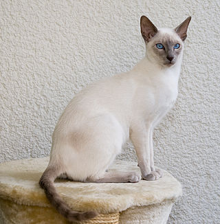
- Origin: Thailand (Siam)
- Appearance: Sleek body, blue almond-shaped eyes, color-pointed (dark face, ears, paws, tail)
- Personality: Very vocal, social, affectionate, intelligent
- Special: One of the oldest recognized breeds
- Vet Note: Prone to respiratory issues, dental disease, amyloidosis
4. Ragdoll

- Origin: USA (California)
- Appearance: Large, blue eyes, semi-long fur, color-pointed like Siamese
- Personality: Goes limp when picked up (hence "Ragdoll"), very gentle, calm, tolerant
- Special: Perfect family and children's cat
- Vet Note: HCM risk, bladder stones
5. Bengal Cat

- Origin: USA (hybrid of Asian Leopard Cat × domestic cat)
- Appearance: Wild leopard-like spots/rosettes on golden/brown coat, muscular body
- Personality: Highly energetic, intelligent, loves to climb and play, very active
- Special: Looks like a mini leopard
- Vet Note: HCM risk, Progressive Retinal Atrophy (PRA)
6. British Shorthair
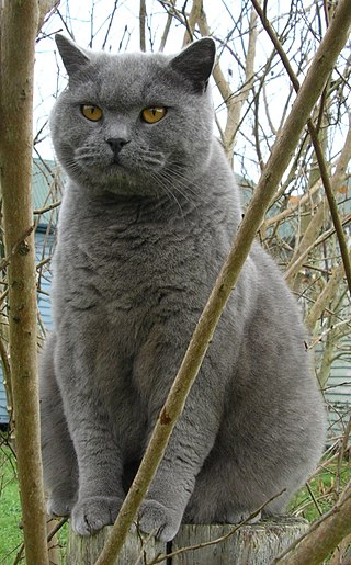
- Origin: United Kingdom
- Appearance: Round face, chubby cheeks, dense plush coat, most famous in blue-grey color
- Personality: Calm, independent, loyal, not very demanding
- Special: The "Cheshire Cat" from Alice in Wonderland is based on this breed
- Vet Note: HCM, obesity prone, polycystic kidney disease
7. Abyssinian Cat

- Origin: Ethiopia (Abyssinia)
- Appearance: Slender, athletic, ticked coat (each hair has multiple color bands), large ears
- Personality: Extremely active, curious, playful, climbs everything
- Special: One of the oldest known cat breeds
- Vet Note: Renal amyloidosis, PRA, dental disease
8. Scottish Fold

- Origin: Scotland
- Appearance: Forward-folded ears (due to cartilage mutation), round owl-like face
- Personality: Sweet, gentle, adaptable, loves human company
- Special: Became globally famous after Taylor Swift's cats
- Vet Note: Osteochondrodysplasia (painful bone/cartilage disease in folded-ear cats) — controversial breed
9. Sphynx Cat

- Origin: Canada
- Appearance: Hairless (due to mutation), wrinkled skin, large ears, peach-fuzz texture
- Personality: Very affectionate, warm, loves to cuddle (needs warmth)
- Special: Not truly hairless — has fine downy fuzz
- Vet Note: HCM very common, skin infections, sunburn risk, hypothermia risk
10. Russian Blue

- Origin: Russia
- Appearance: Short, dense, blue-grey double coat with shimmering silver tips, green eyes
- Personality: Shy with strangers, very loyal to family, intelligent
- Special: Hypoallergenic (produces less Fel d 1 allergen)
- Vet Note: Generally healthy breed; obesity, bladder stones
11. Norwegian Forest Cat

- Origin: Norway
- Appearance: Large, thick double waterproof coat, tufted paws and ears, bushy tail
- Personality: Independent, loves to climb, outdoor-oriented, friendly
- Special: Featured in Norse mythology
- Vet Note: HCM, Glycogen Storage Disease Type IV (GSD IV)
12. Burmese Cat

- Origin: Burma (Myanmar)
- Appearance: Compact, muscular, short glossy coat, golden-yellow eyes
- Personality: Highly social, dog-like, follows owners everywhere, very talkative
- Special: Called "the brick wrapped in silk"
- Vet Note: Hypokalemia, diabetes mellitus, craniofacial defect in some lines
13. Himalayan Cat

- Origin: USA/UK (cross between Persian and Siamese)
- Appearance: Long fur like Persian, color-pointed like Siamese, blue eyes
- Personality: Calm, gentle, loves lap sitting
- Special: Often considered a sub-breed of Persian
- Vet Note: Same as Persian — PKD, brachycephalic issues
14. Birman Cat

- Origin: Burma/France
- Appearance: Semi-long fur, color-pointed, blue eyes, characteristic white "gloves" on paws
- Personality: Gentle, social, quiet, loves being held
- Special: Known as the "Sacred Cat of Burma"
- Vet Note: HCM, kidney disease
15. Turkish Angora

- Origin: Turkey (Ankara)
- Appearance: Slender, long silky coat (usually white), odd-colored eyes common (one blue, one amber)
- Personality: Playful, energetic, intelligent, loves attention
- Special: One of the oldest natural breeds
- Vet Note: Deafness (especially white cats with blue eyes), HCM
16. Devon Rex

- Origin: England (Devon)
- Appearance: Curly/wavy short coat, large ears, pixie-like face
- Personality: Mischievous, active, loves warmth and cuddling
- Special: Called "poodle cat" due to wavy fur
- Vet Note: Hypertrophic myopathy, blood type B common
17. Cornish Rex

- Origin: England (Cornwall)
- Appearance: Very thin, wavy fur (only down layer — no guard hairs), large ears, arched body
- Personality: Extremely active, kitten-like behavior throughout life
- Special: Feels like velvet to touch
- Vet Note: Hypotrichosis, cold sensitivity
18. Tonkinese Cat

- Origin: USA/Canada (Siamese × Burmese cross)
- Appearance: Medium build, aqua/blue-green eyes, color-pointed, short coat
- Personality: Playful, social, vocal, loves people
- Special: Best of both worlds — Siamese + Burmese
- Vet Note: Amyloidosis (from Siamese side)
19. Chartreux

- Origin: France
- Appearance: Robust, blue-grey woolly double coat, orange/copper eyes
- Personality: Quiet (barely vocalizes), observant, intelligent, loyal
- Special: National cat of France — very rare breed
- Vet Note: Luxating patella, kidney disease
20. Manx Cat

- Origin: Isle of Man (UK)
- Appearance: Tailless (or very short tail), round body and head, double coat
- Personality: Playful, dog-like, loyal, good hunters
- Special: Taillessness is due to a natural genetic mutation
- Vet Note: Manx Syndrome (spina bifida-like spinal defects), bowel/bladder issues
🐶 TOP 20 FAMOUS DOG BREEDS
Most Famous → Least Famous Sequence
1. 🥇 Labrador Retriever

- Origin: Canada/UK
- Colors: Yellow, Black, Chocolate
- Personality: Friendly, gentle, eager to please, great with children
- Uses: Guide dogs, therapy dogs, search & rescue, family pets
- Vet Note: Obesity prone, hip/elbow dysplasia, exercise-induced collapse (EIC)
2. German Shepherd

- Origin: Germany
- Personality: Loyal, intelligent, confident, protective
- Uses: Police, military, search & rescue, guide dogs
- Vet Note: Hip dysplasia (very common), degenerative myelopathy, bloat (GDV)
3. Golden Retriever

- Origin: Scotland/UK
- Personality: Gentle, kind, playful, excellent with families and children
- Uses: Therapy dogs, guide dogs, family pets
- Vet Note: Cancer risk highest of all breeds (60%), hip dysplasia, HCM
4. French Bulldog

- Origin: France/England
- Personality: Playful, affectionate, low energy, great apartment dog
- Special: Most popular breed in USA and UK currently
- Vet Note: Severe brachycephalic issues (BOAS), spinal problems (IVDD), skin fold infections
5. Bulldog (English Bulldog)

- Origin: England
- Personality: Calm, stubborn, gentle, loves to sleep
- Special: National symbol of England
- Vet Note: BOAS, cherry eye, skin fold pyoderma, hip dysplasia, heart issues
6. Poodle

- Origin: Germany/France
- Sizes: Standard, Miniature, Toy
- Personality: Highly intelligent (second smartest breed), active, elegant
- Uses: Circus dogs, truffle hunters, water retrievers
- Vet Note: Addison's disease, bloat (standard), progressive retinal atrophy
7. Beagle

- Origin: England
- Personality: Curious, merry, loves to follow scents, great with kids
- Uses: Scent detection (airports), hunting, family pets
- Vet Note: Epilepsy, obesity, hip dysplasia, ear infections (floppy ears)
8. Rottweiler
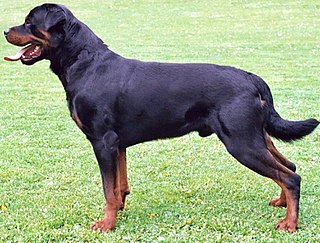
- Origin: Germany
- Personality: Confident, loyal, protective, calm with family
- Uses: Guard dogs, police dogs, herding
- Vet Note: Hip dysplasia, osteosarcoma (bone cancer), heart disease, vWD
9. Yorkshire Terrier (Yorkie)

- Origin: England (Yorkshire)
- Personality: Bold, feisty, affectionate, loves attention
- Special: Small size but big personality — great lap dog
- Vet Note: Tracheal collapse, Legg-Calvé-Perthes disease, hypoglycemia (puppies)
10. Dachshund

- Origin: Germany
- Appearance: Long body, short legs — "sausage dog"
- Personality: Stubborn, brave, playful, loyal
- Uses: Originally bred for hunting badgers
- Vet Note: IVDD (Intervertebral Disc Disease) — most common and serious issue, obesity worsens it
11. Siberian Husky

- Origin: Siberia, Russia
- Appearance: Blue or multi-colored eyes, thick double coat, wolf-like appearance
- Personality: Energetic, mischievous, independent, loves to run
- Uses: Sled dogs, search & rescue
- Vet Note: Hip dysplasia, eye conditions (PRA, cataracts), zinc deficiency
12. Boxer

- Origin: Germany
- Personality: Playful, energetic, loyal, excellent with children
- Special: One of the most playful large breeds
- Vet Note: Heart disease (cardiomyopathy, aortic stenosis), cancer, hip dysplasia, BOAS
13. Dobermann Pinscher

- Origin: Germany
- Personality: Loyal, alert, fearless, highly intelligent
- Uses: Guard dogs, police dogs, military dogs
- Vet Note: Dilated cardiomyopathy (DCM) — major concern, vWD (Type I very common ~70%)
14. Great Dane

- Origin: Germany
- Appearance: Tallest dog breed — up to 86 cm at shoulder
- Personality: Gentle giant — calm, friendly, loving
- Vet Note: Bloat/GDV (most life-threatening), dilated cardiomyopathy, joint issues, short lifespan (6–8 years)
15. Shih Tzu

- Origin: China/Tibet
- Personality: Affectionate, playful, outgoing, loves being with people
- Special: Bred for Chinese royalty
- Vet Note: BOAS (brachycephalic), eye diseases (proptosis), renal dysplasia, ear infections
16. Chihuahua

- Origin: Mexico
- Appearance: World's smallest dog breed
- Personality: Bold, loyal, feisty, devoted to one person
- Vet Note: Tracheal collapse, patellar luxation, hypoglycemia, hydrocephalus, cryptorchidism
17. Border Collie

- Origin: UK (Scottish/English border)
- Personality: Most intelligent dog breed, intense, energetic, workaholic
- Uses: Sheep herding, agility competitions, dog sports
- Vet Note: Hip dysplasia, Collie eye anomaly (CEA), epilepsy
18. Maltese

- Origin: Malta (Mediterranean)
- Appearance: Pure white long silky hair, black eyes and nose
- Personality: Playful, gentle, affectionate, great lap dog
- Vet Note: Tracheal collapse, dental disease, patellar luxation, tear staining
19. Pomeranian

- Origin: Germany/Poland (Pomerania region)
- Appearance: Fluffy double coat, fox-like face, small size
- Personality: Bold, lively, curious, loves attention
- Vet Note: Tracheal collapse, patellar luxation, Alopecia X ("black skin disease"), dental issues
20. Saint Bernard

- Origin: Switzerland
- Appearance: Massive, heavy dog with dense coat, brown/white markings
- Personality: Gentle, patient, calm, great with families
- Uses: Alpine mountain rescue dogs (famous for rescue with brandy barrel)
- Vet Note: Hip/elbow dysplasia, bloat, dilated cardiomyopathy, short lifespan
🦜 FAMOUS PARROT SPECIES/BREEDS
Most Famous → Least Famous Sequence
1. 🥇 African Grey Parrot

- Origin: Central/West Africa
- Appearance: Grey feathers, bright red tail
- Personality: Most intelligent parrot — can learn 1000+ words, understand concepts
- Two Types: Congo African Grey (larger) & Timneh African Grey (smaller, darker)
- Lifespan: 40–60 years
2. Macaw (Blue-and-Gold Macaw)

- Origin: South America
- Appearance: Brilliant blue and yellow/gold feathers — most stunning parrot
- Personality: Loud, playful, affectionate, very social
- Species: Blue-and-Gold, Scarlet Macaw, Green-winged Macaw, Hyacinth Macaw (largest)
- Lifespan: 30–50 years
3. Cockatiel

- Origin: Australia
- Appearance: Grey body, yellow face, orange cheek patches, prominent crest
- Personality: Gentle, affectionate, loves to whistle and sing, good for beginners
- Lifespan: 15–25 years
4. Cockatoo

- Origin: Australia/Indonesia
- Appearance: White with yellow or pink crest, impressive crest display
- Personality: Extremely affectionate, needy, loud, bond strongly with one person
- Species: Sulphur-crested, Umbrella, Moluccan, Black Palm Cockatoo
- Lifespan: 40–80 years
5. Budgerigar (Budgie / Parakeet)

- Origin: Australia
- Appearance: Small, green/yellow (wild) or many color mutations in captivity
- Personality: Social, cheerful, can talk, easy to tame, great for beginners
- Special: Most popular pet bird in the world
- Lifespan: 5–15 years
6. Amazon Parrot

- Origin: South/Central America, Caribbean
- Appearance: Mostly green with colorful head markings (species-dependent)
- Personality: Loud, talkative, opinionated, excellent talkers, love music
- Species: Yellow-naped Amazon, Blue-fronted Amazon, Double Yellow-headed Amazon
- Lifespan: 40–70 years
7. Lovebird

- Origin: Africa/Madagascar
- Appearance: Small, brightly colored — red, green, peach, blue
- Personality: Bond very strongly with mate or owner, playful, active
- Special: Famous for sitting very close to their partner
- Lifespan: 10–15 years
8. Conure (Sun Conure)

- Origin: South America
- Appearance: Brilliant orange-yellow-red — one of most colorful parrots
- Personality: Very loud, affectionate, playful, attention-seeking
- Species: Sun Conure, Green-cheeked Conure, Jenday Conure
- Lifespan: 15–30 years
9. Eclectus Parrot

- Origin: Solomon Islands, New Guinea, Australia
- Appearance: Male = bright green with red/blue. Female = red with blue/purple — so different they were once thought to be different species
- Personality: Calm, gentle, intelligent, excellent talkers
- Lifespan: 30–40 years
10. Caique Parrot

- Origin: South America (Amazon basin)
- Appearance: White belly, green wings, orange/yellow head — very colorful, stocky
- Personality: Most playful and energetic parrot, loves to wrestle, hop, roll on back
- Special: Called the "clown of parrots"
- Lifespan: 20–30 years
11. Pionus Parrot

- Origin: Central/South America
- Personality: Calm, quiet, gentle — great for apartment living
- Species: Blue-headed Pionus, Bronze-winged Pionus, Maximilian's Pionus
- Lifespan: 25–40 years
12. Lorikeet (Rainbow Lorikeet)

- Origin: Australia
- Appearance: Rainbow-colored — blue head, red/orange chest, green back
- Personality: Very active, playful, acrobatic, loves nectar and fruit
- Special: Brush-tipped tongue for feeding on nectar
- Lifespan: 15–25 years
13. Kakapo

- Origin: New Zealand
- Appearance: Large, green, owl-like face, cannot fly
- Personality: Nocturnal, gentle, curious
- Special: World's only flightless parrot, world's heaviest parrot, critically endangered (only ~250 left)
- Lifespan: 40–80+ years
14. Alexandrine Parakeet

- Origin: South Asia (Pakistan, India)
- Appearance: Large green parakeet, red shoulder patch, males have black/pink neck ring
- Personality: Intelligent, independent, can talk well
- Special: Named after Alexander the Great who brought them to Europe
- Lifespan: 25–30 years
15. Ring-necked Parakeet (Rose-ringed Parakeet)

- Origin: Africa and South Asia (very common in Pakistan/India)
- Appearance: Bright green, males have black/pink neck ring, long tail
- Personality: Independent, clever, good talker
- Special: Lives wild in many cities of Pakistan and India — most commonly seen parrot in South Asia
- Lifespan: 25–30 years
16. Senegal Parrot

- Origin: West Africa
- Appearance: Green back, grey head, yellow/orange V-shaped chest
- Personality: Quiet, affectionate, bonds well with one person, good for beginners
- Lifespan: 25–50 years
17. Quaker Parrot (Monk Parakeet)

- Origin: South America
- Appearance: Green with grey face/chest, medium size
- Personality: Bold, talkative, intelligent, build large stick nests
- Special: Only parrot that builds large communal stick nests (not tree cavities)
- Lifespan: 20–30 years
18. Hyacinth Macaw

- Origin: Brazil
- Appearance: Entirely cobalt blue — most beautiful and largest flying parrot in the world
- Personality: Gentle giant — affectionate, playful, gentle despite large size
- Special: Endangered — fewer than 5,000 in wild, most expensive pet parrot
- Lifespan: 50–60 years
19. Black Palm Cockatoo

- Origin: New Guinea, Northern Australia
- Appearance: All black with red cheek patches, enormous beak, dramatic crest
- Personality: Shy, gentle, bond strongly with one person
- Special: Males drum sticks against hollow trees — only non-human to use tools for music
- Lifespan: 40–90 years
20. Kea

- Origin: New Zealand (mountain regions)
- Appearance: Mostly olive-green with orange underwings
- Personality: Most intelligent and mischievous parrot — problem-solving ability remarkable
- Special: Only alpine parrot in the world — lives in snow mountains, opens car locks, steals food from people
- Lifespan: 20–30 years
📊 QUICK SUMMARY
| Category | Total Breeds Listed | Range |
|---|---|---|
| 🐱 Cat Breeds | 20 | Persian (most popular) → Manx (least of 20) |
| 🐶 Dog Breeds | 20 | Labrador (most popular) → Saint Bernard (least of 20) |
| 🦜 Parrot Species | 20 | African Grey (most famous) → Kea (least of 20) |
Note: Images are from Wikimedia Commons (public domain). Har breed most famous se least famous sequence mein diya gaya hai. Har breed ke saath veterinary relevance bhi note ki gayi hai takay ek vet ke liye yeh guide dobly useful ho.