
difference between metastatic node and deposit ct
metastatic lymph node vs tumor deposit CT imaging difference radiology
tumor deposit vs metastatic lymph node CT colorectal cancer
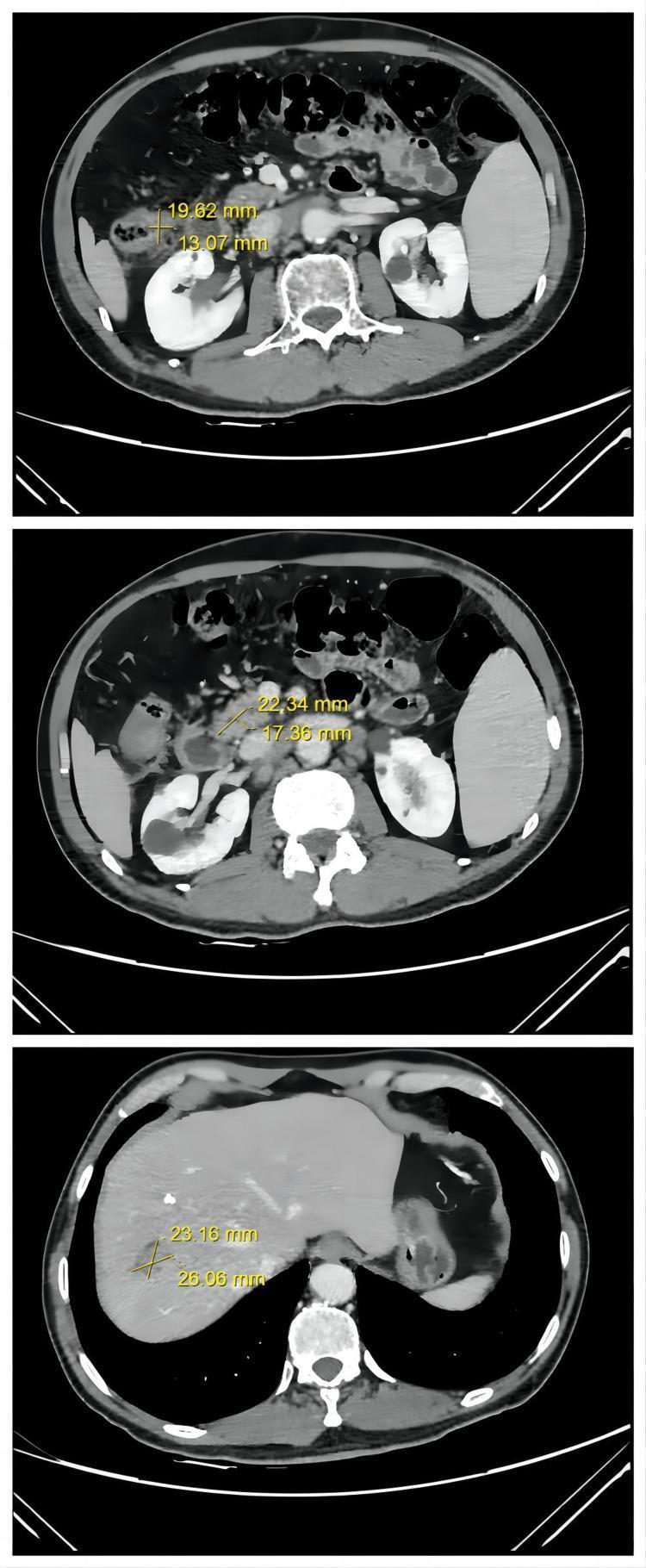
A series of three axial contrast-enhanced Computed Tomography (CT) images of the abdomen, demonstrating the therapeutic response of metastatic colorectal cancer (mCRC). The images utilize caliper measurements to quantify lesion dimensions. The top frame shows a thickened intestinal wall in the ascending colon representing the primary colon tumor. The middle frame highlights retroperitoneal lymph node metastases, characterized as a low-density soft tissue mass adjacent to the major abdominal vessels, measuring approximately 22.34 mm x 17.36 mm. The bottom frame focuses on the liver, revealing a hypo-attenuating, nodular metastasis in the right lobe with dimensions of approximately 23.16 mm x 26.06 mm. These diagnostic images track the regression of malignancy, showing a partial response (PR) following treatment. Key educational landmarks include the liver parenchyma, kidneys, vertebral body, and intestinal loops. The clinical focus is on staging and monitoring secondary malignancies, specifically liver and lymph node involvement in advanced colorectal adenocarcinoma.
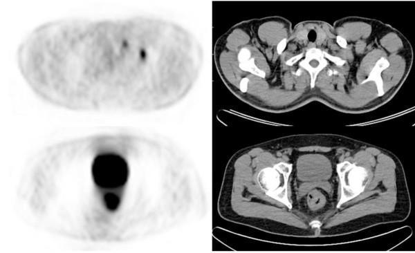
This diagnostic image set consists of paired axial PET and CT scans demonstrating metastatic colorectal cancer. The top row displays the neck region, where the PET scan reveals focal areas of increased metabolic activity (hypermetabolism) in the left supraclavicular region, anatomically correlating with supraclavicular lymph node metastasis on the adjacent CT. The bottom row shows the pelvic region, with the PET scan highlighting a prominent area of intense FDG uptake in the rectum, identifying the primary tumor site. The corresponding axial CT scan provides anatomical localization within the pelvis, showing the rectum, bladder, and iliac structures. These images illustrate the clinical utility of PET/CT in oncology for systemic staging, specifically identifying distant nodal metastases that may alter surgical and chemotherapeutic management strategies for rectal cancer. Key educational concepts include PET/CT fusion for oncologic staging, metabolic characterization of primary tumors, and the identification of regional versus distant lymphadenopathy.
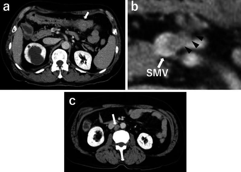
This composite diagnostic image consists of three axial contrast-enhanced abdominal CT scans (a, b, c) demonstrating advanced transverse colon cancer and associated metastatic spread. Panel A shows a primary irregular, enhancing soft tissue mass in the transverse colon (white arrow). Panel B provides a magnified view of the mesenteric region, identifying metastatic lymphadenopathy (black arrowheads) in close proximity to and invading the superior mesenteric vein (SMV). Panel C illustrates further regional progression with a prominent paraaortic lymph node metastasis (white arrow) located near the inferior vena cava and aorta. The primary tumor demonstrates asymmetric bowel wall thickening and heterogenous enhancement, while metastatic nodes appear as rounded, enhancing structures. These images are clinically significant for the staging of colorectal malignancy, specifically highlighting vascular invasion (SMV) and distant lymphatic spread (paraaortic) which classify the disease as advanced stage.
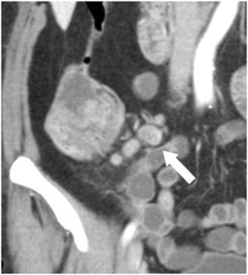
This coronal reformation CT image, taken during the portal phase with IV contrast, demonstrates an enlarged mesocolic lymph node (white arrow) in a patient with an ascending colon tumor. The lymph node measures approximately 10 x 15 mm and is located within the anatomical region most adjacent to the tumor (Region 1). Visually, the node exhibits internal heterogeneity, characterized by mixed attenuation and varying gray-scale intensities within the nodal parenchyma, which is a key morphological predictor for metastatic involvement. Despite this internal complexity, the lymph node maintains a relatively well-defined outer border. Adjacent to the lymph node cluster, the primary colonic mass is visible on the left side of the frame as a large, heterogeneous, and lobulated structure. This image serves as a clinical example of using morphological criteria, such as internal heterogeneity and border definition, rather than size alone, to evaluate lymph node status and predict stage III disease in colorectal cancer patients.
Metastatic Lymph Node vs. Tumor Deposit on CT
Definitions
| Feature | Metastatic Lymph Node (MLN) | Tumor Deposit (TD) |
|---|---|---|
| Definition | Cancer cells spread to and replacing a regional lymph node | A discrete nodule of cancer in the pericolic/perirectal fat or adjacent mesentery, within the lymphatic drainage area of the primary tumor |
| Residual lymph node architecture | Present (residual lymphoid tissue, capsule) | Absent - no identifiable lymph node tissue, vascular structures, or perineural structures |
| Origin | Lymphogenic spread through afferent lymphatics | Thought to arise from lymphovascular invasion (LVI) or perineural invasion (PNI) - not necessarily from a node |
| Key criterion | Must have recognizable nodal tissue | By exclusion - nodule in lymph drainage territory without nodal tissue |
- Sleisenger and Fordtran's Gastrointestinal and Liver Disease: "A tumor deposit is a discrete nodule of cancer present in the pericolic/perirectal fat or in the adjacent mesentery within the lymphatic drainage area of the primary tumor, but without identifiable lymph node tissue or vascular structures; most such deposits are thought to be due to lymphovascular or perineural invasion and carry a poor prognosis."
TNM Staging Implications (AJCC 8th Edition)
| Entity | TNM Code | Clinical Meaning |
|---|---|---|
| 1-3 metastatic nodes | N1a / N1b | Stage III |
| 4+ metastatic nodes | N2a / N2b | Stage III |
| Tumor deposit(s), no nodal metastasis | N1c | T1/T2 with TDs but no LN mets - still Stage III |
| TDs + metastatic nodes | Both counted separately | TDs worsen prognosis independently |
CT Appearances
Metastatic Lymph Node on CT
- Size: Short-axis diameter >8-10 mm (abdominal/pelvic), though small nodes (<5 mm) can still be metastatic in CRC
- Shape: Round rather than oval (L/S ratio <1.5)
- Border: Irregular, spiculated, or ill-defined
- Internal: Loss of fatty hilum, heterogeneous attenuation, central necrosis with ring enhancement (classic in squamous cell mets)
- Distribution: Clustered rather than scattered
- Enhancement pattern: Heterogeneous or rim-enhancing
Tumor Deposit on CT
- A soft-tissue nodule in the pericolonic/perirectal fat or mesentery, separate from the primary tumor edge
- Irregular contour is more characteristic (vs. a well-defined oval node)
- May appear as an ill-defined plaque-like or spiculated density in the mesenteric fat
- No fatty hilum (not expected, since it's not a node)
- Size varies widely; can be >20 mm
- CT cannot distinguish TDs from MLNs with sufficient certainty - this is a known limitation
Why CT Cannot Reliably Distinguish Them
- Irregular border (more common in TDs)
- Lower ADC values (more restricted diffusion)
- Higher Ktrans on DCE-MRI
Prognostic Significance
- Both are poor prognostic signs
- The presence of TDs in addition to MLNs confers a worse prognosis than either alone - their effect is independent and additive
- Patients with TDs alone (N1c) have a prognosis similar to N1 or N2 disease
- TDs are associated with higher rates of distant metastasis and local recurrence
Quick Summary Table
| Feature | Metastatic Lymph Node | Tumor Deposit |
|---|---|---|
| Residual nodal architecture | Yes | No |
| Location | Within lymph node | Pericolonic/mesenteric fat |
| CT shape | Round/oval | Often irregular, plaque-like |
| CT size criterion used | Yes (short axis >8-10 mm) | Not size-based |
| Distinguishable on CT alone | Partially | No - requires histology |
| TNM code | N1a, N1b, N2a, N2b | N1c (if no LN mets present) |
| Thought to arise from | Lymphogenic spread | LVI or perineural invasion |
| Prognostic impact | Poor | Poor (additive with LN mets) |
Help me learn upper limb angiograpahy for the FRCR exam anatomy
What would be most helpful for your FRCR prep? · Which part of the FRCR are you preparing for?
upper limb angiography subclavian axillary brachial artery anatomy labeled
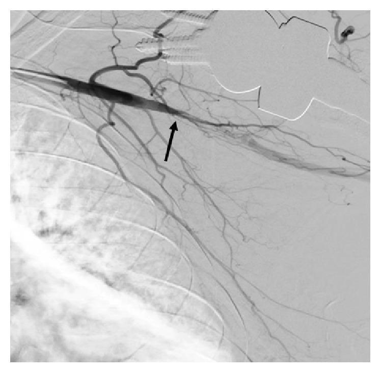
This diagnostic image is a digital subtraction angiogram (DSA) of the left upper extremity, showcasing the vascular anatomy of the subclavian and axillary regions. The primary pathological finding is a focal total occlusion of the axillary artery, indicated by a black arrow. This segment, approximately 4-5 cm in length, shows a complete absence of contrast opacification, representing a vascular blockage. Proximal to the occlusion, the subclavian artery appears patent with normal contrast flow. Several collateral vessels are visible, attempting to bypass the obstructed segment to provide distal perfusion. The image demonstrates key concepts in vascular interventional radiology, specifically the diagnostic identification of arterial occlusive disease and the assessment of collateral circulation. The contrast filling distal to the site of occlusion suggests the presence of distal patency in the brachial artery supplied by these collaterals. This material is highly relevant for medical education focusing on peripheral artery disease (PAD), acute limb ischemia, and diagnostic angiography interpretation.
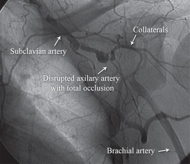
This diagnostic image is a pre-procedural digital subtraction angiography (DSA) of the left upper extremity, captured in an anteroposterior view. It illustrates critical vascular pathology following blunt trauma. The image demonstrates a continuous left subclavian artery that transitions into a severely disrupted axillary artery. There is a clear region of total occlusion in the axillary segment where the column of contrast agent abruptly terminates. To compensate for the lack of primary distal flow, multiple smaller, tortuous collateral vessels are visible, branching off the patent proximal segments in an attempt to perfuse the distal limb. The brachial artery is faintly visible distally, receiving minimal and delayed flow via these collateral pathways. This visual is used to teach the angiographic appearance of traumatic arterial dissection and complete vascular occlusion, highlighting the physiological development of collateral circulation in response to acute ischemic events in vascular surgery and interventional radiology.
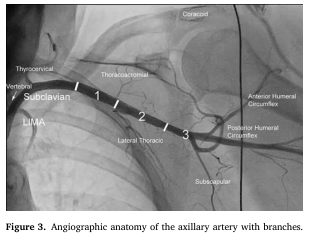
Summary : This figure presents an angiographic image showing the anatomy of the axillary artery and its branches, with clear labeling of major arteries and anatomical landmarks in the shoulder and upper chest region. photo: Scene Overview : • Main subject is the axillary artery and its branches, visualized via angiography in the upper thorax and shoulder region. • Perspective is anteroposterior, showing the left side of the chest and shoulder. • Black-and-white contrast typical of angiographic imaging. Technical Details : • On-image labels identify key arteries: Subclavian, LIMA (Left Internal Mammary Artery), Vertebral, Thyrocervical, Thoracoacromial, Lateral Thoracic, Subscapular, Anterior Humeral Circumflex, Posterior Humeral Circumflex. • Anatomical landmark "Coracoid" is labeled. • The axillary artery is divided into three numbered segments (1, 2, 3) along its course. • No scale bar or magnification is provided. Spatial Relationships : • The subclavian artery transitions into the axillary artery, which is divided into three segments. • Branches arise from each segment: – Segment 1: Thoracoacromial branch. – Segment 2: Lateral thoracic branch. – Segment 3: Subscapular, anterior humeral circumflex, and posterior humeral circumflex branches. • The LIMA and vertebral arteries are shown branching proximally from the subclavian artery. Analysis : • The image clearly delineates the branching pattern of the axillary artery, with each major branch labeled and spatially related to the three anatomical segments. • The figure is useful for understanding vascular anatomy relevant to surgical or interventional procedures in the shoulder and upper chest.
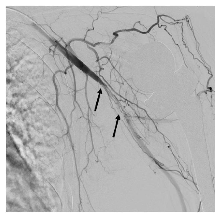
This diagnostic image is a digital subtraction angiogram (DSA) of the left upper extremity, demonstrating vascular pathology in the axillary and brachial arteries. Two black arrows highlight key findings. The superior arrow points to an area of total occlusion in the distal axillary artery, characterized by an abrupt termination of contrast column filling. The inferior arrow indicates the proximal brachial artery, where an irregular, linear contrast pattern suggests a dissection, likely occurring in a subintimal plane. Proximally, the subclavian artery appears patent with smooth contrast flow. Distal to the site of dissection, the brachial artery reconstitutes and remains patent, leading to normally opacified radial and ulnar arteries. The image is clinically significant for diagnosing acute or procedural-related vascular injury, such as arterial dissection and occlusion, requiring interventional management like angioplasty or stenting. It serves as an educational example of upper limb arterial anatomy and common angiographic findings in vascular surgery and radiology.
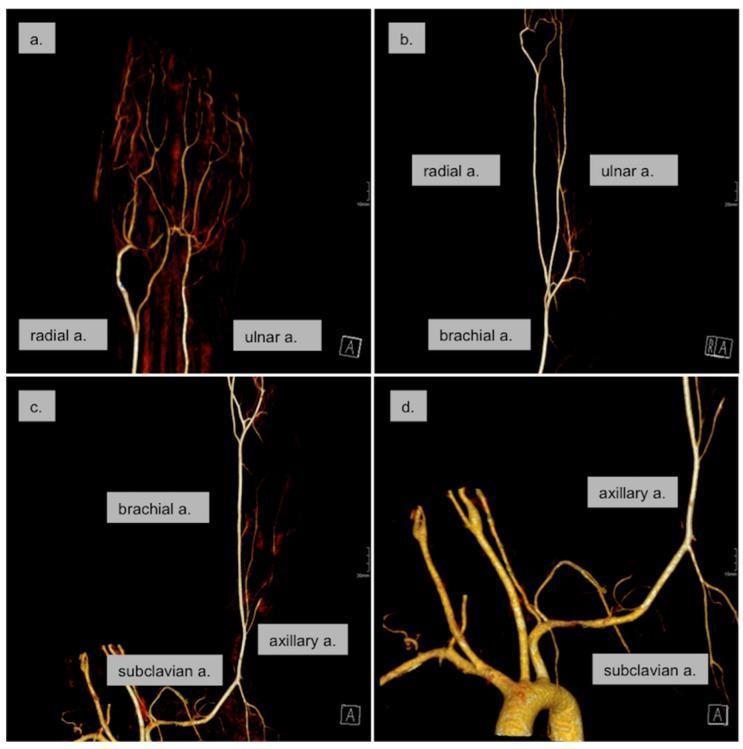
This diagnostic image consists of four panels (a–d) showing a 3D reconstruction of a Computed Tomography Angiography (CTA) of the left upper extremity and aortic arch. The series illustrates the arterial anatomy from the distal hand to the proximal chest. Panel (a) focuses on the hand, showing the superficial and deep palmar arches and digital arteries. Panel (b) depicts the forearm, highlighting the radial and ulnar arteries branching from the brachial artery. Panel (c) shows the mid-to-upper arm including the brachial, axillary, and distal subclavian arteries. Panel (d) displays the aortic arch with the origins of the great vessels, specifically focusing on the left subclavian and axillary arteries. The imaging demonstrates normal arterial diameters, smooth vessel walls, and continuous contrast opacification throughout the entire vascular tree. There is no evidence of thrombi, atherosclerotic plaques, or residual vasospasm. This CTA series is used to evaluate peripheral perfusion and rule out proximal embolic sources in clinical scenarios such as acute limb ischemia or Raynaud's phenomenon.
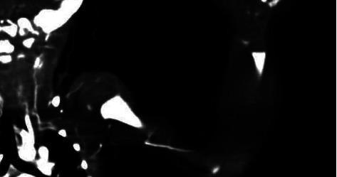
This diagnostic image is a Computed Tomography Angiography (CTA) focusing on the aortic arch and the left upper extremity vasculature. The scan reveals a significant contrast filling defect in the left subclavian artery, originating approximately 2 cm from its takeoff from the aortic arch, diagnostic of a thrombus. Distal to the occlusion, the proximal axillary artery shows faint opacification, likely supplied by collateral circulation. However, there is a subsequent segment of contrast cutoff extending through the distal axillary and proximal brachial regions. Contrast opacification briefly resumes in the midarm until the level of the elbow, where a final complete cutoff occurs, indicating a total lack of distal arterial perfusion. This presentation illustrates a complex pattern of multifocal arterial occlusion and limb ischemia, suitable for medical education regarding neonatal vascular emergencies and radiographic interpretation of thromboembolic events.
FRCR part 1 upper limb angiography anatomy high yield subclavian axillary brachial radial ulnar
https://www.ncbi.nlm.nih.gov/books/NBK27410
brachial artery radial ulnar artery forearm hand palmar arch angiography anatomy
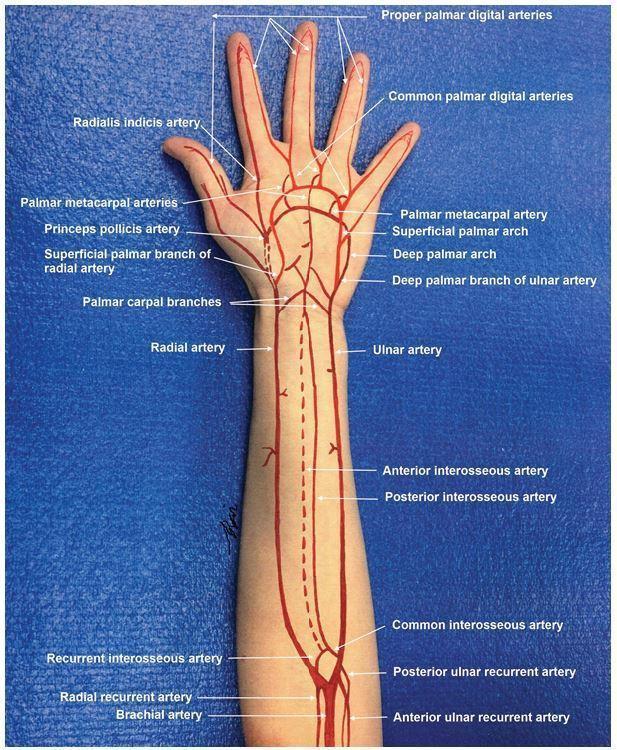
Anatomical diagram superimposed on a photograph of a human arm, illustrating the arterial circulation from the distal humerus to the fingertips. The brachial artery is shown bifurcating into the radial and ulnar arteries. Key forearm vessels are labeled, including the radial and ulnar recurrent arteries, common interosseous artery, and the anterior and posterior interosseous arteries. In the hand, the illustration details the complex vascular anastomoses: the superficial palmar arch (primarily from the ulnar artery) and the deep palmar arch (primarily from the radial artery). Specific terminal branches are identified, such as the princeps pollicis artery, radialis indicis artery, palmar metacarpal arteries, common palmar digital arteries, and proper palmar digital arteries supplying each digit. This educational graphic serves as a guide for understanding upper extremity vascular anatomy, essential for surgical planning, trauma assessment, and performing procedures like the Allen's test.
This diagnostic image consists of four panels (a–d) showing a 3D reconstruction of a Computed Tomography Angiography (CTA) of the left upper extremity and aortic arch. The series illustrates the arterial anatomy from the distal hand to the proximal chest. Panel (a) focuses on the hand, showing the superficial and deep palmar arches and digital arteries. Panel (b) depicts the forearm, highlighting the radial and ulnar arteries branching from the brachial artery. Panel (c) shows the mid-to-upper arm including the brachial, axillary, and distal subclavian arteries. Panel (d) displays the aortic arch with the origins of the great vessels, specifically focusing on the left subclavian and axillary arteries. The imaging demonstrates normal arterial diameters, smooth vessel walls, and continuous contrast opacification throughout the entire vascular tree. There is no evidence of thrombi, atherosclerotic plaques, or residual vasospasm. This CTA series is used to evaluate peripheral perfusion and rule out proximal embolic sources in clinical scenarios such as acute limb ischemia or Raynaud's phenomenon.
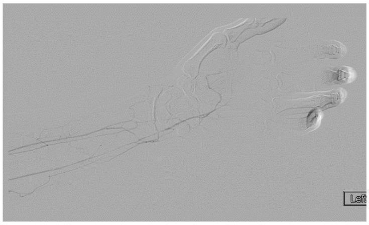
This diagnostic image is a digital subtraction angiogram (DSA) of the left forearm and hand, showcasing the arterial vasculature. The imaging reveals the ulnar artery as the primary source of distal perfusion, extending to form the superficial and deep palmar arches. Notable pathology includes the total occlusion of the radial artery, which is consistent with the patient's history of Arterial Thoracic Outlet Syndrome (ATOS) and distal thromboembolism. The palmar arch shows opacification with contrast leading to the common digital arteries. While there is visible flow to the index, middle, ring, and little fingers via the digital branches, there is a marked absence of contrast opacification in the thumb (princeps pollicis artery), indicating localized ischemia or occlusion. The image demonstrates the clinical consequences of a proximal brachial thrombus causing distal arterial compromise and the importance of assessing collateral flow through the palmar arch in cases of upper extremity arterial disease.
**Imaging Modality:** Digital Subtraction Angiography (DSA) / Arteriography. **Anatomical Region:** Left forearm and hand. **Observed Pathology:** Arteriogram demonstrating significant vascular anomalies of the distal upper extremity. The ulnar artery appears atretic, failing to provide significant contribution to the palmar circulation. The deep palmar arch shows filling predominantly via the radial artery, indicating a lack of normal dual arterial supply. **Characteristic Visual Features:** - **Atretic Ulnar Artery:** Absence or severe hypoplasia of the ulnar artery as it approaches the wrist and hand. - **Palmar Arch Hypoplasia:** The palmar arches are hypoplastic, particularly on the ulnar side. - **Digital Artery Compromise:** The 4th and 5th digital arteries originate from a diminutive deep palmar arch, exhibiting markedly reduced contrast opacification and poor distal blood flow compared to the radial-side digits. - **Collateralization:** Visual evidence of compensatory filling of the deep arch from the radial system. **Key Diagnostic Features:** Congenital or acquired ulnar artery atresia resulting in an incomplete palmar arch and distal digital ischemia of the ulnar-sided phalanges. Relevant for surgical planning and evaluation of peripheral vascular disease.
FRCR Part 1 - Upper Limb Angiography Anatomy
The Big Picture: Transition Points (MUST KNOW)
| Transition | Landmark | From | To |
|---|---|---|---|
| 1 | Lateral border of 1st rib | Subclavian | Axillary |
| 2 | Inferior border of teres major | Axillary | Brachial |
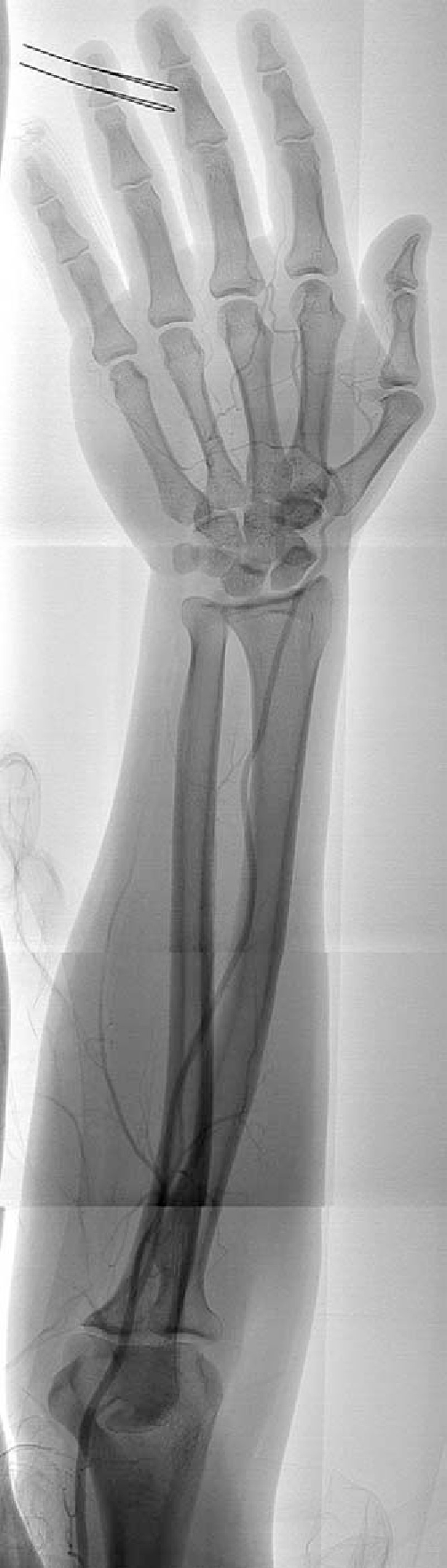
| 3 | Neck of radius (cubital fossa, just below elbow) | Brachial | Radial + Ulnar |
1. SUBCLAVIAN ARTERY
- Right: branch of the brachiocephalic (innominate) trunk, which also gives the right common carotid
- Left: arises directly from the aortic arch (3rd branch)
| Branch | Notes |
|---|---|
| Vertebral artery | 1st and largest branch; runs through foramina transversaria C6-C1 |
| Internal thoracic artery | Descends behind costal cartilages; gives anterior intercostals and musculophrenic; used for CABG |
| Thyrocervical trunk | 3 branches: inferior thyroid, suprascapular, transverse cervical |
| Costocervical trunk | 2 branches: superior intercostal (1st & 2nd post intercostals), deep cervical |
| Dorsal scapular artery | Inconstant; may arise from subclavian directly or from thyrocervical |
2. AXILLARY ARTERY
- Part 1 = proximal to pec minor
- Part 2 = posterior to pec minor
- Part 3 = distal to pec minor
| Part | Branch | Notes |
|---|---|---|
| 1 (1 branch) | Superior thoracic | Supplies upper chest wall; small |
| 2 (2 branches) | Thoraco-acromial | Short trunk; divides into pectoral, deltoid, clavicular, acromial branches |
| 2 | Lateral thoracic | Runs along lateral chest wall; supplies breast in females |
| 3 (3 branches) | Subscapular | Largest branch; divides into circumflex scapular + thoracodorsal |
| 3 | Anterior circumflex humeral | Smaller; loops around surgical neck of humerus anteriorly |
| 3 | Posterior circumflex humeral | Larger; runs through quadrilateral space with axillary nerve |
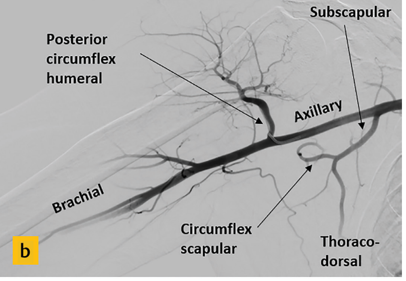
- Posterior circumflex humeral passes through the quadrilateral space (bounded by: teres minor, teres major, long head of triceps, surgical neck of humerus) - with the axillary nerve
- The subscapular artery is the largest branch of the axillary artery
- Subscapular artery subdivides into circumflex scapular (goes through triangular space) and thoracodorsal
3. BRACHIAL ARTERY
| Branch | Notes |
|---|---|
| Profunda brachii (deep brachial) | First and largest branch; accompanies radial nerve in spiral groove; gives middle collateral + radial collateral arteries |
| Superior ulnar collateral | Accompanies ulnar nerve through medial intermuscular septum |
| Inferior ulnar collateral | Supplies elbow anastomosis |
| Nutrient artery to humerus | |
| Muscular branches |
- Profunda brachii accompanies the radial nerve in the radial/spiral groove - injury in mid-shaft humeral fracture
- Brachial bifurcation is the most common site for emboli to lodge in the upper limb
- High bifurcation (in the arm or axilla) occurs in 15-20% - this is a very common variant question
4. RADIAL ARTERY
- Smaller terminal branch of brachial
- Runs along the radial (lateral) side of the forearm
- Palpable at the wrist, lateral to flexor carpi radialis
- Passes through the anatomical snuffbox (between extensor pollicis longus and extensor pollicis brevis/abductor pollicis longus)
- Crosses the floor of the snuffbox (scaphoid and trapezium) and dips between the heads of 1st dorsal interosseous into the palm
- Forms the deep palmar arch
- Radial recurrent (at elbow - part of elbow anastomosis)
- Palmar carpal branch
- Superficial palmar branch (contributes to superficial palmar arch)
- Princeps pollicis (thumb artery)
- Radialis indicis (to index finger)
- Deep palmar arch branches
5. ULNAR ARTERY
- Larger terminal branch of brachial (supplies more of the hand)
- Runs along the ulnar (medial) side of the forearm, deep to flexor carpi ulnaris
- Passes superficial to the flexor retinaculum at the wrist (through Guyon's canal)
- Forms the superficial palmar arch
- Anterior and posterior ulnar recurrent (elbow anastomosis)
- Common interosseous artery (gives anterior and posterior interosseous arteries - supplies deep forearm muscles)
- Deep palmar branch
- Superficial palmar arch branches
6. HAND ARTERIES - Palmar Arches
| Arch | Formed mainly by | Completed by | Branches |
|---|---|---|---|
| Superficial palmar arch | Ulnar artery | Superficial palmar branch of radial | Common palmar digital arteries → proper palmar digital arteries |
| Deep palmar arch | Radial artery | Deep palmar branch of ulnar | Palmar metacarpal arteries |
- Superficial arch = primarily ulnar; Deep arch = primarily radial (remembering this differentiates them)
- An incomplete superficial palmar arch occurs in ~20% of individuals - clinically important before radial artery harvest (Allen's test)
- Princeps pollicis (thumb) arises from the radial artery or the deep palmar arch
- Radialis indicis supplies the radial side of the index finger
7. HIGH-YIELD ANATOMICAL VARIANTS (Very Common in MCQs)
| Variant | Frequency | Why it Matters |
|---|---|---|
| High bifurcation of brachial - bifurcates in axilla or arm | 15-20% | Can cause slow dialysis fistula maturation; looks odd on angiogram |
| High origin of radial artery from axillary artery | ~15% | Radial artery seen surprisingly high |
| Aberrant right subclavian artery (arteria lusoria) | ~0.5-2% | Arises as last branch of aortic arch; crosses behind oesophagus → dysphagia lusoria |
| Left vertebral artery direct from aortic arch | ~5% | Third branch of aortic arch instead of from subclavian |
| Incomplete superficial palmar arch | ~20% | Critical before radial artery harvest (CABG, radial access) |
| Subscapular artery from 2nd part of axillary | 29-36% | Most common axillary branch variant |
| Common trunk of subscapular + posterior circumflex humeral | 12-42% | Common variant of axillary branching |
| Superficial brachial artery | Rare | Early splitting of axillary into superficial and deep brachial |
8. COLLATERAL PATHWAYS AT THE ELBOW
- Profunda brachii → radial collateral (to radial recurrent)
- Profunda brachii → middle collateral (to interosseous recurrent)
- Superior ulnar collateral (to posterior ulnar recurrent)
- Inferior ulnar collateral (to anterior ulnar recurrent)
9. ANGIOGRAPHY TECHNIQUE - FRCR BASICS
- Standard examination runs from proximal subclavian → wrist
- Catheter access typically via femoral artery (common); brachial or radial access also used
- Digital subtraction angiography (DSA) removes background bone and soft tissue
- Hand arteries (palmar arches, digital arteries) are imaged selectively with hand in pronation, using reactive hyperaemia (warm water/sphygmomanometer cuff occlusion release) to increase distal flow
- On a standard AP angiogram: radial artery lies laterally (thumb side), ulnar artery lies medially
10. QUICK MCQ DRILL
| Question | Answer |
|---|---|
| Where does subclavian → axillary? | Lateral border of 1st rib |
| Where does axillary → brachial? | Inferior border of teres major |
| What divides axillary into 3 parts? | Pectoralis minor muscle |
| How many branches per part? | 1, 2, 3 (increasing distally) |
| Largest branch of axillary artery? | Subscapular |
| What artery accompanies the radial nerve? | Profunda brachii |
| Which palmar arch is mainly ulnar? | Superficial |
| Which palmar arch is mainly radial? | Deep |
| Where do emboli most commonly lodge in upper limb? | Brachial bifurcation (cubital fossa) |
| What passes through the quadrilateral space? | Posterior circumflex humeral artery + axillary nerve |
| Arteria lusoria - what is it? | Aberrant right subclavian from aortic arch |
| How does radial artery reach the palm? | Through anatomical snuffbox |
| What is Guyon's canal? | Ulnar tunnel at wrist - ulnar artery + nerve pass through |
| High brachial bifurcation rate? | ~15-20% |