
Frozen shoulder, dds for sgoulder pain
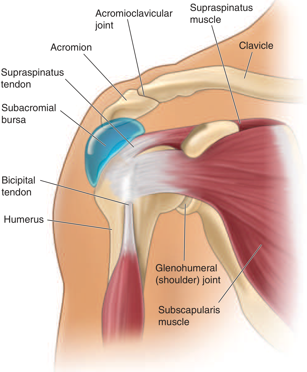
frozen shoulder adhesive capsulitis anatomy diagram
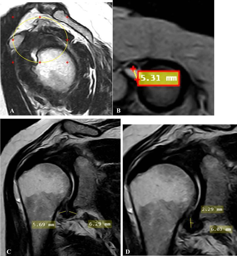
A multi-panel MRI study of the shoulder demonstrating radiological signs of adhesive capsulitis (frozen shoulder). Image A (oblique sagittal T2-weighted) shows the complete obliteration of the subcoracoid fat triangle within the rotator interval (encircled). Image B (oblique axial fat-suppressed PD-weighted) identifies a significant effusion (5.31 mm) within the long head of the biceps tendon sheath, exceeding the normal threshold. Images C and D (oblique coronal T2-weighted) focus on the axillary recess. Image C measures the pathological thickening of the joint capsule at the humeral (5.69 mm) and glenoid (6.29 mm) aspects. Image D provides measurements for the maximal height (2.29 mm) and width (6.03 mm) of the axillary recess space. This composite image illustrates key diagnostic criteria including rotator interval fat replacement, synovial effusion, and capsular thickening, which are critical for the radiological staging of adhesive capsulitis in a clinical setting.
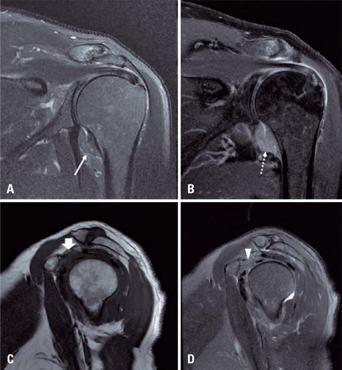
This diagnostic image set consists of four MRI views of a human shoulder illustrating the classic findings of adhesive capsulitis (frozen shoulder). Image A (coronal T2-weighted with fat suppression) and Image B (coronal T1-weighted post-gadolinium with fat suppression) demonstrate significant thickening of the glenohumeral joint capsule and pericapsular edema, particularly within the axillary recess. The post-contrast image (B) shows intense enhancement of the thickened capsule, indicating active synovitis and hypervascularity. Images C (sagittal T1-weighted) and D (sagittal T2-weighted with fat suppression) focus on the rotator interval. They reveal the characteristic obliteration of the normal subcoracoid fat pad by hypointense inflammatory tissue on T1 (arrow in C) and hyperintense edema on T2 (arrowhead in D). These imaging features are essential for confirming adhesive capsulitis in patients presenting with clinical shoulder stiffness and pain, allowing for the differentiation from other pathology such as rotator cuff tears or labral injuries.
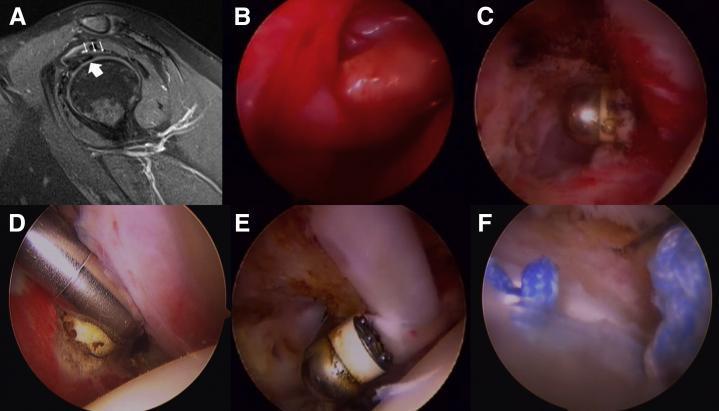
This composite image illustrates the diagnostic and surgical management of adhesive capsulitis (frozen shoulder). Panel A is a sagittal proton density fat-saturated MRI of the shoulder, demonstrating a thickened coracohumeral ligament (CHL, indicated by thin arrows) positioned superiorly to the long head of the biceps tendon (LHBT, indicated by a thick arrow), a characteristic radiological sign of this condition. Panels B through F provide a sequential arthroscopic view of the surgical intervention. Panel B shows severe synovitis with erythematous and thickened fibrous tissue obscuring the rotator interval. Panels C and D visualize the arthroscopic release of the rotator interval and the inferior glenohumeral ligament using specialized instruments. Panel E depicts the release of intra-articular adhesions surrounding the biceps tendon. Panel F shows the final procedural stage: a biceps tenodesis performed using a suture anchor technique, with visible blue sutures and the anchor site. The sequence highlights the progression from diagnostic imaging of ligamentous pathology to the intraoperative debridement and reconstructive stabilization of shoulder structures.
Frozen Shoulder (Adhesive Capsulitis)
Definition & Epidemiology
- Miller's Review of Orthopaedics 9th Ed, p. 1143-1157
- Goldman-Cecil Medicine, p. 2777
Pathophysiology
- Miller's Review of Orthopaedics, p. 1151-1154
Risk Factors / Associations
| Category | Examples |
|---|---|
| Systemic disease | Diabetes mellitus (most important), thyroid disease |
| Inflammatory | Rheumatoid arthritis |
| Post-procedure | Chest or breast surgery, prolonged immobilization |
| Psychological | Depression and anxiety worsen symptoms |
| Most cases | Idiopathic |
Clinical Features
- Insidious onset of pain followed by selective loss of external rotation - the earliest and most characteristic finding
- Later: global ROM loss (all planes)
- Active ROM = Passive ROM (unlike rotator cuff tear where active is worse than passive)
- Night pain is common
Imaging
- X-ray: Must be done first to exclude glenohumeral OA and locked posterior dislocation (both cause selective external rotation loss)
- Arthrography: Shows reduced joint capsule volume / loss of axillary recess
- MRI: Thickening of capsule along axillary pouch, thickening of CHL, obliteration of subcoracoid fat triangle, rotator interval synovitis - but none are pathognomonic
Treatment
- NSAIDs
- Corticosteroid injections: 1 mL of methylprednisolone 40 mg/mL into the glenohumeral joint AND subacromial bursa
- Physical therapy: pendulum exercises → wall-climbing → active ROM → strengthening; ice packs, ultrasound, TENS
- Oral steroids improve pain/ROM short term
- Distension arthrography occasionally used
- Arthroscopic capsular release (risk to axillary nerve; selective vs. complete release)
- Manipulation under anesthesia + steroid injection (equivalent efficacy to arthroscopic release in trials, but release carries highest procedural risk)
- Goldman-Cecil Medicine, p. 2777; Miller's Review of Orthopaedics
Differential Diagnosis of Shoulder Pain
INTRINSIC (Periarticular / Articular)
1. Rotator Cuff Tendinitis / Impingement Syndrome
- Most common cause overall; ~30% of elderly with shoulder pain
- Pain on active abduction (not passive) - key differentiator
- Pain over lateral deltoid, night pain
- Neer impingement test positive (pain before 180° of forced forward flexion)
- Involves the supraspinatus most commonly (supraspinatus, infraspinatus, teres minor, subscapularis)
2. Rotator Cuff Tear
- Active ROM worse than passive ROM (opposite of frozen shoulder)
- Weakness on testing
- Supraspinatus most commonly torn
- Full-thickness tears: may palpate synovial effusion, suggest acute tear
3. Subacromial / Subdeltoid Bursitis
- Very common; pain with lateral deltoid pressure
- Often coexists with rotator cuff disease
- Pain on active abduction
4. Adhesive Capsulitis (Frozen Shoulder)
- As described above - global loss of both active AND passive motion
5. Bicipital Tendinitis
- Pain with direct pressure over bicipital groove
- Tenderness as humerus is rotated internally/externally
- Speed's test, Yergason's test positive
6. Acromioclavicular (AC) Joint Disease
- OA or RA commonly involves AC joint
- Local pain, bony hypertrophy, or synovial swelling at AC joint
- Pain with cross-body adduction
7. Glenohumeral Osteoarthritis
- Pain on movement in ALL planes (like frozen shoulder)
- Associated with trauma, osteonecrosis, occupational causes
- X-ray shows joint space narrowing
- Can cause selective loss of external rotation (must exclude with X-ray before diagnosing frozen shoulder)
8. Glenohumeral Inflammatory Arthritis (RA, crystal arthropathy)
- Synovial effusion palpable (rare in shoulder - suggests infection, RA, amyloidosis, acute cuff tear)
9. Locked Posterior Shoulder Dislocation
- Also causes selective loss of external rotation
- Must be excluded radiographically before diagnosing frozen shoulder
10. Labral Tear (SLAP, Bankart)
- Usually post-traumatic or overhead athletes
- Clicking, catching, instability
11. Glenohumeral Instability
- Anterior/posterior subluxation
- Apprehension test positive
12. Calcific Tendinitis
- Calcium hydroxyapatite deposits in rotator cuff
- Acute intense pain; visible on X-ray
EXTRINSIC (Referred Pain to Shoulder)
| Cause | Clues |
|---|---|
| Cervical spine disease (C5-C6 disc herniation, DDD) | Most common extrinsic source; radicular pain, neck tenderness, dermatomal sensory/motor findings; mimics rotator cuff disease |
| Brachial plexus neuritis (Parsonage-Turner) | Sudden severe pain → rapid weakness and atrophy; post-viral/post-immunization |
| Thoracic outlet syndrome | More common in women; pain to medial forearm, numbness/tingling of ring/small fingers; positive provocative tests |
| Suprascapular nerve entrapment | Infraspinatus atrophy, weak external rotation |
| Pancoast tumor | Apical lung tumor compresses brachial plexus; shoulder/arm pain + Horner syndrome |
| Axillary artery thrombosis | Repetitive trauma; ischemic symptoms |
| Referred visceral pain | Gallbladder/liver → right shoulder; diaphragmatic irritation → shoulder tip; cardiac ischemia → left arm/shoulder |
| Fibromyalgia | Diffuse periarticular pain, multiple tender points, sleep disturbance |
- Tintinalli's Emergency Medicine, p. 1935; Harrison's Principles of Internal Medicine 22E, p. 2941-2954
Key Distinguishing Features at a Glance
| Condition | Active ROM | Passive ROM | Key Feature |
|---|---|---|---|
| Frozen shoulder | Reduced | Equally reduced | Global restriction, active = passive |
| Rotator cuff tear | Reduced | Near-normal | Weakness; active < passive |
| Rotator cuff tendinitis | Painful arc (60-120°) | Normal | Impingement signs positive |
| GH osteoarthritis | Reduced (all planes) | Reduced | X-ray changes; loss of ER |
| Cervical radiculopathy | Normal shoulder ROM | Normal | Neck exam positive; neurological signs |
| Bursitis | Painful arc | Normal | Point tenderness at bursa |
Clinical pearl: Always obtain shoulder X-rays before diagnosing frozen shoulder - glenohumeral OA and locked posterior dislocation both cause selective external rotation loss and must be excluded first. (Miller's Review of Orthopaedics)
Age manageme
What aspect of age management are you asking about?
No acute gastro-enteritis