
AO Classification of lower end of femur
Reading File
The AO classification divides distal femur fractures into three main types (A, B, C) based on the extent of articular involvement. Let me search the medical library for detailed content.
Searching sources...
Finding Sources
Finding Sources
Reading File
Reading File
There's a classification diagram image. Let me fetch it and also read more of the text for the detailed subtypes.
Loading Image
Reading File
I now have the full OTA/AO classification diagram and detailed text from Rockwood & Green's. Here is the complete answer:
AO/OTA Classification of Distal Femur Fractures (Segment 33)
The AO/OTA system codes the distal femur as segment 33 (bone 3 = femur, segment 3 = distal). The classification is based on the fundamental principle of articular involvement and fracture complexity, dividing all fractures into three types and nine groups.
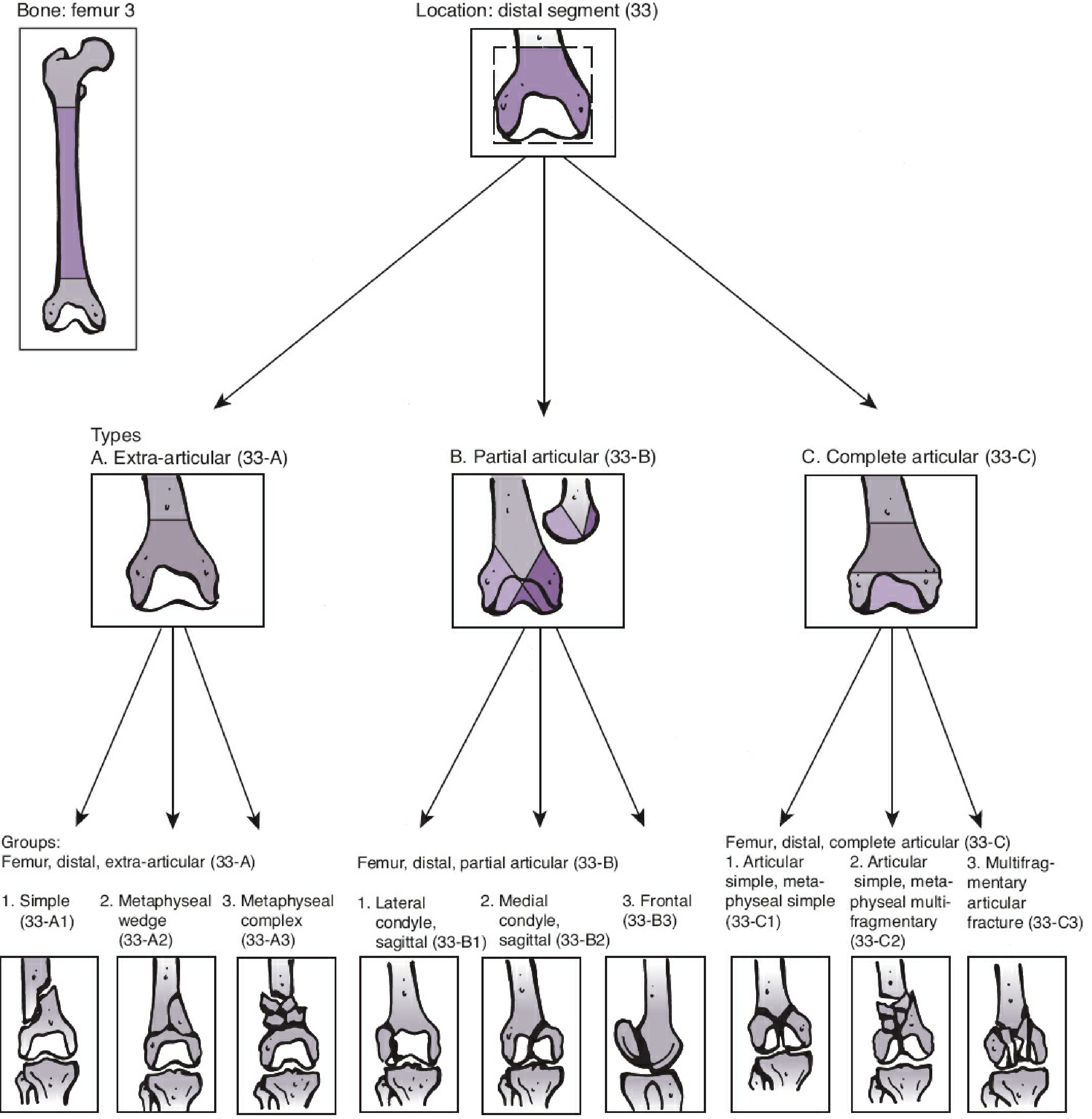
Figure: OTA/AO classification of distal femur fractures (43A-C) - Rockwood and Green's Fractures in Adults, 10th ed. 2025
Type A - Extra-articular (33-A)
The articular surface is completely intact. The fracture involves only the metaphysis/diaphysis.
| Group | Code | Description |
|---|---|---|
| A1 | 33-A1 | Simple - single metaphyseal fracture line (transverse or oblique) |
| A2 | 33-A2 | Metaphyseal wedge - butterfly/wedge fragment present, main fragments maintain contact |
| A3 | 33-A3 | Metaphyseal complex (comminuted) - severe comminution of the metaphysis, no contact between main fragments |
Type B - Partial Articular (33-B)
Part of the articular surface is fractured, but the remaining articular segment stays in continuity with the diaphysis. These are unicondylar fractures.
| Group | Code | Description |
|---|---|---|
| B1 | 33-B1 | Lateral condyle, sagittal plane - fracture runs in the sagittal plane splitting the lateral condyle |
| B2 | 33-B2 | Medial condyle, sagittal plane - sagittal split of the medial condyle |
| B3 | 33-B3 | Frontal plane (Hoffa fracture) - fracture runs in the coronal plane, detaching a posterior condylar fragment; often missed on plain X-rays (40% missed rate noted in studies) |
Type C - Complete Articular (33-C)
The articular surface is completely disrupted and separated from the diaphysis. These are bicondylar (intercondylar/T- or Y-shaped) fractures - the most severe type.
| Group | Code | Description |
|---|---|---|
| C1 | 33-C1 | Articular simple + Metaphyseal simple - single intercondylar split (Y or T pattern) with simple metaphyseal component |
| C2 | 33-C2 | Articular simple + Metaphyseal multifragmentary - simple condylar split but comminuted metaphysis |
| C3 | 33-C3 | Multifragmentary articular fracture - comminuted both at the articular level and the metaphysis; most complex, worst prognosis |
Key Principles
- Severity increases from A to C and from 1 to 3 within each type
- Treatment planning: Type A - primarily metaphyseal fixation (plate/nail); Type B - anatomic condylar fixation (lag screws +/- buttress plate); Type C - combined articular reconstruction + metaphyseal bridging
- B3 (Hoffa) fractures are easily missed on AP/lateral X-rays and require CT for identification
- The OTA/AO system does not capture "fracture personality" factors (soft tissue injury, osteoporosis, vascular injury) which also influence management - these must be assessed separately
Coding Structure
Bone (3) - Segment (3) - Type (A/B/C) - Group (1/2/3)
       ↓
   33-A1, 33-B3, 33-C2, etc.
- Rockwood and Green's Fractures in Adults, 10th ed. 2025, Chapter 58 (Classification of Distal Femur Fractures)
This is a shared conversation. Sign in to Orris to start your own chat.