
tell me about it
python3 -c " import pdfplumber pdf_path = '/home/daytona/workspace/attachments/1919cc61-d4f2-4052-989c-ef0bf2a66ef9/CamScanner 06-30-2026 16.29 (1).pdf' with pdfplumber.open(pdf_path) as pdf: print(f'Pages: {len(pdf.pages)}') for i, page in enumerate(pdf.pages): text = page.extract_text() print(f'--- Page {i+1} ---') print(text) print() "
python3 -c " import subprocess # Convert PDF to image first result = subprocess.run(['pdftoppm', '-r', '300', '-png', '/home/daytona/workspace/attachments/1919cc61-d4f2-4052-989c-ef0bf2a66ef9/CamScanner 06-30-2026 16.29 (1).pdf', '/home/daytona/workspace/attachments/1919cc61-d4f2-4052-989c-ef0bf2a66ef9/page'], capture_output=True, text=True) print('stdout:', result.stdout) print('stderr:', result.stderr) print('returncode:', result.returncode) import os files = os.listdir('/home/daytona/workspace/attachments/1919cc61-d4f2-4052-989c-ef0bf2a66ef9/') print('Files:', files) "
python3 -c " import pytesseract from PIL import Image img = Image.open('/home/daytona/workspace/attachments/1919cc61-d4f2-4052-989c-ef0bf2a66ef9/page-1.png') text = pytesseract.image_to_string(img) print(text) "
MRI Right Knee Joint - Report Explained
What the MRI Sequences Mean
- T1WI - shows anatomy and bone structure well
- T2WI / FS T2WI - sensitive to fluid and soft tissue injury (bright = fluid/swelling)
- STIR - suppresses fat signal, very sensitive for edema and bone bruises
- PD Fat Sat - proton density with fat suppression, excellent for meniscus and cartilage
Findings - Breaking Each Down
1. Articular Cartilage
- The articular cartilage (the smooth coating over bone ends) shows very mild changes just beneath the surface (subchondral).
- This is a minor finding, likely related to the trauma. It means there is slight stress/bruising under the cartilage surface but no significant cartilage damage.
2. Meniscus - Grade I injury to lateral meniscus posterior horn
- Normal menisci are uniformly dark (low signal) on all MRI sequences.
- Increased signal inside the meniscus but not reaching the articular surface = Grade I or II (intrasubstance degeneration or early injury).
- The impression calls this Grade I - meaning the signal change is entirely within the substance of the meniscus and does NOT break through to the surface. This is considered an internal degeneration/contusion rather than a true tear.
- A tear is only diagnosed on MRI when the high signal extends to the articular (joint) surface of the meniscus. (Grainger & Allison's Diagnostic Radiology)
- The peripheral meniscocapsular signal may reflect a small amount of fluid or edema at the capsular junction of the meniscus.
- The medial meniscus is completely normal.
3. Ligaments - Grade II ACL injury
- Grade II ACL injury = partial tear. Some fibers are intact, but there is abnormal signal (edema/hemorrhage within the ligament), thickening, and loss of normal taut fiber alignment.
- A complete (Grade III) ACL tear would show full fiber discontinuity and retraction.
- On MRI, partial tears/sprains are recognized by "altered signal and/or laxity in the presence of continuity of some fibers." (Grainger & Allison's Diagnostic Radiology)
- High signal at the femoral and tibial attachments confirms the injury extends to both insertion points.
4. Joint Effusion
- There is intra-articular fluid (effusion) - i.e., fluid inside the knee joint capsule. This appears bright (hyperintense) on T2WI.
- The suprapatellar recess is the pouch above the kneecap - a common place for fluid to pool after knee injury.
- Effusion after trauma is expected and indicates the joint has responded to injury with inflammation and/or bleeding (hemarthrosis).
5. Bones - Osseous Contusions (Bone Bruises)
- This is a classic bone bruise (osseous contusion) pattern - dark on T1, bright on T2/STIR.
- Histologically this represents "cancellous bone microfractures as well as edema and hemorrhage within the fatty marrow." (Rockwood & Green's Fractures in Adults, 10th ed.)
- The contusions are at the lateral femoral condyle and bilateral tibial condyles - this is the classic "pivot shift" contusion pattern seen with ACL injuries, where the lateral femoral condyle and posterolateral tibial plateau clash during the injury mechanism.
- Importantly, there are no compression fractures - meaning the bony injury is at the micro/marrow level only, with intact bone surfaces.
- These bone bruises typically resolve within months with conservative care.
Summary: What Shadman Has
| Finding | Grade/Severity | Significance |
|---|---|---|
| Lateral meniscus posterior horn signal change | Grade I | Intrasubstance, no surface tear - conservative management |
| ACL injury | Grade II | Partial tear - some fibers intact but significant ligament damage |
| PCL | Normal | No injury |
| MCL / LCL | Normal | No injury |
| Joint effusion | Present | Expected after trauma |
| Bone bruises (tibial + lateral femoral condyles) | Present, no fracture | Classic ACL injury pattern, will resolve |
| Articular cartilage | Minimal subchondral change | Minor finding |
What Happens Next
- Conservative (non-surgical): Physiotherapy, quadriceps strengthening, bracing - often sufficient for Grade II partial tears, especially if the patient does not require high-level pivoting sports.
- Surgical (ACL reconstruction): May be needed if the knee remains unstable or if he wishes to return to demanding physical activity. Typically considered more strongly for complete (Grade III) tears.
- The meniscal Grade I injury and bone bruises are managed conservatively and monitored.
Find recent research on Grade II ACL injury outcomes with conservative vs surgical treatment
"anterior cruciate ligament"[MeSH Terms] AND "conservative treatment" AND "partial tear"
"anterior cruciate ligament injuries"[MeSH Terms] AND ("conservative treatment" OR "rehabilitation") AND ("surgical treatment" OR "reconstruction")
Grade II partial ACL tear conservative vs surgical treatment outcomes 2023 2024 2025
PMID: 40603829
"anterior cruciate ligament" AND "partial tear" AND (conservative OR nonoperative OR "non-operative")
https://pmc.ncbi.nlm.nih.gov/articles/PMC11027445
PMID: 35395764
PMID: 41908101
PMID: 35779101
Recent Research: Grade II ACL Injury - Conservative vs. Surgical Treatment
Key Papers Found
| Evidence Tier | Year | Study | PMID |
|---|---|---|---|
| Systematic Review + Meta-Analysis (Tier 1) | 2025 | Filbay et al. - ACL reconstruction vs rehabilitation alone: RTS rates | 40603829 |
| Systematic Review (Tier 1) | 2022 | Giummarra et al. - Non-surgical management of partial ACL tears | 35395764 |
| Cohort Study | 2023 | Rai et al. - Progression risk of partial ACL tears in young patients | 35779101 |
| Retrospective Cohort | 2026 | Hannon et al. - Nonoperative outcomes in pediatric partial ACL tears | 41908101 |
1. Return to Sport: Surgery vs. Rehabilitation Alone
- No significant difference in return-to-sport (RTS) rates between ACL reconstruction and rehabilitation alone: OR 1.5 (95% CI 0.76-2.97).
- A small difference in Tegner Activity Scale scores favored surgery (mean difference 0.7), but this did not exceed the minimal detectable change and disappeared after excluding high-bias studies.
- 10 of 15 studies had high risk of confounding bias (often favoring surgery due to group allocation and different RTS advice between groups).
- Overall evidence certainty: low to very low due to heterogeneity and bias.
- Bottom line from the best current evidence: ACL reconstruction is not clearly superior to supervised rehabilitation for return-to-sport rates after ACL injury.
2. Does a Partial (Grade II) ACL Tear Progress to Complete Rupture?
- 47.3% of conservatively managed partial ACL tears progressed to a complete tear.
- Risk factors for progression:
- Age under 35 years (Shadman is 21 - this applies directly)
- Rigorous physical activity / pivoting contact sports
- High ACL-RSI (return-to-sport after injury) score in early rehab (i.e., rushing back)
- Early return to activity before adequate healing
- Patients who progressed had IKDC scores drop from 95.7 to 52.4 and Tegner scores drop from 7.6 to 5.7 at 24 months - meaningful functional decline.
- The 52.7% who remained stable did well conservatively.
- 25% progressed to ACL insufficiency with a median survival time of 11.4 months after return to sport.
- No single clinical or MRI factor reliably predicted who would progress - this makes patient counseling difficult.
- For a young patient (21 years), this progression risk is clinically important.
3. Efficacy of Non-Surgical Physiotherapy for Partial Tears
- Only 2 RCTs met eligibility criteria (Tai Chi and Pilates programs).
- Both improved muscle peak torque strength; Tai Chi also significantly reduced pain and improved proprioception.
- The authors concluded that physiotherapy is effective for pain and strength, but high-quality, long-term RCTs are lacking for partial ACL tears specifically.
- "Full scale, high-quality randomised studies are required with long term outcomes."
4. What the 2025 Clinical Picture Looks Like
- Conservative management delivers comparable long-term functional outcomes to surgery in non-athletes and patients with partial tears.
- Surgery offers superior mechanical stability and higher return-to-sport rates in high-demand athletes (pivoting sports, contact disciplines).
- MRI and arthroscopy agreed on tear location in only 43% of cases in a 2025 prospective study - meaning surgical planning based on MRI alone has meaningful uncertainty.
What This Means for Shadman (21-year-old, Grade II ACL, trauma)
| Factor | Implication |
|---|---|
| Age 21 | Higher risk of progression (Rai et al.) - strongest risk factor |
| Grade II (partial tear) | Conservative is a reasonable first-line approach |
| Concurrent lateral meniscus Grade I + bone contusions | These should resolve conservatively; meniscus monitoring needed |
| No collateral ligament injury | Favorable prognostic sign |
| Activity level & sport | The key deciding factor - his orthopedic surgeon will assess this |
| Joint effusion | Will guide early rehabilitation pace |
Evidence Gaps
Create a decision flowchart comparing conservative vs surgical pathways for Grade II ACL injury 20days ago and he has decided not go for surgery. So what do you say? should he return to Football (goalkeeping) and badminton without surgery?
"anterior cruciate ligament" AND "return to sport" AND "partial tear" AND nonoperative
"anterior cruciate ligament" AND "return to sport" AND criteria AND rehabilitation
goalkeeper football badminton ACL partial tear conservative return to sport criteria timeline evidence 2024 2025
A detailed clinical decision flowchart for Grade II ACL (Anterior Cruciate Ligament) partial tear management. The flowchart starts at the top with "Grade II ACL Injury (Partial Tear) - Confirmed on MRI" and splits into two main branches: LEFT branch "Conservative (Non-Surgical) Pathway" and RIGHT branch "Surgical (ACL Reconstruction) Pathway". LEFT branch steps: 1. Acute Phase (0-6 weeks): RICE, hinged knee brace, pain control, reduce effusion, gentle ROM. 2. Rehabilitation Phase (6-12 weeks): Quadriceps/hamstring strengthening, proprioception, neuromuscular training, no pivoting. 3. Functional Phase (3-5 months): Sport-specific drills, agility, single-leg hop tests. 4. Return to Sport Gate (4-6 months): Decision diamond "Functional criteria met? Limb Symmetry Index >85%, no instability, ACL-RSI >65". If YES arrow to "Graduated Return to Sport". If NO arrow loops back to Rehabilitation. Below that another diamond "Knee still unstable or progression to Grade III?" - YES arrow crosses to surgical pathway, NO stays conservative. RIGHT branch steps: 1. Pre-surgery optimization (2-4 weeks): Reduce swelling, restore ROM, prehabilitation. 2. ACL Reconstruction surgery (autograft/allograft). 3. Post-op Rehabilitation (0-9 months): 6 phases from swelling control to full sport. 4. Return to Sport Gate (9-12 months): Criteria LSI >90%, hop tests, psychological readiness ACL-RSI >90%. 5. Full Return to Competition. Bottom section: Risk factors for progression box (red): Age under 35, pivoting sports, early return to activity, high activity demands. Color scheme: conservative pathway in blue/green, surgical in orange/red, decision diamonds in yellow, risk box in red. Clean medical infographic style with clear arrows.
https://pmc.ncbi.nlm.nih.gov/articles/PMC12582233
Decision Flowchart: Grade II ACL - Conservative vs Surgical Pathway
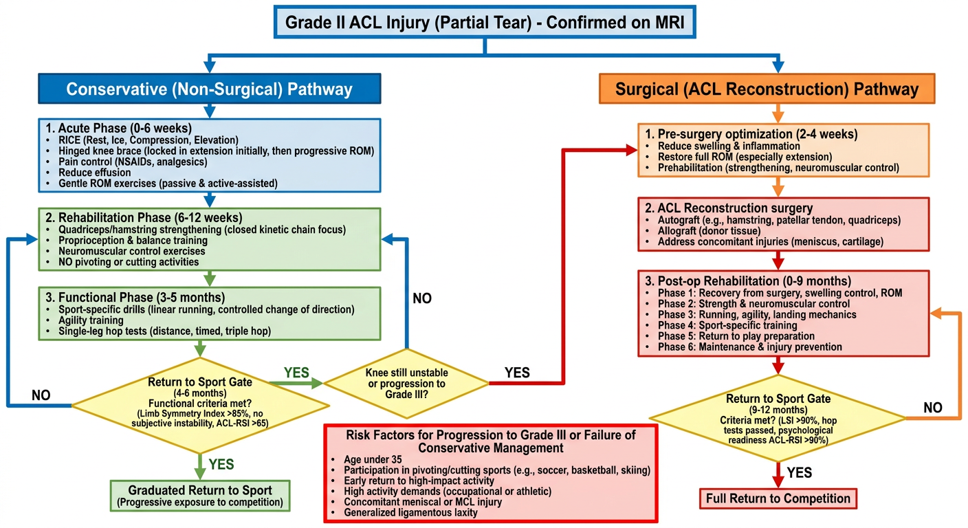
Should Shadman Return to Football (Goalkeeping) and Badminton?
Where He Is Right Now (Day 20)
- Active joint effusion (fluid in the knee)
- Bone marrow edema in both tibial and lateral femoral condyles
- Partial ACL fibers under mechanical stress - not yet healed or stabilized
Sport Risk Analysis: Goalkeeping vs Badminton
| Factor | Goalkeeping (Football) | Badminton |
|---|---|---|
| ACL demand | Moderate - less running, but explosive dives, jumps, landing, sudden direction changes | HIGH - rapid lateral lunges, split steps, explosive rotational jumps, sudden deceleration |
| Pivot/cutting load | Moderate | Very high |
| Knee valgus stress | Moderate (diving saves) | High (lunge shots, smash landings) |
| Contact risk | Yes (collisions, aerial duels) | None |
| Sport classification | Moderate-high demand | High demand / "pivot sport" |
| Conservative ACL evidence | Very limited (Krutsch et al., 2025 - KSTA) | No specific evidence |
The Evidence-Based Verdict
"Non-surgical treatment of ACL ruptures in football is generally restricted to indications such as minimal partial ACL ruptures... players who intend to retire." "Rehabilitation should include gradual build-up and sport-specific tests of neuromotor skills and leg axis stability before returning to the field."
- 47.3% of partial ACL tears progressed to complete rupture when managed conservatively
- Age under 35 and early return to activity are the two biggest independent risk factors for progression
- Shadman hits both - he is 21, and likely eager to return
Conservative rehab can match surgery for return-to-sport rates - but only when rehabilitation is completed properly with functional milestone criteria met, not based on calendar time.
The Functional Criteria Shadman Must Meet Before Returning to Sport
- Full pain-free range of motion (0-135°)
- No joint effusion (swelling resolved)
- Quadriceps strength ≥70% of the uninjured side (by dynamometry or single-leg squat test)
- Single-leg balance ≥30 seconds without compensation
- Single-leg hop test ≥85% of contralateral side
- Triple hop test ≥85%
- 10 controlled drop jumps with stable landing mechanics
- No pain or instability during straight-line running
- Limb Symmetry Index (LSI) >85-90%
- T-test change of direction without compensation
- Ability to decelerate and pivot without instability
- ACL-RSI score ≥65 (psychological readiness scale)
- No effusion, no giving-way episodes, Lachman negative or near-normal
Realistic Timeline for Shadman
| Milestone | Approximate Timeframe |
|---|---|
| Injury | Day 0 (June 10, 2026) |
| Current status | Day 20 (June 30) - acute phase |
| Resolve effusion, regain ROM | Weeks 4-6 (~late July) |
| Begin strengthening rehab | Weeks 4-8 |
| Begin jogging/light football | Month 3-4 (September) |
| Goalkeeping training (no dives/aerial) | Month 4-5 (October) |
| Conservative return to goalkeeping | Month 4-6 (Oct-Dec 2026) - if criteria met |
| Return to badminton | Month 5-7 (Nov-Jan 2027) - later than football due to higher ACL demand |
| Full competition | Month 6+ with functional criteria fully passed |
What Shadman Should Do Right Now
-
See Dr. Md. Kamrul Alam (his referring orthopedic surgeon) for a clinical exam - Lachman, pivot shift, anterior drawer tests will tell whether the partial ACL is functionally stable. If pivot shift is positive, the knee is already functionally unstable and the conservative path becomes riskier.
-
Start structured physiotherapy immediately - not rest, but progressive guided rehab. The longer he waits without guided rehab, the more quadriceps atrophy and proprioception loss occurs.
-
Wear a hinged knee brace during all activity for at least 6-8 weeks.
-
Do not self-test return to sport - no "let me just try a few kicks" - one uncontrolled landing or cutting move at this stage could convert the Grade II into a Grade III tear.
-
The 25% progression-to-insufficiency data (Hannon et al. 2026) and 47% progression data (Rai et al. 2023) are real risks for him given his age and sports. If his knee gives way even once during rehab, surgery should be reconsidered.
Bottom Line
Goalkeeping: Possible conservatively, but not before 4-6 months and only after passing all functional criteria. Goalkeeping is moderate-demand for the ACL compared to outfield football, but dives and aerial challenges must be reintroduced very gradually last.
Badminton: Higher risk than goalkeeping. The explosive lateral lunge is one of the most ACL-loading movements in sport. Return should be later (month 5-7), and only after the knee has demonstrated full stability in football first. If he plays badminton competitively (smashes, net play, cross-court lunges), this is the sport most likely to rupture the remaining fibers if returned to too soon.
Surgery should stay on the table as an option - his decision is respected, but he and his family should know that if he fails conservative rehab or the knee gives way during return-to-sport, reconstruction at that point carries the same surgical risk but with a less favorable outcome due to secondary cartilage and meniscus damage that accumulates with instability episodes.
It have been 32 days since the injury. Now I am recovering very well. I can walk almost properly. I am doing cycling to go to my class and doing daily life activities while talking medicines prescribed by doctor. My question is what types of exercise should I start doing? For gaining full bending, should I push hard? Tell me all the things I should do and not to do.
"anterior cruciate ligament" AND rehabilitation AND "range of motion" AND "knee flexion" AND exercises
partial ACL tear conservative rehabilitation week 4-8 exercises knee flexion ROM should you force bending 2024 2025
https://www.laurenredlermd.com/pdf/partial-acl-non-op.pdf
Your Grade II ACL Rehab Guide - Day 32 (Week 5)
The Big Question: Should You PUSH Hard to Get Full Bending?
- Your partial ACL fibers are still healing - the ligament heals over 6-12 weeks from injury. You are at week 5.
- Your lateral meniscus (Grade I), bone contusions, and joint capsule are also still recovering.
- Forceful bending creates excessive intra-articular pressure and can stretch or re-rupture the healing ACL fibers.
- Bending that causes sharp pain or makes the knee swell up = a warning signal, not something to push through.
"Working on knee flexion might be uncomfortable, but it should never be unbearable. This is not something you have to force." - E3 Rehab ACL Protocol
Your Current Phase: Week 4-8 (Strengthening Phase)
DAILY (Every Day)
1. Heel Slides - for knee bending ROM
- Sets/Reps: 2 sets x 20 reps, 1-2 times per day
- Tip: You can loop a towel or belt around your ankle and gently pull to assist the last few degrees of bend. Never force it to pain.
2. Quad Sets (Quadriceps Activation) - most important exercise right now
- Sets/Reps: 3 sets x 20 reps, several times per day (you can do these while watching TV, studying)
- Why: The quad is the primary protector of the ACL. Every day without activating it causes atrophy and instability.
3. Cycling (You are already doing this!)
- Adjust your seat so the knee does not fully straighten OR deeply bend - mid-range cycling is perfect.
- Gradually lower the seat height over weeks to increase the bend, as tolerated.
- Keep resistance low for now. Focus on smooth rotation, not speed or power.
- Duration: 15-20 min/day is sufficient at this stage.
3-5 TIMES PER WEEK (Strengthening Program)
4. Straight Leg Raises (SLR)
- Sets/Reps: 3 sets x 15-20 reps
- Progress: Add a light ankle weight (0.5-1 kg) when 20 reps feel easy.
5. Hip Abduction & Adduction
- Sets/Reps: 3 sets x 15 reps each direction
- Why: Hip strength controls knee alignment and reduces ACL stress significantly.
6. Mini Wall Squats (30-45°)
- Sets/Reps: 3 sets x 10 reps
- Important: Do NOT go past 60° for now. Keep knees in line with toes - do not let knees cave inward.
7. Step-Ups (Forward)
- Sets/Reps: 3 sets x 10-15 reps
- Progress: Increase step height to 20 cm in week 6-7.
8. Hamstring Curls (Standing)
- Sets/Reps: 3 sets x 15 reps
- Progress: Add ankle weight (0.5-1 kg) when easy.
9. Heel Raises (Calf)
- Sets/Reps: 3 sets x 20 reps
10. Glute Bridges
- Sets/Reps: 3 sets x 15 reps
- Progress: Single-leg bridge when double feels easy.
3 TIMES PER WEEK (Balance & Proprioception - Critical for ACL Healing)
11. Single-Leg Balance
- Progression ladder:
- Week 5-6: Eyes open, flat floor, 30 sec
- Week 6-7: Eyes closed, flat floor, 30 sec
- Week 7-8: Eyes open, on a folded towel/pillow (unstable surface), 30 sec
- Week 8+: Eyes closed, unstable surface
12. Patellar (Kneecap) Mobilization
- Why: Prevents scar tissue from limiting kneecap movement, which affects the last degrees of both bending and straightening.
Full Weekly Schedule (Sample)
| Day | What to Do |
|---|---|
| Monday | Quad sets + Heel slides + Cycling (15 min) + Strength session (exercises 4-10) + Balance |
| Tuesday | Quad sets + Heel slides + Cycling + Rest from strength |
| Wednesday | Quad sets + Heel slides + Strength session + Balance |
| Thursday | Quad sets + Heel slides + Cycling (slightly longer) + Rest |
| Friday | Quad sets + Heel slides + Strength session + Balance |
| Saturday | Quad sets + Heel slides + Cycling + Light walking |
| Sunday | Rest or very gentle cycling only |
THINGS TO DO ✅
| ✅ | Continue cycling - it is ideal rehab at this stage |
| ✅ | Wear your hinged brace during all walks outside and any uneven surface |
| ✅ | Ice the knee for 15 min after every exercise session if it feels warm or achy |
| ✅ | Elevate the leg when resting if any swelling appears |
| ✅ | Walk on flat, even surfaces - stairs are fine going up (injured leg leads going up, good leg leads going down) |
| ✅ | Gentle stretching: calf, hamstring, quad stretch while lying prone (face down) - hold 30 seconds each, 3x daily |
| ✅ | Take your prescribed medicines as directed (likely NSAIDs for inflammation - do not stop early) |
| ✅ | See a physiotherapist in person if at all possible - a hands-on assessment of your current range of motion will guide this program more precisely |
THINGS NOT TO DO ❌
| ❌ | Do not force bending past pain - ever |
| ❌ | Do not squat past 60-70° (deep squats, Asian squat position) for now |
| ❌ | Do not run, jog, or sprint - zero running until at least month 3 and functional criteria are met |
| ❌ | Do not jump or hop on the injured leg |
| ❌ | Do not pivot, twist, or cut - no sudden change of direction on the knee |
| ❌ | Do not sit cross-legged (floor sitting with knee rotated out and bent) |
| ❌ | Do not kneel directly on the knee for prolonged periods |
| ❌ | Do not play any sport - football, badminton, or any recreational sport |
| ❌ | Do not ignore swelling - if the knee swells significantly after any exercise, you have done too much. Rest 1-2 days and restart at a lower intensity |
| ❌ | Do not stop the brace prematurely - continue wearing it during outdoor activities until your physio or surgeon clears you |
| ❌ | Do not cycle on hilly terrain or rough roads - flat cycling only; standing up on the pedals puts rotational stress on the knee |
Warning Signs - Stop Exercise and See Your Doctor If:
- Knee gives way (buckling) during walking or any exercise
- Sudden increase in swelling (significantly more than baseline)
- Sharp locking pain (knee gets stuck and won't move)
- New clicking or popping sensation with pain
- Numbness or tingling in the leg
What Comes Next (Week 8-12 Preview)
- Light leg press machine (double then single leg)
- Increased squat depth (up to 90°)
- Begin straight-line jogging on flat ground
- More advanced proprioception (perturbation training, balance board)
Its 7th weak now. Im improving good. But the problem is I didn’t do the Exercises for knee extension range of motion good. As a result, the knee extension range of motion is weak. I am trying to do it now but knee hurts. I have recovered my quads well. Everything is fine now. But the improvement has stopped a little and in some cases i am facing new pains. What should i do
knee extension deficit week 6-8 ACL conservative rehab arthrofibrosis extension lag treatment exercises 2024
"knee extension" AND deficit AND "anterior cruciate ligament" AND rehabilitation AND arthrofibrosis
https://e3rehab.com/acl-rehab-phase-1-0-to-8-weeks
Week 7 - Extension Deficit + New Pains: What Is Happening & What To Do
First - Understand What Is Going On
- Walking with a bent knee puts 3x more stress on the kneecap joint (patellofemoral pain = one of your "new pains")
- It forces the hamstrings and calf to work overtime - causing tightness and aching behind the knee
- It is the number one risk factor for arthrofibrosis (permanent scar tissue stiffness) if not corrected now
- Extension deficit at week 7 in a non-operative ACL is a strong reason to see a physiotherapist in person - this needs manual work
The "New Pains" - What They Likely Are
| Pain Location | Likely Cause |
|---|---|
| Front of knee / kneecap | Patellofemoral stress from walking with bent knee |
| Behind the knee (popliteal) | Tight hamstrings, posterior capsule tightness |
| Inner or outer knee | Compensatory overload from abnormal gait |
| General aching after exercise | Pushing too hard on extension stretches, causing inflammatory flare |
What NOT To Do (Critical)
The Extension Recovery Plan - Start Today
Exercise 1: Heel Prop (Passive Extension) - THE MOST IMPORTANT ONE
- Lie flat on your back on a firm surface (floor is better than a soft bed)
- Place a rolled towel, a water bottle, or a firm pillow under your heel - NOT under your knee
- Let gravity slowly pull the back of the knee downward toward the floor
- Stay in this position for 10-15 minutes continuously
- Do this 4-6 times per day - aim for at least 1 hour of total time daily
Exercise 2: Quad Sets Combined With Heel Prop
- Reps: 3 sets x 10 reps, during each heel prop session
- Why: The quad is the active extensor. With good quads (which you have), activating them during the stretch accelerates extension recovery.
Exercise 3: Prone (Face-Down) Hang
- Duration: 10-15 minutes, 2-3 times per day
- Add: Place a light weight around the ankle after 1 week if gravity alone is not enough.
Exercise 4: Standing Terminal Knee Extension (TKE) with Resistance Band
- Tie a resistance band (or use a folded belt/cloth) around a fixed object at knee height
- Place the band behind your knee, step forward slightly so the band pulls the knee forward
- Start with your knee slightly bent (~20°)
- Squeeze your quad and straighten the knee fully - press through that last bit of extension
- Hold fully straight 2 seconds, slowly allow it to bend back
- Sets/Reps: 3 sets x 15 reps
- Why: This directly trains terminal knee extension - the range you are missing - under gentle resistance. It is one of the most targeted exercises for extension deficit.
Exercise 5: Backward Walking
- Sessions: 3-4 times per day, even just in your room
- Why: Backward walking forces the knee to go into terminal extension with every step. It is used specifically in ACL rehab to reinforce full straightening in a weight-bearing, functional way. It also retrains normal gait mechanics.
Exercise 6: Patellar (Kneecap) Mobilization - Do Not Skip
- Sit with leg straight and relaxed (quad not contracted)
- Use your fingers and thumbs to push the kneecap slowly: up toward thigh, down toward shin, left, right
- Hold each direction 5-10 seconds, gently
- Sets: 2 sets x 20 gentle pushes in each direction
- Frequency: Daily, ideally before your extension exercises
Managing the New Pains
- Apply a warm compress (warm damp towel, 5-10 minutes) BEFORE extension stretching to loosen the soft tissue
- Apply ice (15 min) AFTER exercise if the knee feels warm, achy, or swollen afterward
- Do NOT ice before - you want the tissue warm and pliable for stretching, cold and numb for recovery after
- Patellar mobilization (Exercise 6) directly addresses this
- Avoid prolonged sitting with the knee bent >90° (e.g. cross-legged, sitting on heels)
- When sitting on a chair, occasionally straighten the leg fully and hold for a few seconds
- Gentle calf stretch: stand, place injured foot slightly behind you, heel flat, lean forward gently - hold 30 seconds x 3 repetitions
- Gentle hamstring stretch: lie on back, bend hip to 90°, then slowly straighten the knee as far as comfortable - hold 20 seconds
When to Stop and See a Doctor URGENTLY
- The knee cannot straighten within 10° of the other leg (e.g. other leg is 0°, injured leg is stuck at 15° or more)
- Extension has been getting worse, not better over the past 2 weeks
- You have swelling that is worsening, not stable
- Locking - the knee gets stuck and physically cannot move past a point (this may indicate the Grade I meniscal injury has progressed)
- Pain is becoming more severe rather than improving
Your Revised Daily Priority Order (Week 7 Onwards)
| Priority | Task | Frequency |
|---|---|---|
| 🔴 1st | Heel prop (passive extension) | 4-6x/day, 10-15 min each |
| 🔴 2nd | Quad sets during heel prop | Every heel prop session |
| 🔴 3rd | Patellar mobilization | Daily |
| 🟡 4th | Prone hang | 2-3x/day |
| 🟡 5th | Terminal knee extension with band | 3x/week |
| 🟡 6th | Backward walking | 3-4x/day, 1-2 minutes |
| 🟢 7th | Continue quad strengthening | 3x/week |
| 🟢 8th | Cycling (keep doing it) | Daily |
| 🟢 9th | Single-leg balance | 3x/week |
Its hurt while doing heel prop. The pain is like where the articular cartilage or lateral meniscus is situated. On the lateral side of kneecap. This area also hurts sometime when walking or other exercises
lateral knee pain ACL rehab week 7 lateral meniscus bone contusion heel prop extension hurt lateral patellofemoral
lateral knee anatomy meniscus iliotibial band patellofemoral diagram
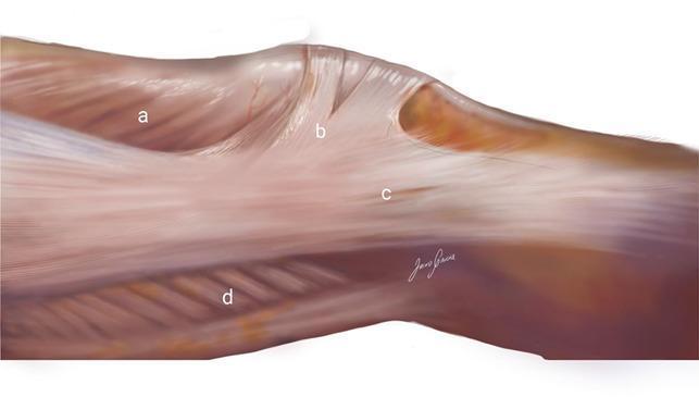
Anatomical illustration of the lateral aspect of the human knee, specifically depicting the superficial fascia and musculature. The diagram uses alphabetical labels to identify key structures: (a) refers to the vastus lateralis muscle, showing its characteristic pennate fiber arrangement; (b) indicates the superficial oblique retinaculum, also known as arciform fibers, which appear as distinct, vertically oriented translucent bands crossing toward the lateral patella; (c) denotes the superficial layer of the iliotibial band (ITB), a dense, longitudinally oriented fibrous sheet that provides lateral stability and inserts onto Gerdy’s tubercle; and (d) identifies the biceps femoris muscle on the posterior-lateral border. The illustration highlights the complex integration between the ITB, the quadriceps extensor mechanism, and the lateral patellar retinaculum, emphasizing the functional anatomy of the lateral patellofemoral complex relevant for orthopedics and sports medicine education.
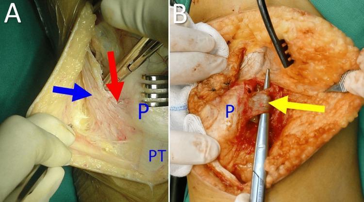
This surgical clinical photograph, divided into two panels (A and B), demonstrates the anatomical structures of the lateral patellar retinaculum during a knee procedure. In Panel A, a blue arrow identifies the iliotibial tract (ITB), shown as a broad fibrous band. A red arrow indicates the iliopatellar band (IPB), which branches from the ITB and inserts obliquely onto the lateral aspect of the patella (labeled 'P'). The patellar tendon ('PT') is visible inferiorly. Panel B shows a deeper dissection where a yellow arrow points to the lateral patellofemoral ligament (LPFL), isolated by a surgical instrument. The LPFL is characterized as a distinct capsular thickening located deep to the IPB, originating near the lateral femoral epicondyle and inserting on the middle third of the lateral patella. These images are intended for educational instruction on patellofemoral joint stability and the surgical anatomy relevant to treating patellar dislocation or instability.
⚠️ This Pain Is a Warning Sign - Please Read This Carefully
Where Exactly Is the Pain? (This Matters a Lot)
| Structure | Where exactly | Why it would hurt now |
|---|---|---|
| Lateral joint line / lateral meniscus | Below and beside the kneecap, in the groove between femur and tibia | Your MRI showed Grade I lateral meniscus injury - it may be progressing or being compressed during extension |
| Iliotibial band (ITB) / lateral retinaculum | Running along the outer side of the kneecap down to the outer shinbone | Very commonly irritated in ACL rehab - tight from disuse and compensatory gait |
| Lateral patellofemoral joint | Where the outer edge of the kneecap articulates with the femur | Your MRI showed articular cartilage changes + bone contusions here - the kneecap may be tracking incorrectly |
Why Heel Prop Specifically Hurts Here
- The kneecap is pulled laterally by the iliotibial band as the knee approaches full extension - this is normal anatomy, but in a stiff post-injury knee it causes excessive lateral patellar compression
- The lateral compartment (where your lateral meniscus and bone contusions are) gets compressed as the joint surfaces come together at full extension
- If there is still residual effusion (fluid) in the joint, that fluid gets pressurized at end-range extension and causes lateral compartment pain
The Most Concerning Possibility: Lateral Meniscus Progression
- Pain specifically at the lateral joint line (below and beside the outer kneecap)
- Pain with walking, especially going down stairs or slopes
- Pain with extension - particularly the last few degrees
- Clicking, popping, or a "catching" sensation on the lateral side
- Intermittent swelling after activity
What You Must Do Right Now
🔴 STOP these immediately:
- Stop heel prop - it is specifically provoking the lateral compartment. Do not do it until the pain source is identified.
- Stop any exercise that reproduces this lateral pain - if it hurts, it means compression or tension is being applied to a structure that is not ready. Continuing through that pain is not safe with your injury profile.
- Stop any deep squats or step-downs for now.
🟡 MODIFY your cycling:
- Keep cycling ONLY if it is pain-free. If you feel the lateral pain while pedalling, stop.
- Make sure your seat is high enough so the knee does not bend past 70-80° at the bottom of each stroke.
- No resistance/hills - flat, easy spinning only.
🟢 SAFE things you can still do:
- Quad sets (lying flat, tightening the front thigh) - as long as these do not cause the lateral pain
- Straight leg raises - no knee bend, so usually safe
- Ankle pumps (moving foot up/down) - keeps circulation going
- Gentle patellar mobilization - pushing kneecap gently in all directions while the leg is relaxed and flat (this actually helps lateral patellar compression)
- Ice the lateral side for 15 minutes after any activity, 2-3x/day
- Elevate the leg when resting
Alternative Extension Exercise (Safer Than Heel Prop Right Now)
- Sit on the floor with your back straight against a wall and both legs out in front
- Do NOT prop the heel - instead, just let the leg rest flat on the floor
- Gently tighten the quad and try to press the back of the knee toward the floor
- Hold 10 seconds, relax
- The floor provides cushioning and there is no gravity-assisted compression on the lateral compartment
The Most Important Message
- You have a known lateral meniscus injury from your MRI that can progress
- You have lateral bone contusions that are still healing
- You now have new lateral pain that is reproducible with specific movements
- Your progress has plateaued and you are getting new symptoms, not fewer
- Palpate the exact location of the pain (joint line? retinaculum? patellar edge?)
- Do a McMurray test to assess the meniscus
- Check your patellar tracking
- Decide whether a repeat MRI is needed
- Clear you for which exercises are safe to continue
My MRI says that the injury is in posterior horn of lateral meniscus. So the pain should be in the back/outer side of the knee. But my pain is in the front side. It seems like on the area where the anterior horn is located. Not far from kneecap. And the pain Doesn't feel Everytime. Also it's not unbearable. Another fact is, this pain was not happening before 6th week. I am very confused.
anterior lateral knee pain new onset week 6-7 ACL rehab fat pad impingement lateral retinaculum ITB anterior horn meniscus
Your Confusion Makes Complete Sense - Here Is the Answer
Anatomy Quick Reference
         KNEECAP (Patella)
              |
    [Lateral  |  Medial]
    side      |  side
              |
        Patellar Tendon
              |
    ← Fat Pad sits here (under tendon, beside it)
    ← Anterior horn of lateral meniscus: outer side,
      just below the joint line, near the patellar tendon
    ← ITB / lateral retinaculum: running along the
      outer edge of the kneecap downward
The Three Most Likely Causes (In Order of Probability)
1. 🥇 Most Likely: Infrapatellar Fat Pad Irritation (Hoffa's Syndrome)
- During the first 4-5 weeks you were protected and loading little
- As you started doing more exercises, step-ups, cycling with more resistance, and extension work, the fat pad began getting pinched between the kneecap and the femur during terminal extension (the last 10-20° of straightening)
- Heel prop specifically compresses the fat pad as the knee is pushed toward full extension - this perfectly explains why heel prop provokes it
- The fat pad was also likely irritated during the original trauma (your bone contusions were at the lateral femoral condyle - very close to the fat pad attachment)
- Front of knee, on either or both sides of the patellar tendon
- Worse at end-range extension (full straightening)
- Worse going down stairs, after exercise, sometimes after sitting
- Intermittent - not constant
- Starts at week 5-7 when loading increases
- Not unbearable - matches your description exactly
2. 🥈 Second Most Likely: Lateral Plica Irritation
- Increased activity at week 6-7 after a protected phase is a classic trigger
- The original trauma likely irritated the synovial lining, and the plica has thickened during healing
- Cycling is a known irritant of plica (repetitive flexion-extension)
- Lateral side of kneecap, anterolateral
- Intermittent catching, snapping, or aching - sometimes a clicking sensation
- Worse with cycling (repetitive movement) and going up/down stairs
- Comes and goes - not constant
3. 🥉 Third: ITB / Lateral Retinaculum Tightness
- Your gait has been slightly altered for 7 weeks due to the extension deficit
- The ITB "impingement zone" occurs at exactly 20-30° of knee flexion - right where you spend a lot of time walking and during exercises
- Cycling can irritate it because of repetitive knee extension
- Lateral side of kneecap and slightly above/along the outer thigh
- Pain with cycling (especially going from bent to straight - the "down-pedal" motion)
- Worse going down stairs
- Sometimes a burning quality
Putting It All Together
| Feature you described | What it suggests |
|---|---|
| Front-lateral, near kneecap | Fat pad, plica, or ITB - NOT posterior meniscus |
| Started at week 6-7, not before | Classic timing for fat pad / plica irritation from increased loading |
| Hurts during heel prop | Fat pad gets pinched at terminal extension - hallmark of fat pad irritation |
| Intermittent, not constant | Fat pad or plica (mechanical, positional pain - not structural damage) |
| Not unbearable | Consistent with irritation, not tear or significant structural damage |
| Also during walking and exercise | ITB and fat pad both fit |
What To Do For This Specifically
For Fat Pad Irritation:
- For heel prop: place a very thin folded towel under the lower thigh (not the knee, not the calf) - this creates a tiny angle that unloads the fat pad from compression at terminal extension. Try this instead of the full gravity hang.
- Stop quad sets if they cause the front pain - they compress the fat pad directly. Do them very gently if at all.
For ITB/Lateral Retinaculum Tightness:
- Lie on your side, place a foam roller or firm cylindrical object under the outer thigh
- Slowly roll from hip to just above the knee
- 60-90 seconds each side, once daily
- Do NOT roll directly over the knee itself
- Stand, cross your injured leg behind your good leg
- Lean sideways away from the injured side, pressing your hip outward
- Hold 30 seconds x 3 repetitions
- Should feel a gentle pull along the outer thigh and hip
Should You Worry?
- Gets worse over the next 1-2 weeks despite modifying exercises
- Starts becoming constant rather than intermittent
- You develop swelling in the front of the knee
- You feel a sharp catch or lock during movement
Will I ever be able to go back to my beloved football pitch again?
"anterior cruciate ligament" AND "partial tear" AND "return to sport" AND football AND conservative
partial ACL tear Grade II conservative treatment return to football goalkeeper prognosis young patient success rate 2024 2025
Will You Get Back to Football? Yes - Here Is the Honest Truth.
The Short Answer
What the Evidence Actually Says
- ~52% return to their previous sport level without surgery (Burnham et al.)
- ~39-47% of partial tears in patients under 30 progress to complete rupture if they return to pivoting sports too early (PMC study, n=young active patients)
- Age ≤20 years + pivoting contact sports are the two biggest independent risk factors for progression - you are 21, right on that boundary
- Players who returned to pivoting sports prematurely had 6.3× higher odds of progressing to complete rupture
Why Goalkeeping Is Actually Your Best Friend Here
| Demand | Outfield Player | Goalkeeper |
|---|---|---|
| Sprinting & cutting | Constant, unpredictable | Rare, short bursts |
| Pivoting/turning under load | Very high | Moderate |
| Total distance per match | 10-12 km | 5-6 km |
| Aerial duels with impact | Frequent | Present but controlled |
| Explosive lateral movement | Frequent | Present (dives) |
| ACL stress pattern | Very high | Moderate |
The Real Timeline for You
| Phase | Target | What Needs to Happen First |
|---|---|---|
| Controlled ball work (stationary) | Month 4-5 (Oct-Nov 2026) | Full extension restored, no swelling, single-leg balance solid |
| Light goalkeeping - footwork drills, no diving | Month 5-6 (Nov-Dec 2026) | Quad symmetry >70%, straight-line jogging pain-free |
| Full goalkeeping training - diving, lateral movement | Month 6-8 (Dec 2026 - Feb 2027) | Hop tests >85%, no instability, physio clearance |
| Return to match goalkeeping | Month 7-9 (Jan-Mar 2027) | All functional criteria met |
The Three Paths From Here
What Stands Between You and the Pitch Right Now
- Extension deficit - your most urgent task right now
- Fat pad irritation - manageable, will settle with load modification
- Bone contusions - still healing, need roughly 3 more months to fully remodel
- Lateral meniscal Grade I - monitoring only, no active problem right now
- Neuromuscular control - your knee's proprioception (position sense) needs retraining, which is the work of months 2-4
One Thing to Hold Onto
Generate a printable home exercise rehabilitation program PDF for weeks 7-12
ls /tmp/workspace/ 2>/dev/null || mkdir -p /tmp/workspace/shadman-rehab && echo "ready"
mkdir -p /tmp/workspace/shadman-rehab && cat > /tmp/workspace/shadman-rehab/generate_pdf.py << 'PYEOF' from reportlab.lib.pagesizes import A4 from reportlab.lib import colors from reportlab.lib.units import mm from reportlab.lib.styles import getSampleStyleSheet, ParagraphStyle from reportlab.platypus import (SimpleDocTemplate, Paragraph, Spacer, Table, TableStyle, HRFlowable, KeepTogether) from reportlab.lib.enums import TA_CENTER, TA_LEFT, TA_JUSTIFY from reportlab.platypus import PageBreak from reportlab.lib.colors import HexColor import datetime # ── Colour palette ────────────────────────────────────────────────────────── C_NAVY = HexColor('#1a3a5c') C_TEAL = HexColor('#0d7377') C_LTEAL = HexColor('#14a085') C_AMBER = HexColor('#e67e22') C_RED = HexColor('#c0392b') C_GREEN = HexColor('#27ae60') C_LGREY = HexColor('#f4f6f8') C_MGREY = HexColor('#dee2e6') C_DGREY = HexColor('#495057') C_WHITE = colors.white C_BLACK = colors.black OUTPUT = '/tmp/workspace/shadman-rehab/Shadman_ACL_Rehab_Weeks7-12.pdf' PAGE_W, PAGE_H = A4 MARGIN = 18 * mm doc = SimpleDocTemplate( OUTPUT, pagesize=A4, leftMargin=MARGIN, rightMargin=MARGIN, topMargin=22*mm, bottomMargin=18*mm, title="Grade II ACL Rehabilitation Program – Weeks 7-12", author="Orris Clinical AI", subject="Home Exercise Rehabilitation Program for Shadman Mahmud" ) styles = getSampleStyleSheet() def S(name, **kw): return ParagraphStyle(name, **kw) sTitle = S('sTitle', fontSize=22, textColor=C_WHITE, spaceAfter=2, fontName='Helvetica-Bold', alignment=TA_CENTER) sSub = S('sSub', fontSize=11, textColor=HexColor('#cce8e8'), fontName='Helvetica', alignment=TA_CENTER, spaceAfter=0) sH1 = S('sH1', fontSize=13, textColor=C_WHITE, fontName='Helvetica-Bold', leading=16) sH2 = S('sH2', fontSize=11, textColor=C_NAVY, fontName='Helvetica-Bold', spaceBefore=6, spaceAfter=3) sH3 = S('sH3', fontSize=10, textColor=C_TEAL, fontName='Helvetica-Bold', spaceBefore=4, spaceAfter=2) sBody = S('sBody', fontSize=9, textColor=C_DGREY, fontName='Helvetica', leading=14, spaceAfter=2, alignment=TA_JUSTIFY) sBullet = S('sBullet', fontSize=9, textColor=C_DGREY, fontName='Helvetica', leading=13, leftIndent=12, spaceAfter=1) sSmall = S('sSmall', fontSize=8, textColor=C_DGREY, fontName='Helvetica-Oblique', leading=11) sWarn = S('sWarn', fontSize=9, textColor=C_WHITE, fontName='Helvetica-Bold', leading=12, alignment=TA_LEFT) sLabel = S('sLabel', fontSize=8, textColor=C_NAVY, fontName='Helvetica-Bold', leading=11) sCell = S('sCell', fontSize=8.5,textColor=C_DGREY, fontName='Helvetica', leading=12) sCellB = S('sCellB', fontSize=8.5,textColor=C_NAVY, fontName='Helvetica-Bold', leading=12) sFooter = S('sFooter', fontSize=7.5,textColor=HexColor('#868e96'), fontName='Helvetica-Oblique', alignment=TA_CENTER) story = [] W = PAGE_W - 2*MARGIN # usable width # ═══════════════════════════════════════════════════════════════════════════ # HEADER BANNER # ═══════════════════════════════════════════════════════════════════════════ banner_data = [[ Paragraph("HOME REHABILITATION PROGRAM", sTitle), Paragraph("Grade II ACL Partial Tear • Conservative Management • Weeks 7–12", sSub), Paragraph("Shadman Mahmud | DOB: 21 yrs | Injury: 10 Jun 2026 | Generated: 07 Aug 2026", sSub), ]] banner = Table(banner_data, colWidths=[W]) banner.setStyle(TableStyle([ ('BACKGROUND', (0,0),(0,-1), C_NAVY), ('ROWPADDING', (0,0),(-1,-1), 10), ('BOX', (0,0),(-1,-1), 0, C_NAVY), ('VALIGN', (0,0),(-1,-1), 'MIDDLE'), ])) story.append(banner) story.append(Spacer(1, 6)) # ═══════════════════════════════════════════════════════════════════════════ # PATIENT INFO ROW # ═══════════════════════════════════════════════════════════════════════════ def info_card(label, value, bg=C_LGREY): return Table( [[Paragraph(label, sSmall)], [Paragraph(value, S('iv', fontSize=9, fontName='Helvetica-Bold', textColor=C_NAVY, leading=11))]], colWidths=[(W-12)/5] ), TableStyle([ ('BACKGROUND',(0,0),(-1,-1), bg), ('ROWPADDING',(0,0),(-1,-1), 5), ('BOX', (0,0),(-1,-1), 0.5, C_MGREY), ('ROUNDEDCORNERS', [3]), ]) cards = [] for lbl, val in [ ("Diagnosis", "Grade II ACL + Grade I Lateral Meniscus"), ("Management", "Conservative (Non-Surgical)"), ("Phase", "Strengthening & Functional"), ("Weeks", "7 – 12 (Aug – Sep 2026)"), ("Goal", "Return to Goalkeeping Training"), ]: t, ts = info_card(lbl, val) t.setStyle(ts) cards.append(t) info_row = Table([cards], colWidths=[(W-12)/5]*5, hAlign='LEFT') info_row.setStyle(TableStyle([ ('VALIGN',(0,0),(-1,-1),'TOP'), ('LEFTPADDING',(0,0),(-1,-1),3), ('RIGHTPADDING',(0,0),(-1,-1),3), ])) story.append(info_row) story.append(Spacer(1, 8)) # ═══════════════════════════════════════════════════════════════════════════ # WARNING BOX # ═══════════════════════════════════════════════════════════════════════════ warn_text = ("⚠ IMPORTANT: You currently have a knee extension deficit and lateral fat pad irritation. " "PRIORITY #1 for Weeks 7–8 is restoring full knee extension before advancing strengthening. " "Stop any exercise that produces lateral front-knee pain and apply ice for 15 min afterward.") warn_tbl = Table([[Paragraph(warn_text, sWarn)]], colWidths=[W]) warn_tbl.setStyle(TableStyle([ ('BACKGROUND',(0,0),(-1,-1), C_AMBER), ('ROWPADDING',(0,0),(-1,-1), 9), ('BOX', (0,0),(-1,-1), 0, C_AMBER), ])) story.append(warn_tbl) story.append(Spacer(1, 8)) # ═══════════════════════════════════════════════════════════════════════════ # PHASE OVERVIEW TABLE # ═══════════════════════════════════════════════════════════════════════════ story.append(Paragraph("PHASE OVERVIEW", sH2)) story.append(HRFlowable(width=W, thickness=1.5, color=C_TEAL, spaceAfter=4)) phase_header = ['Phase', 'Weeks', 'Dates (approx)', 'Main Focus', 'Key Milestone to Advance'] phase_rows = [ ['2B', '7–8', 'Aug 7–21', 'Extension recovery, fat pad management, base strengthening', 'Full knee extension (<5° deficit); lateral pain resolved'], ['3', '9–10', 'Aug 22 – Sep 4', 'Progressive strengthening, begin jogging prep, proprioception', 'Single-leg hop ≥80% symmetry; jog 10 min pain-free'], ['4', '11–12', 'Sep 5–18', 'Functional strength, agility intro, running program', 'Limb Symmetry Index >85%; T-test change of direction'], ] phase_data = [phase_header] + phase_rows phase_col_w = [W*0.07, W*0.07, W*0.13, W*0.44, W*0.29] phase_tbl = Table(phase_data, colWidths=phase_col_w) phase_tbl.setStyle(TableStyle([ ('BACKGROUND', (0,0),(-1,0), C_TEAL), ('TEXTCOLOR', (0,0),(-1,0), C_WHITE), ('FONTNAME', (0,0),(-1,0), 'Helvetica-Bold'), ('FONTSIZE', (0,0),(-1,-1), 8), ('ROWPADDING', (0,0),(-1,-1), 5), ('ROWBACKGROUNDS',(0,1),(-1,-1),[C_WHITE, C_LGREY]), ('GRID', (0,0),(-1,-1), 0.5, C_MGREY), ('VALIGN', (0,0),(-1,-1), 'TOP'), ('TEXTCOLOR', (0,1),(-1,-1), C_DGREY), ('FONTNAME', (0,1),(0,-1), 'Helvetica-Bold'), ('TEXTCOLOR', (0,1),(0,-1), C_NAVY), ])) story.append(phase_tbl) story.append(Spacer(1, 10)) # ═══════════════════════════════════════════════════════════════════════════ # HELPER: exercise table builder # ═══════════════════════════════════════════════════════════════════════════ def ex_table(rows): """rows = list of (Exercise, Sets×Reps, Freq, Notes)""" header = ['Exercise', 'Sets × Reps', 'Frequency', 'Key Notes / Technique'] data = [header] + rows cw = [W*0.28, W*0.13, W*0.13, W*0.46] t = Table(data, colWidths=cw) t.setStyle(TableStyle([ ('BACKGROUND', (0,0),(-1,0), C_NAVY), ('TEXTCOLOR', (0,0),(-1,0), C_WHITE), ('FONTNAME', (0,0),(-1,0), 'Helvetica-Bold'), ('FONTSIZE', (0,0),(-1,-1), 8), ('ROWPADDING', (0,0),(-1,-1), 5), ('ROWBACKGROUNDS',(0,1),(-1,-1),[C_WHITE, C_LGREY]), ('GRID', (0,0),(-1,-1), 0.4, C_MGREY), ('VALIGN', (0,0),(-1,-1), 'TOP'), ('TEXTCOLOR', (0,1),(-1,-1), C_DGREY), ('FONTNAME', (0,1),(0,-1), 'Helvetica-Bold'), ('TEXTCOLOR', (0,1),(0,-1), C_TEAL), ])) return t def section_header(text, color=C_NAVY): bg = Table([[Paragraph(text, sH1)]], colWidths=[W]) bg.setStyle(TableStyle([ ('BACKGROUND',(0,0),(-1,-1), color), ('ROWPADDING',(0,0),(-1,-1), 7), ('BOX', (0,0),(-1,-1), 0, color), ])) return bg # ═══════════════════════════════════════════════════════════════════════════ # PHASE 2B – WEEKS 7–8 # ═══════════════════════════════════════════════════════════════════════════ story.append(section_header("PHASE 2B | WEEKS 7–8 (Aug 7 – Aug 21) — Extension Recovery + Base Strengthening")) story.append(Spacer(1, 5)) story.append(Paragraph("DAILY PRIORITY: KNEE EXTENSION RESTORATION", sH2)) story.append(Paragraph( "Extension deficit is your #1 problem right now. Do these EVERY day, multiple times. " "Target: extension within 5° of the good leg by end of Week 8.", sBody)) ext_rows = [ ["Heel Prop (Modified)\n– thin towel under lower thigh", "4–6 sessions\n10–15 min each", "Daily\n(aim 1 hr total)", "Lie flat on FIRM floor. Heel on rolled towel. Thin towel under lower thigh to reduce fat pad compression. Let gravity gently pull knee straight. Mild ache = OK. Sharp lateral pain = STOP."], ["Quad Sets during Heel Prop", "3 × 10\n(10-sec holds)", "Every heel\nprop session", "While in heel prop position: tighten front thigh (quad), press back of knee toward floor. Hold 10 sec. Relaxes. This trains active extension."], ["Prone Hang\n– face down off bed edge", "2–3 sessions\n10–15 min each", "Daily", "Lie face-down, lower legs hanging off bed from knee down. Let foot weight extend knee passively. Add 0.5 kg ankle weight after 5 days if tolerated."], ["Terminal Knee Extension\n– resistance band", "3 × 15 reps", "Daily", "Band behind knee at knee height. Step forward, band pulls knee. Start slightly bent ~20°. Squeeze quad, push to FULL straight. Hold 2 sec. Slowly return. Works the last 15–20° of extension specifically."], ["Backward Walking", "3–4 × 1 min", "Daily\n(in room)", "Walk backward on flat surface. Forces terminal extension with every step. Retrains normal gait. Safe for lateral knee."], ["Patellar Mobilisation", "2 × 20 reps\neach direction", "Daily\nbefore exercises", "Leg flat & relaxed. Push kneecap gently: up, down, left, right. Hold 5–10 sec each. Do NOT contract quad during this. Prevents scar adhesions limiting both extension and flexion."], ] story.append(ex_table(ext_rows)) story.append(Spacer(1, 8)) story.append(Paragraph("STRENGTHENING (3–5 × per week)", sH2)) strength_w78_rows = [ ["Quad Sets (seated/lying)", "3 × 20", "3–5×/week", "Seated or lying, straighten leg fully, tighten quad for 10 sec. STOP if lateral front knee pain."], ["Straight Leg Raises (SLR)", "3 × 20", "3–5×/week", "Lying flat. Good leg bent. Tighten quad on straight leg, lift to 45°. Hold 2 sec. Lower slowly. Add 0.5 kg ankle weight when easy."], ["Hip 4-Way (Abduction / Adduction / Extension / Flexion)", "3 × 15 each", "3×/week", "Lying on side or face-down. Slow controlled movement. Hip strength controls knee alignment and ACL stress."], ["Glute Bridge (Double Leg)", "3 × 15\n(5-sec hold)", "3×/week", "Lie on back, feet flat, knees bent. Push hips up to straight line. Squeeze glutes at top. Progress to single-leg bridge from Week 9."], ["Wall Squat (30–45°)", "3 × 10\n(10-sec hold)", "3×/week", "Back against wall, feet 40 cm out. Slide down to 30–45° ONLY. Knees track over toes. No knee cave inward. Do NOT go past 60° yet."], ["Step-Ups Forward (15 cm step)", "3 × 12", "3×/week", "Injured leg leads stepping up. Control the step-DOWN slowly. Increase step height to 20 cm in Week 8 if pain-free."], ["Hamstring Curls (standing)", "3 × 15", "3×/week", "Hold chair for balance. Bend injured knee toward buttock slowly. Slow lowering is more important than lifting. Add ankle weight when easy."], ["Heel Raises (double then single leg)", "3 × 20", "3×/week", "Rise onto toes, hold 2 sec, lower SLOWLY. Progress to single-leg when double is easy. Calf strength critical for landing mechanics later."], ] story.append(ex_table(strength_w78_rows)) story.append(Spacer(1, 8)) story.append(Paragraph("CARDIO & PROPRIOCEPTION (3 × per week)", sH2)) cardio_w78_rows = [ ["Cycling (stationary / flat road)", "20–25 min", "Daily", "Keep seat HIGH so knee stays 70–80° max bend. ZERO resistance/hills. Stop if lateral front-knee pain during pedalling."], ["Single-Leg Balance (flat floor)", "3 × 30–45 sec", "3×/week", "Week 7: eyes open. Week 8: progress to eyes closed. Stand on injured leg, hold without touching anything. Good leg slightly lifted."], ["Weight Shifts (side to side)", "3 × 30 sec", "3×/week", "Stand feet shoulder-width. Slowly shift all weight onto injured leg for 3 sec, back to both. Prepares for single-leg loading."], ] story.append(ex_table(cardio_w78_rows)) story.append(Spacer(1, 8)) # Fat pad box fat_warn = Table([[Paragraph( "FAT PAD IRRITATION MANAGEMENT (Weeks 7–8) | " "Apply WARM compress 5–10 min BEFORE extension stretching. " "Apply ICE 15 min AFTER any exercise that provokes lateral front-knee pain. " "Do patellar mobilisation DAILY. " "Avoid sitting with knee bent >90° for prolonged periods. " "Gentle calf stretch 3 × 30 sec daily.", sWarn)]], colWidths=[W]) fat_warn.setStyle(TableStyle([ ('BACKGROUND',(0,0),(-1,-1), C_LTEAL), ('ROWPADDING',(0,0),(-1,-1), 8), ])) story.append(fat_warn) story.append(Spacer(1, 6)) # Milestone gate W8 gate_w8 = Table([[Paragraph( "✅ END OF WEEK 8 MILESTONE GATE — Do NOT advance to Phase 3 until ALL are met:\n" "□ Knee extension within 5° of uninjured side □ No lateral front-knee pain at rest or walking " "□ Single-leg balance 45 sec eyes open □ Step-ups 20 cm pain-free □ No swelling after exercise", sWarn)]], colWidths=[W]) gate_w8.setStyle(TableStyle([ ('BACKGROUND',(0,0),(-1,-1), C_GREEN), ('ROWPADDING',(0,0),(-1,-1), 8), ])) story.append(gate_w8) # ═══════════════════════════════════════════════════════════════════════════ # PAGE BREAK # ═══════════════════════════════════════════════════════════════════════════ story.append(PageBreak()) # ═══════════════════════════════════════════════════════════════════════════ # PHASE 3 – WEEKS 9–10 # ═══════════════════════════════════════════════════════════════════════════ story.append(section_header("PHASE 3 | WEEKS 9–10 (Aug 22 – Sep 4) — Progressive Strengthening + Jogging Prep", C_TEAL)) story.append(Spacer(1, 5)) story.append(Paragraph( "Extension should now be near-full. Strengthening intensifies. Jogging begins ONLY when functional criteria are met " "(see gate). Proprioception training advances to dynamic balance.", sBody)) story.append(Spacer(1, 5)) story.append(Paragraph("STRENGTHENING (3–4 × per week)", sH2)) strength_w910_rows = [ ["Leg Press (double limb)", "3 × 15\nlow-moderate load", "3×/week", "Feet shoulder-width on platform. Press through 0–90° range. Do NOT lock knee at full extension forcefully. Increase load weekly."], ["Leg Press (single limb) – progress from double", "3 × 12\nmoderate load", "3×/week", "Begin week 10. Injured leg only. Same 0–90° range. Reveals side-to-side strength imbalance."], ["Step-Ups (20–25 cm step)", "3 × 12–15", "3×/week", "Increase step height from Week 8. Slow controlled descent is the key. Add light hand weight (1 kg) when 15 reps easy."], ["Single-Leg Deadlift (bodyweight)", "3 × 10 each leg", "3×/week", "Stand on injured leg, hinge forward at hip while non-standing leg extends behind. Back flat. Feel the hamstring/glute load. Excellent for proprioception + posterior chain."], ["Squat Progression to 70–80°", "3 × 12", "3×/week", "Increase depth from 45° to 70–80°. Heels on ground. Knees track toes. No pain. Full depth (90°+) is Phase 4."], ["Side-Step with Band (lateral monster walk)", "3 × 15 each direction", "3×/week", "Resistance band around ankles. Step sideways in squat position. Controlled. Activates hip abductors critical for ACL protection."], ["Single-Leg Bridge", "3 × 15\n(5-sec hold)", "3×/week", "Progress from double-leg bridge. Unsupported leg straight. Push hips high. Hold. Lower slowly. Glute + hamstring."], ["Nordic Hamstring Curl (assisted)", "2 × 8–10", "2×/week", "Kneel on mat, feet anchored under sofa/bed. Slowly lower body forward (resist with hamstrings). Assist return with hands. Strongest hamstring exercise for ACL rehab."], ] story.append(ex_table(strength_w910_rows)) story.append(Spacer(1, 8)) story.append(Paragraph("CARDIO & PROPRIOCEPTION (3–5 × per week)", sH2)) cardio_w910_rows = [ ["Cycling (progressive resistance)", "25–30 min", "Daily", "Begin lowering seat slightly to increase knee bend. Add light resistance. Stop if any knee pain."], ["Single-Leg Balance on Unstable Surface", "3 × 30–45 sec", "3×/week", "Week 9: eyes open on folded towel. Week 10: eyes closed on folded towel. Progress to foam pad."], ["Perturbation Training", "3 × 30 sec", "3×/week", "Stand on injured leg. Have someone gently push shoulders in random directions. Resist without losing balance. Retrains ligament-independent knee stability."], ["Straight-Line Jogging (begin Week 10 if gate met)", "10–15 min\n(walk-jog)", "3×/week", "Start with walk 3 min / jog 1 min × 5 cycles on FLAT surface. No turns. No inclines. Stop immediately if any instability or pain."], ] story.append(ex_table(cardio_w910_rows)) story.append(Spacer(1, 8)) story.append(Paragraph("STRETCHING (Daily, after warm-up)", sH2)) stretch_rows = [ ["Hamstring Stretch", "3 × 30 sec", "Daily", "Lying on back. Bend hip to 90°, slowly straighten knee toward ceiling as far as comfortable. Hold at end range."], ["Calf / Gastroc Stretch", "3 × 30 sec", "Daily", "Stand at wall. Injured foot back, heel flat, lean forward. Then slightly bend the back knee for soleus stretch."], ["Quad Stretch (standing or side-lying)", "3 × 30 sec", "Daily", "Stand, bend injured knee behind. Hold ankle. Gentle pull only. Hips level throughout."], ["ITB / Hip Stretch (lateral lean)", "3 × 30 sec", "Daily", "Stand, cross injured leg behind good leg. Lean sideways away from injured side. Feel outer thigh pull. Reduces lateral patellar tension."], ["Foam Roll – Outer Thigh (ITB)", "60–90 sec", "Daily", "Lie on side. Roller under outer thigh (hip to just ABOVE knee). Slow rolling. Do NOT roll over the knee itself."], ] story.append(ex_table(stretch_rows)) story.append(Spacer(1, 6)) gate_w10 = Table([[Paragraph( "✅ END OF WEEK 10 MILESTONE GATE — Do NOT advance to Phase 4 until ALL are met:\n" "□ Full knee extension equal to uninjured side □ Single-leg hop ≥80% of uninjured side (distance) " "□ Jog 10 min continuously pain-free □ Single-leg squat × 10 controlled, no knee cave □ No swelling after jogging", sWarn)]], colWidths=[W]) gate_w10.setStyle(TableStyle([ ('BACKGROUND',(0,0),(-1,-1), C_GREEN), ('ROWPADDING',(0,0),(-1,-1), 8), ])) story.append(gate_w10) # ═══════════════════════════════════════════════════════════════════════════ # PAGE BREAK # ═══════════════════════════════════════════════════════════════════════════ story.append(PageBreak()) # ═══════════════════════════════════════════════════════════════════════════ # PHASE 4 – WEEKS 11–12 # ═══════════════════════════════════════════════════════════════════════════ story.append(section_header("PHASE 4 | WEEKS 11–12 (Sep 5 – Sep 18) — Functional Strength + Agility Introduction", C_AMBER)) story.append(Spacer(1, 5)) story.append(Paragraph( "This is the bridge between rehabilitation and sport-specific training. " "Strength work intensifies, agility is introduced cautiously, and running volume increases. " "The knee should feel stable in all daily activities by this point.", sBody)) story.append(Spacer(1, 5)) story.append(Paragraph("STRENGTHENING (3–4 × per week)", sH2)) strength_w1112_rows = [ ["Single-Leg Leg Press (increased load)", "4 × 10\nhigh load", "3×/week", "Target: equal load to uninjured leg within this phase. This is your primary quadriceps strength measure."], ["Bulgarian Split Squat", "3 × 10 each leg", "3×/week", "Rear foot elevated on chair ~40 cm. Front foot (injured) forward. Lower until front knee ~80–90°. Knee tracks over toes. Excellent unilateral knee strength builder."], ["Step-Down (eccentric focus, 25–30 cm)", "3 × 10", "3×/week", "Stand on injured leg on step edge. Slowly lower good foot toward floor (3–4 sec). Touch floor, return. Slow eccentric quad control = ACL's best protection."], ["Lateral Step-Up (side approach)", "3 × 12", "3×/week", "Stand beside 20 cm step. Step up sideways with injured leg. Controlled step down. Loads hip abductors + quad simultaneously."], ["Nordic Hamstring Curl (progressing)", "3 × 8", "2×/week", "Same as Phase 3 but with less arm assistance. Target: lower body unsupported for first 30–40° of descent by Week 12."], ["Single-Leg Squat (pistol prep, to box)", "3 × 8–10", "3×/week", "Injured leg only. Lower onto a chair/box (45–50 cm height). Control descent. Stand back up single-leg. Increase depth progressively."], ["Plyometric – Double-Leg Jump and Land", "3 × 10", "2×/week", "Two-foot jump forward (50 cm). Land SOFTLY – bend knees and hips on landing. Hold landing position 2 sec. No valgus (knees caving in). Introduce Week 11 only."], ["Plyometric – Double-Leg Lateral Jump", "3 × 10", "2×/week", "Jump sideways over a line. Land both feet, hold 2 sec. Controlled. Begin Week 12. Progression to single-leg plyometrics is Phase 5."], ] story.append(ex_table(strength_w1112_rows)) story.append(Spacer(1, 8)) story.append(Paragraph("RUNNING & AGILITY (3 × per week)", sH2)) run_rows = [ ["Running Program (progressive)", "20–25 min total", "3×/week", "Week 11: jog 4 min / walk 1 min × 5. Week 12: jog 5 min / walk 1 min × 5. Flat surface only. No turns."], ["Backward Running", "3 × 30 m", "2×/week", "Continue from Phase 3. Increase pace slightly. Reinforces terminal knee extension under load."], ["Agility Ladder (straight patterns only)", "3 sets", "2×/week", "Forward linear patterns first. No lateral shuffles or cross-step patterns until Week 13+. Focus on foot placement and knee position."], ["Figure-of-8 Walking (large loops, 10 m diameter)", "3 × 2 min", "2×/week", "WALKING only – no running. Gradually introduces directional change load. Monitor for any lateral instability. This is the first gentle pivot introduction."], ] story.append(ex_table(run_rows)) story.append(Spacer(1, 8)) story.append(Paragraph("PROPRIOCEPTION (3 × per week)", sH2)) prop_rows = [ ["Single-Leg Balance on Foam Pad (eyes closed)", "3 × 45 sec", "3×/week", "Should be achievable by Week 12. Ultimate proprioception benchmark for this phase."], ["Perturbation – with ball throwing", "3 × 45 sec", "3×/week", "Stand on injured leg. Throw and catch a ball with another person while maintaining balance. Challenges neuromuscular control under distraction."], ["Mini Trampoline or Wobble Board (if available)", "3 × 45 sec", "3×/week", "Stand on injured leg on unstable surface. Progress to catching a ball. Excellent for ligament proprioception re-education."], ] story.append(ex_table(prop_rows)) story.append(Spacer(1, 8)) gate_w12 = Table([[Paragraph( "✅ END OF WEEK 12 MILESTONE GATE — Criteria to advance to Goalkeeper-Specific Training:\n" "□ Limb Symmetry Index (hop tests) ≥85% □ Single-leg squat × 10 with full depth, no pain " "□ Jog 25 min continuously pain-free □ Figure-of-8 walking pain-free & stable " "□ Double-leg landing controlled with no valgus □ ACL-RSI psychological readiness score ≥65 " "□ No swelling after any session □ Cleared by physiotherapist/surgeon", sWarn)]], colWidths=[W]) gate_w12.setStyle(TableStyle([ ('BACKGROUND',(0,0),(-1,-1), C_GREEN), ('ROWPADDING',(0,0),(-1,-1), 8), ])) story.append(gate_w12) story.append(Spacer(1, 8)) # ═══════════════════════════════════════════════════════════════════════════ # WEEKLY SCHEDULE TEMPLATE # ═══════════════════════════════════════════════════════════════════════════ story.append(PageBreak()) story.append(section_header("WEEKLY SCHEDULE TEMPLATE (Weeks 7–12)", C_NAVY)) story.append(Spacer(1, 5)) sched_header = ['', 'Monday', 'Tuesday', 'Wednesday', 'Thursday', 'Friday', 'Saturday', 'Sunday'] sched_w78 = [ ['Wks 7–8', 'Heel prop × 4\nQuad sets\nCycling 20min\nStrength A', 'Heel prop × 4\nCycling\nBalance', 'Heel prop × 4\nQuad sets\nCycling\nStrength B', 'Heel prop × 4\nCycling\nBalance', 'Heel prop × 4\nQuad sets\nStrength A', 'Heel prop × 3\nCycling\nLight walk', 'Rest\n(Heel prop only)'], ['Wks 9–10', 'Strength A\nCycling 25min\nBalance', 'Cycling\nStretch only', 'Strength B\nJog prep\nBalance', 'Cycling\nStretch', 'Strength A\nJog/walk\nBalance', 'Cycling\nLight activity', 'Rest'], ['Wks 11–12', 'Strength A\nRunning\nAgility', 'Cycling\nStretch\nFoam roll', 'Strength B\nRunning\nPlyometrics', 'Rest or\nlight cycling', 'Strength A\nRunning\nAgility', 'Figure-8\nwalk drills', 'Rest'], ] sched_data = [sched_header] + sched_w78 day_w = (W - W*0.09) / 7 sched_cw = [W*0.09] + [day_w]*7 sched_tbl = Table(sched_data, colWidths=sched_cw) sched_tbl.setStyle(TableStyle([ ('BACKGROUND', (0,0),(-1,0), C_TEAL), ('TEXTCOLOR', (0,0),(-1,0), C_WHITE), ('FONTNAME', (0,0),(-1,0), 'Helvetica-Bold'), ('BACKGROUND', (0,1),(0,-1), C_NAVY), ('TEXTCOLOR', (0,1),(0,-1), C_WHITE), ('FONTNAME', (0,1),(0,-1), 'Helvetica-Bold'), ('FONTSIZE', (0,0),(-1,-1), 7.5), ('ROWPADDING', (0,0),(-1,-1), 5), ('GRID', (0,0),(-1,-1), 0.4, C_MGREY), ('ROWBACKGROUNDS',(1,1),(-1,-1), [C_WHITE, C_LGREY]), ('VALIGN', (0,0),(-1,-1), 'TOP'), ('TEXTCOLOR', (1,1),(-1,-1), C_DGREY), ('BACKGROUND', (7,1),(7,-1), HexColor('#fff3e0')), ])) story.append(sched_tbl) story.append(Spacer(1, 10)) # ═══════════════════════════════════════════════════════════════════════════ # DO / DON'T TABLE # ═══════════════════════════════════════════════════════════════════════════ story.append(Paragraph("COMPLETE DO & DON'T GUIDE", sH2)) story.append(HRFlowable(width=W, thickness=1.5, color=C_TEAL, spaceAfter=4)) do_dont_data = [ [Paragraph("✅ DO", S('dh', fontSize=10, fontName='Helvetica-Bold', textColor=C_WHITE, leading=13)), Paragraph("❌ DON'T", S('dh2', fontSize=10, fontName='Helvetica-Bold', textColor=C_WHITE, leading=13))], [Paragraph("• Prioritise extension exercises above everything else\n" "• Wear hinged knee brace outdoors and on uneven surfaces\n" "• Ice lateral knee 15 min after any provocative exercise\n" "• Apply warm compress BEFORE extension stretching\n" "• Progress by criteria, not by date\n" "• Take your prescribed medications as directed\n" "• Keep cycling (seat height appropriate)\n" "• Do patellar mobilisation daily\n" "• Foam roll the outer thigh daily\n" "• Report any 'giving way' (buckling) to your doctor immediately\n" "• Stay consistent – daily gentle work beats occasional hard sessions\n" "• Sleep adequately – tissue healing is maximum during sleep", sBullet), Paragraph("• Do NOT force extension through sharp lateral pain\n" "• Do NOT deep squat (past 80°) until Week 11+\n" "• Do NOT run until jogging prep criteria are met (Week 10)\n" "• Do NOT jump or hop on injured leg until Week 11\n" "• Do NOT pivot, cut, or change direction running until Week 13+\n" "• Do NOT return to football or badminton during this program\n" "• Do NOT sit cross-legged or kneel on hard surface for long periods\n" "• Do NOT ignore new swelling – rest 1–2 days and reassess\n" "• Do NOT remove the brace prematurely without physio clearance\n" "• Do NOT push through a 'giving way' sensation\n" "• Do NOT cycle on hills, rough roads, or with high resistance yet\n" "• Do NOT compare your recovery timeline to others", sBullet)], ] dd_tbl = Table(do_dont_data, colWidths=[W/2 - 3, W/2 - 3]) dd_tbl.setStyle(TableStyle([ ('BACKGROUND', (0,0),(0,0), C_GREEN), ('BACKGROUND', (1,0),(1,0), C_RED), ('ROWPADDING', (0,0),(-1,-1), 7), ('GRID', (0,0),(-1,-1), 0.4, C_MGREY), ('VALIGN', (0,0),(-1,-1), 'TOP'), ('BACKGROUND', (0,1),(0,1), HexColor('#f0fff4')), ('BACKGROUND', (1,1),(1,1), HexColor('#fff5f5')), ])) story.append(dd_tbl) story.append(Spacer(1, 10)) # ═══════════════════════════════════════════════════════════════════════════ # WARNING SIGNS BOX # ═══════════════════════════════════════════════════════════════════════════ warn2 = Table([[Paragraph( "🚨 STOP EXERCISE AND CONTACT DR. KAMRUL ALAM IMMEDIATELY IF: " "Knee gives way (buckling) during walking or exercise • " "Sudden large increase in swelling • " "Knee locks and cannot be straightened • " "Sharp new pop or crack with pain • " "Extension deficit WORSENING over 2 consecutive weeks • " "Numbness or tingling in the leg", sWarn)]], colWidths=[W]) warn2.setStyle(TableStyle([ ('BACKGROUND',(0,0),(-1,-1), C_RED), ('ROWPADDING',(0,0),(-1,-1), 9), ])) story.append(warn2) story.append(Spacer(1, 8)) # ═══════════════════════════════════════════════════════════════════════════ # CONTACT / FOOTER # ═══════════════════════════════════════════════════════════════════════════ footer_data = [[ Paragraph("Referring Surgeon\nDr. Md. Kamrul Alam\nMBBS, MS (Ortho-Surgery)\nAsst. Prof", S('fc', fontSize=8, fontName='Helvetica', textColor=C_DGREY, leading=12)), Paragraph("Original MRI\nMount Adora Diagnostic Center\nAkhalia, Sylhet – 3100\nDate of Injury: 10 June 2026", S('fc', fontSize=8, fontName='Helvetica', textColor=C_DGREY, leading=12)), Paragraph("Program Duration\nWeeks 7–12\n(7 Aug 2026 – 18 Sep 2026)\nNext Review: Week 12", S('fc', fontSize=8, fontName='Helvetica', textColor=C_DGREY, leading=12)), Paragraph("Generated by Orris Clinical AI\n07 August 2026\n\n" "⚠ This program is for guidance only.\nAlways follow your treating\nphysician's advice.", S('fc', fontSize=7.5, fontName='Helvetica-Oblique', textColor=HexColor('#868e96'), leading=11)), ]] foot_tbl = Table(footer_data, colWidths=[W/4]*4) foot_tbl.setStyle(TableStyle([ ('BACKGROUND', (0,0),(-1,-1), C_LGREY), ('ROWPADDING', (0,0),(-1,-1), 8), ('GRID', (0,0),(-1,-1), 0.4, C_MGREY), ('VALIGN', (0,0),(-1,-1), 'TOP'), ])) story.append(foot_tbl) # ═══════════════════════════════════════════════════════════════════════════ # BUILD # ═══════════════════════════════════════════════════════════════════════════ doc.build(story) print(f"PDF generated: {OUTPUT}") PYEOF echo "Script written successfully"
cd /tmp/workspace/shadman-rehab && python3 generate_pdf.py
python3 -c " from pypdf import PdfReader r = PdfReader('/tmp/workspace/shadman-rehab/Shadman_ACL_Rehab_Weeks7-12.pdf') print(f'Pages: {len(r.pages)}') import os size = os.path.getsize('/tmp/workspace/shadman-rehab/Shadman_ACL_Rehab_Weeks7-12.pdf') print(f'File size: {size/1024:.1f} KB') "
Shadman ACL Rehab Weeks7 12
PDF Document · PDF
- Your personal patient info header (name, injury date, diagnosis)
- Amber warning box highlighting your current extension deficit and fat pad issue
- Phase overview table with dates, focus areas, and milestone gates for each phase
- Full extension recovery exercise table (heel prop, quad sets, prone hang, terminal knee extension, backward walking, patellar mobilisation)
- Base strengthening table (SLR, hip 4-way, bridges, wall squats, step-ups, hamstring curls, heel raises)
- Cardio and proprioception section
- Teal fat pad management box
- Green milestone gate checklist for advancing to Phase 3
- Progressive strengthening (leg press, single-leg deadlift, Nordic curls, lateral monster walk, split squat)
- Jogging introduction protocol (walk-jog progression)
- Full stretching program (hamstring, calf, quad, ITB, foam rolling)
- Green milestone gate for advancing to Phase 4
- Functional strength (Bulgarian split squat, step-down eccentric, plyometric double-leg jumps)
- Running and agility introduction (running program, agility ladder, figure-of-8 walking)
- Advanced proprioception drills
- Final milestone gate checklist for goalkeeper training clearance
- 3-phase weekly schedule template (Monday through Sunday for all phases)
- Full DO / DON'T guide in green/red colour coded table
- Red emergency warning signs box
- Footer with Dr. Kamrul Alam's info, MRI details, and program dates
I want to start plyometrics . Give me guideline
"anterior cruciate ligament" AND plyometric AND rehabilitation AND criteria AND "return to sport"
ACL partial tear conservative plyometric rehabilitation criteria readiness assessment 2024 2025 guidelines progression
https://pmc.ncbi.nlm.nih.gov/articles/PMC8169025
Plyometric Training After Grade II ACL Injury: Complete Guidelines
First - Are You Ready Right Now?
| Criteria | Standard | Your Status (Week 9) |
|---|---|---|
| Pain at rest | 0-1 / 10 | ✅ Check if met |
| Pain during daily activities | < 2 / 10 | ✅ Check if met |
| Full knee extension | 0° (equal to other leg) | ⚠️ You had a deficit - must be resolved |
| Knee flexion | > 120° | ✅ Likely met from cycling |
| Double-leg squat (bodyweight) | < 20% asymmetry in loading | Check: both legs feel equal? |
| Quad strength (Limb Symmetry Index) | > 70% of uninjured side | You said quads recovered well - likely met |
| No swelling after exercise | Zero effusion | Must be confirmed |
| Straight-line jogging | 10 min pain-free | Must be achieved first |
The Golden Rules of ACL Plyometric Training
- Ankle - soft landing, heel-toe, not flat-foot or toe-only
- Bend - knees must bend on landing (30-45°), never land stiff-legged
- Check knees - they must track over the toes, NEVER cave inward (valgus). Knee cave during landing is the exact mechanism of ACL injury.
Double-leg → Alternating legs → Single leg Low impact → Moderate → High Vertical → Forward → Lateral → Rotational
- Begin double-leg plyometrics: LSI > 70%
- Begin single-leg plyometrics: LSI > 80%
- Return to sport plyometrics: LSI > 90%
The 4-Stage Plyometric Progression
STAGE 1 - Double-Leg Foundation
1A. Box Step-Down Landing (Not a jump - a landing)
- Sets/Reps: 3 × 10
- Watch for: Knees tracking over toes, silent landing (landing noise = landing too hard), equal weight on both legs
- Why start here: Teaches controlled landing mechanics before any jumping is introduced
1B. Double-Leg Jump in Place
- Sets/Reps: 3 × 8-10
- Cue: "Land like a cat" - silent, soft, controlled
1C. Double-Leg Forward Jump (Broad Jump)
- Sets/Reps: 3 × 8
- Key: Hold each landing. The hold is the training stimulus - it forces the muscles to stabilize rather than bouncing reflexively.
1D. Double-Leg Box Jump (up only, step down)
- Sets/Reps: 3 × 8
- Why step down: Jumping off a box (drop jump) requires LSI >80% and comes in Stage 2.
- All Stage 1 exercises pain-free and swelling-free after sessions
- Landing mechanics controlled: no knee cave, symmetric loading
- Single-leg squat × 10 controlled at 60-70° depth
- LSI estimated > 75%
STAGE 2 - Double-Leg Power + Single-Leg Introduction
2A. Double-Leg Drop Jump (30 cm box)
- Sets/Reps: 3 × 6-8
- Critical: This is the most demanding bilateral plyometric. Watch for knee valgus under the reactive load.
2B. Double-Leg Lateral Jump
- Sets/Reps: 3 × 10 each direction
- Introduces: Lateral force vectors, important for goalkeeping
2C. Alternating Split Jump (Scissor Jump)
- Sets/Reps: 3 × 6 each leg
- Why: Introduces asymmetric loading - each leg handles different forces alternately
2D. Single-Leg Landing (from double-leg jump)
- Sets/Reps: 3 × 8 each leg
- This is the single most important exercise before single-leg plyometrics. If you cannot land safely on the injured leg from a bilateral jump, you are not ready for single-leg takeoffs.
- Drop jump mechanics clean - no knee cave under reactive load
- Single-leg landing control: can hold 3-sec landing on injured leg without wobble or pain
- LSI confirmed > 80%
- No swelling after any session
STAGE 3 - Single-Leg Plyometrics
3A. Single-Leg Hop for Distance (injured leg)
- Sets/Reps: 3 × 6 each leg
- Measure: Mark distance. The goal is injured leg = 80%+ of uninjured leg distance (Limb Symmetry Index for single-leg hop test)
3B. Single-Leg Hop in Place
- Sets/Reps: 3 × 5 sets of 5 hops
3C. Single-Leg Lateral Hop
- Sets/Reps: 3 × 8 each direction each leg
- Introduces: Lateral force loading - directly relevant to goalkeeper diving motion
3D. Triple Hop for Distance
- Sets/Reps: 3 attempts each leg
- Standard: Injured leg ≥ 85% of uninjured leg distance required before advancing
STAGE 4 - Sport-Specific Reactive Plyometrics
- Reactive lateral hops - responding to a visual cue to hop left or right
- Bound and decelerate - sprint 5m, stop explosively on injured leg
- Jump-turn-land - small jump, 45° rotation in air, controlled landing
- Goalkeeper-specific: Diving landing practice on both sides (starting from low heights, progressing)
The "Pain Traffic Light" During Plyometrics
| Signal | Meaning | Action |
|---|---|---|
| 🟢 No pain (0/10), no swelling | Safe | Continue, progress as planned |
| 🟡 Mild ache during (1-2/10), gone within 24 hrs, no swelling | Acceptable | Continue current level, do NOT advance |
| 🟠 Pain 3-4/10 during OR swelling after | Warning | Reduce volume by 50%, rest 48 hrs |
| 🔴 Pain >4/10, swelling, instability, or pain lasting >24 hrs | Stop | Rest, ice, see physiotherapist |
Your Personal Plyometric Roadmap
| Stage | Target Week | Prerequisites |
|---|---|---|
| Stage 1 (double-leg landings) | Week 11-12 | Extension fully restored, fat pad pain resolved, jog 10 min pain-free |
| Stage 2 (double-leg power + SL landing) | Week 13-14 | Stage 1 criteria met, LSI >75% |
| Stage 3 (single-leg hops) | Week 15-16 | Stage 2 criteria met, LSI >80% |
| Stage 4 (sport-specific reactive) | Month 4-5 | All Stage 3 criteria, LSI >90% |
| Goalkeeper diving practice | Month 5-6 | Stage 4 complete, physio clearance |
give me the characteristics or the symptoms if Copers after 2 month of injury
"ACL coper" AND characteristics AND classification AND functional
ACL coper definition characteristics symptoms criteria 2 months post injury classification tests 2023 2024
PMID: 30790527
"ACL coper" AND neuromuscular AND "knee stability" AND "non-coper"
ACL Copers: What They Are, Their Characteristics, and Whether You Might Be One
What Is an ACL "Coper"?
| Category | Definition |
|---|---|
| Coper | Returns to cutting/pivoting sport for ≥1 year with a stable knee, WITHOUT surgery. Neuromuscular adaptations provide functional stability despite ligamentous laxity. |
| Potential Coper | Meets clinical screening criteria at 2-5 months post-injury. Has not yet proven long-term stability but shows the characteristics that predict it. |
| Non-Coper | Persistent functional instability despite rehabilitation. Experiences giving way during daily activities or sport. Usually requires ACL reconstruction for return to sport. |
When Can You Assess Coper Status?
- Full knee range of motion
- Minimal or no joint effusion (swelling)
- Minimal pain (<2/10)
- Quad Limb Symmetry Index ≥70%
The Official Coper Criteria (Fitzgerald / Hurd Protocol)
Criterion 1: Hop Test Limb Symmetry Index ≥80%
| Hop Test | How It Works |
|---|---|
| Single-leg hop for distance | One hop forward on one leg, measure distance |
| 6-metre timed hop | Hop on one leg to cover 6 metres, measure time |
| Triple hop for distance | 3 consecutive hops on one leg, measure total distance |
| Triple crossover hop for distance | 3 hops crossing over a line, measure distance |
Criterion 2: Knee Outcome Survey - Activities of Daily Living Scale (KOS-ADLS) Score ≥80%
- 100% = perfect function, no symptoms
- ≥80% = coper threshold
- <80% = non-coper on this criterion
Criterion 3: Global Rating of Knee Function ≥60%
- ≥60% = meets coper criterion
- <60% = non-coper on this criterion
Criterion 4: ≤1 Episode of "Giving Way" in Recent Weeks
- 0 episodes = strong coper sign
- 1 episode = borderline, still meets criterion
- 2+ episodes = non-coper on this criterion, very likely needs surgery for return to sport
The Broader Characteristics of Copers (Beyond the Formal Tests)
What copers do differently in their muscles and brain:
Coper vs. Non-Coper: Side-by-Side Comparison
| Characteristic | Coper | Non-Coper |
|---|---|---|
| Giving way episodes | 0-1 in recent weeks | 2+ (even during daily activities) |
| Knee stability during daily life | Feels stable | Feels unpredictable, guarded |
| Single-leg hop LSI | ≥80% | <80% |
| KOS-ADLS score | ≥80% | <80% |
| Global knee rating | ≥60% | <60% |
| Hamstring activation | Increased (compensatory) | Not increased |
| Quad strength | Near-normal (≥70-80% LSI) | Significantly reduced |
| Landing mechanics | Controlled, minimal valgus | Often shows valgus/asymmetry |
| Fear of movement (kinesiophobia) | Low to moderate | High |
| Gait pattern | Near-normal by 2 months | Altered, guarded, antalgic |
| Muscle inhibition | Minimal | Significant quad inhibition |
| Proprioception recovery | Good | Poor |
The Important Nuance: Copers Can Still Get Secondary Injuries
Being a coper does NOT mean the knee is fully protected. A functionally compensating knee can still sustain secondary damage to the meniscus and cartilage during sport - especially if pivoting loads are high and repeated over months/years without surgical stabilization.
What Does This Mean for You? (Your Honest Coper Assessment at 2 Months)
| Criterion | Your Situation |
|---|---|
| Giving way episodes | You have not reported any - this is the most encouraging sign |
| Daily activity function | Walking normally, cycling, daily life activities - suggests KOS-ADLS likely ≥80% |
| Global rating | You feel you are recovering well - likely ≥60% |
| Hop tests | Not yet tested - your extension deficit and fat pad irritation mean hop tests cannot be validly done yet |
| Quad strength | You said quads recovered well - likely ≥70% LSI |
| Fear/kinesiophobia | You are asking about return to sport and plyometrics - suggests low fear, a coper characteristic |
What will be the situation of my knee after 20 or more years
"anterior cruciate ligament" AND "osteoarthritis" AND "long-term" AND outcome
"ACL" AND "conservative" OR "nonoperative" AND "osteoarthritis" AND "20 year" OR "long term follow"
ACL injury knee osteoarthritis risk 20 years later conservative vs surgery meniscus long term outcome 2024 2025
PMID: 38193189
PMID: 32736511
PMID: 33852440
PMID: 34423060
Your Knee at 20+ Years: The Honest, Evidence-Based Picture
The Headline Numbers
| Finding at 30-37 years | Result |
|---|---|
| Radiographic osteoarthritis (visible on X-ray) | 65-75% of patients |
| Symptomatic OA (X-ray changes + actual pain/symptoms) | Only 38-50% |
| Good/excellent subjective knee function (Lysholm score) | 65% of patients |
| Total knee replacement needed | ~2-7% |
| Activity level | Drops by about 4 points on a 10-point activity scale, but daily function often stays good |
Does Surgery Change This Outcome?
- ACL injury itself raises OA risk about 7-fold compared to an uninjured knee
- ACL reconstruction surgery raises OA risk about 8-fold
- Surgical treatment showed a HIGHER prevalence of OA at 10+ years follow-up compared to nonsurgical treatment (odds ratio 1.40)
- Early-surgery knees: 50% had radiographic OA
- Never-surgery knees: 75% had radiographic OA
- But symptomatic OA (the kind that actually hurts) was the same in both groups: 50%
What Actually Determines Your Outcome at 20+ Years
What This Realistically Means for You Specifically
- Your baseline risk profile is more favorable than the average patient in these studies, many of whom had complete ACL ruptures with combined meniscal tears
- You are very likely to have some radiographic knee changes by your 50s (this is now close to unavoidable after any ACL injury, per the 7-fold risk figure) - this is common and not a catastrophic outcome
- Whether that becomes painful, function-limiting arthritis depends much more on what you do in the next 10-15 years (protecting the meniscus, maintaining strength, avoiding re-injury, managing weight) than on the surgical decision you already made
- Total knee replacement by 50s-60s is possible but not the likely outcome - only 2-7% in these cohorts, and that includes people with worse initial injuries than yours
The Practical Message
- Lifelong quad/hip strength maintenance (not just until "recovered")
- Staying lean/fit long-term
- Being cautious about high-risk pivoting situations even years from now, especially without a fully stable ACL
- Getting knee pain or swelling checked promptly if it appears years down the line, rather than ignoring it
I got a little inward twist while doing some basic plyometrics. I heard a little pop sound. I didn't fall and I was able to walk that day. After that day I got massive swelling and I was unable to walk. But I did the first phase rehab (RICE) and some medicine. After that, within 2-3 days my swelling was sattled and I was walking properly. I got my range of motion 100% back within 4-5 days. So does the twist refer to Grade 2 to Grade 3 or complete tearm