
Write about clavipectoral facia and structures piercing it 6 marks
I'll look this up in the medical library for an accurate, detailed answer.
Reading File
Now let me search the library for clavipectoral fascia content in parallel:
Finding Sources
Finding Sources
Excellent results. Now let me read the dedicated section from Gray's Anatomy for Students and the THIEME Atlas in parallel:
Reading File
Reading File
Reading File
Now let me fetch the diagram from Gray's Anatomy for Students for a visual:
Loading Image
Loading Image
I now have comprehensive, high-quality information from Gray's Anatomy for Students, Fischer's Mastery of Surgery, and the THIEME Atlas. Here is the complete 6-mark answer:
Clavipectoral Fascia
Definition and Extent
The clavipectoral fascia is a thick sheet of deep connective tissue situated deep to the pectoralis major and the pectoral fascia. It connects the clavicle above to the floor of the axilla below, and encloses two muscles: the subclavius superiorly and the pectoralis minor inferiorly, spanning the gap between them.
(Gray's Anatomy for Students)
Parts of the Clavipectoral Fascia
The fascia is described in three named parts:
| Part | Location | Notes |
|---|---|---|
| Costocoracoid membrane | Between subclavius and pectoralis minor | Pierced by structures entering/leaving axilla |
| Fascia enclosing pectoralis minor | Around pectoralis minor muscle | Forms a sheath around it |
| Suspensory ligament of axilla | Below pectoralis minor, continuous with axillary fascia | Pulls axillary skin upward during arm abduction, forming the axillary fossa |
The clavipectoral fascia also covers the subclavian vein, fusing with its wall - the traction exerted keeps the venous lumen patent, facilitating venous return to the superior vena cava.
(THIEME Atlas of Anatomy; Fischer's Mastery of Surgery)
Structures Piercing the Clavipectoral Fascia
Structures pass through the clavipectoral fascia either between subclavius and pectoralis minor (through the costocoracoid membrane) or inferior to pectoralis minor, travelling between the axilla and the anterior chest wall:
Piercing the costocoracoid membrane (between subclavius and pectoralis minor):
-
Cephalic vein - Ascends in the deltopectoral groove, pierces the fascia, and drains into the axillary vein in the infraclavicular fossa.
-
Thoraco-acromial artery (a branch of the 2nd part of the axillary artery) - Curves around the superior margin of pectoralis minor, penetrates the fascia, and immediately divides into four branches: pectoral, deltoid, clavicular, and acromial branches.
-
Lateral pectoral nerve (C5, C6, C7 - from the lateral cord of the brachial plexus) - Primarily supplies pectoralis major; it pierces the costocoracoid membrane to reach the muscle.
Piercing inferior to pectoralis minor:
- Lateral thoracic artery - Leaves the axilla by passing through the fascia inferior to pectoralis minor.
(Gray's Anatomy for Students; Fischer's Mastery of Surgery)
Diagram
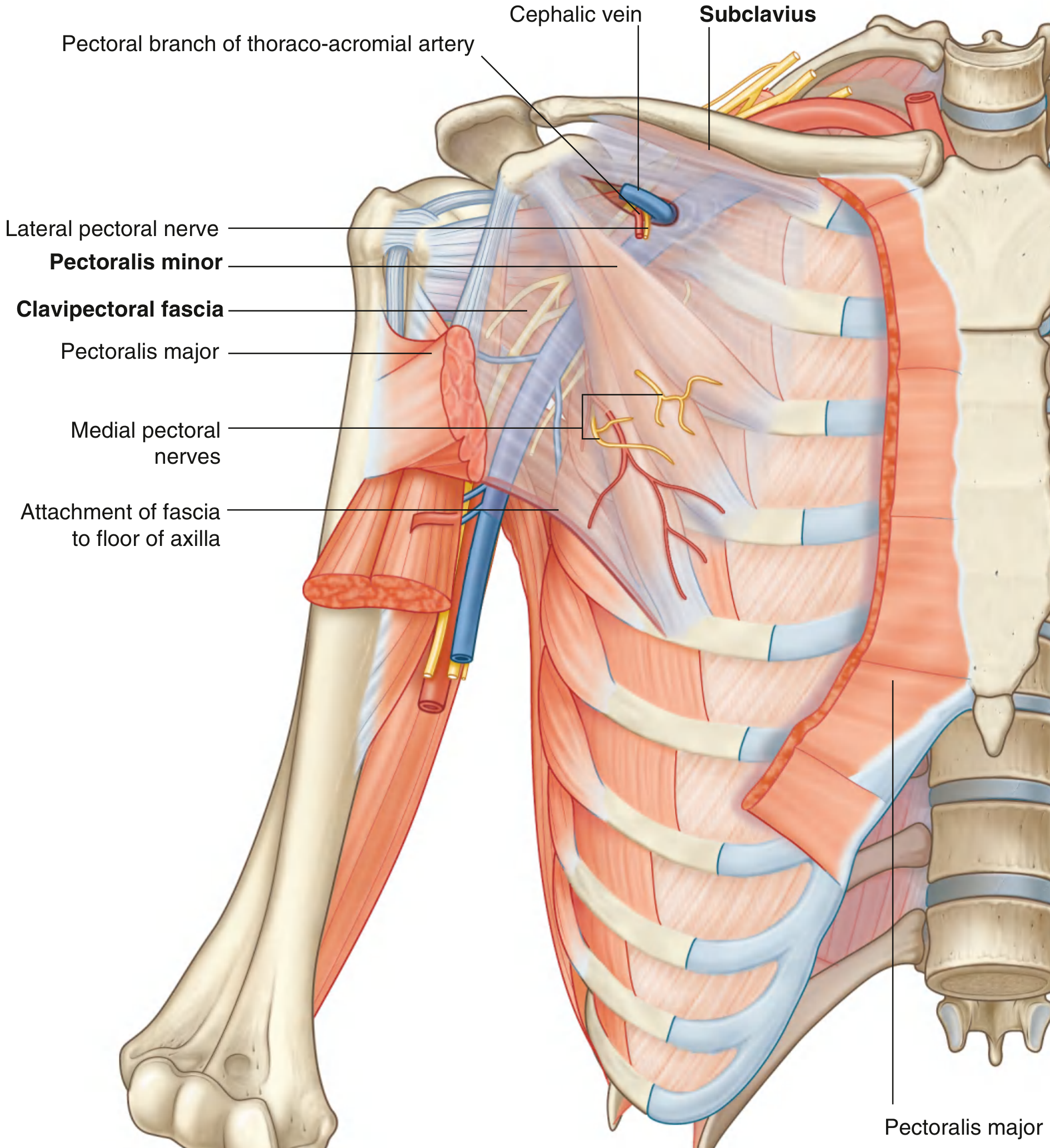
Anterior view of the pectoral region showing the clavipectoral fascia, structures piercing it (cephalic vein, thoraco-acromial artery, lateral pectoral nerve), and the attachment of the fascia to the floor of the axilla. (Gray's Anatomy for Students)
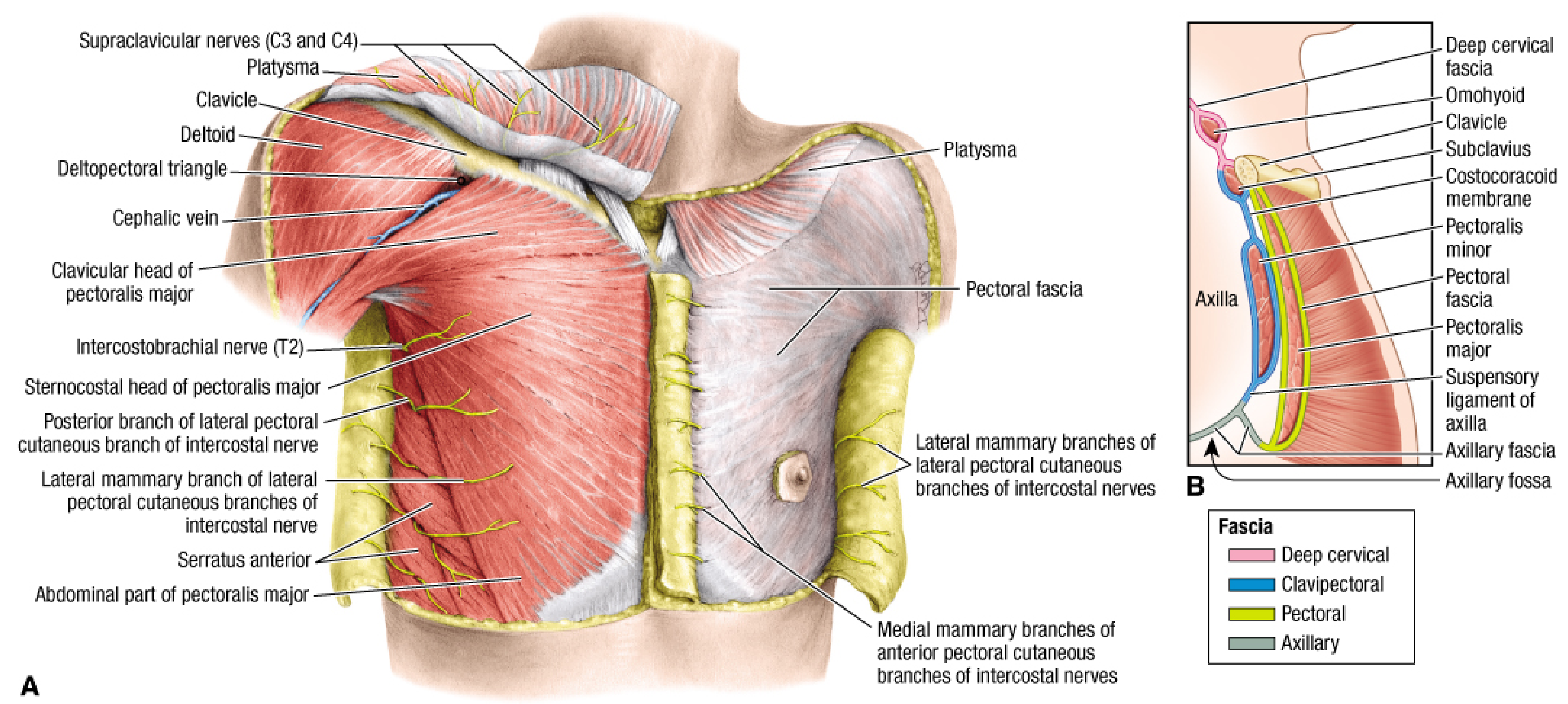
Sagittal section showing the clavipectoral fascia (blue) in relation to the subclavius, costocoracoid membrane, pectoralis minor, suspensory ligament of the axilla, and axillary fascia. (Fischer's Mastery of Surgery)
Summary Mnemonic for Piercing Structures
"C.T.L.L." - Cephalic vein, Thoraco-acromial artery, Lateral pectoral nerve (through costocoracoid membrane) + Lateral thoracic artery (below pectoralis minor).
Note on the medial pectoral nerve: It does not pierce the clavipectoral fascia - it penetrates directly through the pectoralis minor muscle itself (or loops around its lower margin) to reach pectoralis major. This is a common exam distinction.
This is a shared conversation. Sign in to Orris to start your own chat.