
Anaphylaxis
"anaphylaxis"[MeSH Terms] AND management
anaphylaxis pathophysiology mechanism diagram
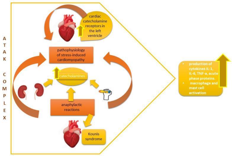
This pathophysiology diagram illustrates the 'ATAK complex', a multifaceted clinical entity involving the interplay between anaphylaxis, Takotsubo syndrome, and Kounis syndrome. The central mechanism highlights the surge of catecholamines, which links anaphylactic reactions to the pathophysiology of stress-induced cardiomyopathy. The visual layout features anatomical illustrations of the heart and kidneys, connected by orange bidirectional arrows indicating systemic feedback loops. Key components include: 1) Cardiac catecholamine receptors in the left ventricle, 2) Kounis syndrome arising from allergic myocardial ischemia, 3) Kidney involvement in systemic stress, and 4) Anaphylactic reactions triggered by external agents (depicted by an epinephrine autoinjector icon). On the right, the diagram emphasizes a massive inflammatory response characterized by increased production of cytokines (IL-1, IL-6, TNF-α), acute phase proteins, and the activation of macrophages and mast cells. This educational schematic is used to demonstrate how hypersensitivity and adrenergic surges converge to cause acute cardiac dysfunction, relevant to cardiology and immunology specialties.
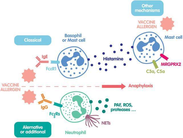
This pathophysiology diagram illustrates three main mechanisms of vaccine-induced anaphylaxis. The 'Classical' pathway (top left) depicts a Vaccine Allergen cross-linking IgE antibodies bound to FcεR1 receptors on a Basophil or Mast cell, triggering degranulation and the release of Histamine. The 'Alternative or additional' pathway (bottom) shows the Vaccine Allergen interacting with IgG antibodies bound to FcγRs on a Neutrophil, resulting in the release of PAF (Platelet-Activating Factor), ROS (Reactive Oxygen Species), proteases, and the formation of NETs (Neutrophil Extracellular Traps). The third section, 'Other mechanisms' (top right), describes non-IgE-mediated Mast cell activation through complement components C3a and C5a receptors, or direct activation of the MRGPRX2 receptor by the vaccine allergen, also leading to histamine release. All three pathways converge on a central axis culminating in Anaphylaxis. The diagram is designed for clinical immunology education, highlighting the diverse cellular and molecular drivers of hypersensitivity reactions.
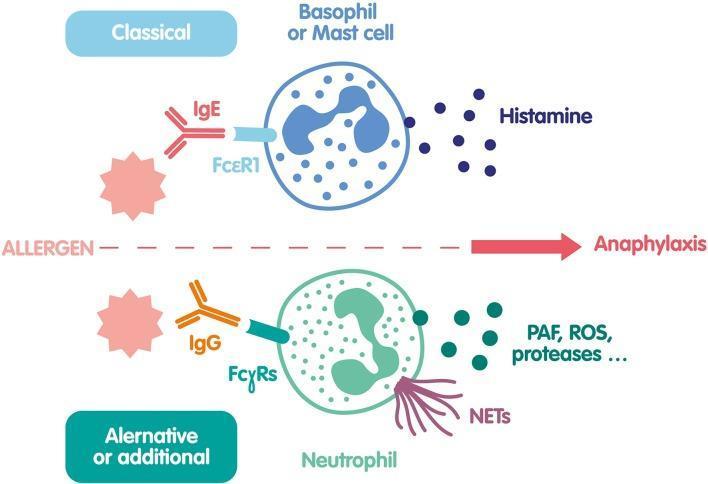
An educational pathophysiology diagram illustrating the 'Classical' and 'Alternative or Additional' cellular pathways leading to anaphylaxis. The top section depicts the classical pathway where an allergen binds to IgE, which is anchored to the FcεRI receptor on a basophil or mast cell. This activation results in degranulation and the release of histamine. The bottom section shows an alternative pathway involving a neutrophil. Here, the allergen interacts with IgG antibodies bound to FcγRs receptors. Neutrophil activation triggers the release of various inflammatory mediators including Platelet-Activating Factor (PAF), Reactive Oxygen Species (ROS), proteases, and Neutrophil Extracellular Traps (NETs). Morphologically, the basophil/mast cell is shown with a bilobed nucleus and blue granules, while the neutrophil is depicted with a characteristic multi-lobed nucleus and purple, web-like NET projections. The diagram highlights different immunological components (IgE vs. IgG) and effector cells contributing to the systemic response of anaphylaxis.
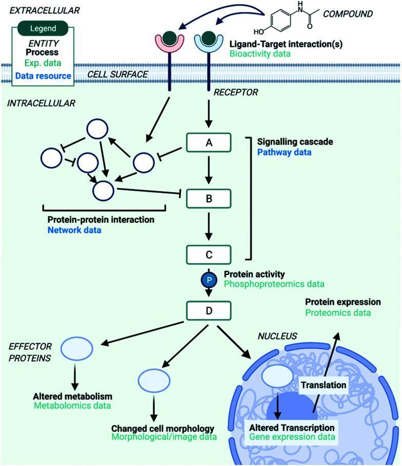
A pathophysiology diagram illustrating the systems-level Mechanism of Action (MoA) of a chemical compound on a human cell, mapping biological processes to specific types of multi-omics data. The diagram is divided into extracellular, cell surface, and intracellular compartments. It begins with a 'Compound' (chemical structure) engaging in a 'Ligand-Target interaction' with a cell surface receptor, labeled as Bioactivity data. Intracellularly, this triggers a 'Signalling cascade' (Pathway data) involving sequential protein nodes (A, B, C, D). Parallel 'Protein-protein interactions' (Network data) are shown interacting with the cascade through activation and inhibition arrows. Downstream effects are categorized by data type: Phosphoproteomics (protein activity/phosphorylation at node C), Proteomics (protein expression at node D), Metabolomics (altered metabolism via effector proteins), and Morphological/image data (changed cell morphology). The cascade terminates in the nucleus, affecting 'Translation' and 'Altered Transcription,' which is identified as Gene expression data. This infographic serves as an educational summary of how various experimental data resources provide biological context for drug interaction studies.
Anaphylaxis
Definition
Pathophysiology
IgE-Mediated Mechanism (Classical)
- Sensitization phase (minimum 1 week): An allergen is processed by antigen-presenting cells, presented to T-helper 2 (TH2) lymphocytes, which then instruct allergen-specific B cells to switch from IgM/IgG to IgE production (driven by IL-4/IL-13 and CD40-CD40L signaling).
- Elicitation phase (subsequent exposure): The allergen cross-links IgE bound to high-affinity FcεRI receptors on mast cell/basophil surfaces, triggering degranulation.
- Mediator release: Histamine, tryptase, leukotrienes (LTC4), prostaglandin D2, platelet-activating factor (PAF), thromboxane, and nitric oxide are released.
Note: Anaphylaxis does not occur on first exposure because specific IgE is not yet formed. Cross-sensitization can allow first-apparent-exposure reactions when a structurally similar allergen was previously encountered.
Non-IgE Mechanisms
Key Mediators and Their Effects
| Mediator | Physiologic Effect | Clinical Manifestation |
|---|---|---|
| Histamine, leukotrienes, thromboxane, prostaglandins, PAF, nitric oxide | Vascular permeability, vasodilation, smooth muscle spasm, mucous secretion, myocardial depression | Urticaria, angioedema, bronchospasm, hypotension, GI cramps |
| Tryptase | Contact system activation → bradykinin production | Enhances severity |
| PAF | Impaired platelet aggregation, vascular permeability | Enhances cardiovascular collapse |
Causes / Triggers
IgE-Mediated
- Insect stings (Hymenoptera)
- Foods: peanuts, tree nuts, sesame, seafood (adults); eggs, cow's milk, wheat, soy (children)
- Drugs: penicillins and other beta-lactam antibiotics, chemotherapy agents (platins), monoclonal antibodies (e.g., rituximab)
- Latex: especially in patients with multiple surgeries or occupational exposure
- Allergen extracts (immunotherapy)
- Alpha-gal syndrome (from Lone Star tick bite - delayed reaction to red meat)
Non-IgE-Mediated
- Radiocontrast media (idiosyncratic, not IgE, not related to "iodine allergy")
- Aspirin / COX-1 inhibitor NSAIDs
- Narcotics (except fentanyl)
- Vancomycin
- Physical triggers: exercise, cold, heat, vibration, pressure
- Complement activation
- Idiopathic (~1/3 of cases have no identifiable cause)
Epidemiology
- Lifetime incidence: 2-8% in adults (higher in children due to food allergies)
- ~1,500-2,000 deaths/year in the US attributed to anaphylaxis
- Fatal anaphylaxis is rare (<1% of cases), but medication-related fatalities are rising in North America
- ~50% of reactions occur at home; ~15% at medical facilities
- Perioperative incidence: 1 in 2,000-10,000 cases (muscle relaxants and antibiotics are the most common triggers in that setting)
- About 1/3 of reactions are biphasic (recurrence 1-72 hours after apparent resolution)
Risk Factors
For Having Anaphylaxis
- Age extremes (pregnant women, infants, teenagers, elderly)
- Parenteral > oral route of administration
- History of atopy or previous anaphylaxis
- Summer/fall season (outdoor allergens)
- Mastocytosis
- Higher socioeconomic status
- Emotional stress, acute infection, physical exertion
For Increased Severity / Mortality
- Cardiovascular disease (especially with beta-blocker or ACE inhibitor use)
- Poorly controlled asthma
- Hereditary alpha-tryptasemia (elevated baseline tryptase, affects up to 5% of population)
- Clonal mast cell disorders (mastocytosis, c-KIT mutation)
- Elderly patients (reduced physiologic reserve)
- Delayed epinephrine administration
- Upright posture at symptom onset (worsens distributive shock)
Clinical Presentation
Diagnostic Criteria (Clinical)
- Acute onset of illness with skin/mucosal involvement (urticaria, flushing, pruritus, angioedema) PLUS at least one of: respiratory compromise OR reduced BP/end-organ dysfunction
- Two or more of the following after exposure to a likely allergen: skin/mucosa symptoms, respiratory symptoms, reduced BP, persistent GI symptoms
- Reduced BP after exposure to a known allergen
Signs and Symptoms by System
| System | Manifestations |
|---|---|
| Cutaneous | Urticaria, angioedema, flushing, diffuse erythema, scalp pruritus |
| Respiratory | Bronchospasm (wheezing), laryngeal edema (stridor), rhinorrhea, cough |
| Cardiovascular | Hypotension, tachycardia, dysrhythmia, cardiac arrest |
| GI | Nausea, vomiting, diarrhea, abdominal cramping (hyperperistalsis) |
| Neurologic | Anxiety, altered consciousness, dizziness, seizures |
Diagnosis
- Primarily clinical - no tests required before treatment
- Serum tryptase: Should be drawn 1-3 hours after symptom onset; elevated acute vs. baseline tryptase supports the diagnosis (and may suggest underlying mast cell disorder if persistently elevated)
- Specific IgE testing (skin prick tests or serum IgE) is done after recovery to identify the trigger
- Elevated urinary histamine metabolites (N-methylhistamine) and prostaglandin D2 can confirm the diagnosis retrospectively
Differential Diagnosis
- Vasovagal syncope (bradycardia, no urticaria/bronchospasm)
- Hereditary angioedema (no urticaria, recurrent, C1-inhibitor deficiency)
- ACE inhibitor-associated angioedema (bradykinin-mediated, no urticaria)
- Carcinoid tumor / Mastocytosis (flushing without allergen trigger)
- Septic shock (fever, slower onset)
- Scombroidosis (fish poisoning - mimics anaphylaxis)
- Panic attack / Vocal cord dysfunction
- Pheochromocytoma
Management
Immediate Measures (Simultaneous)
- Remove the triggering agent
- Place patient in supine position (legs elevated if hypotensive - upright posture worsens outcome)
- Cardiac monitoring, pulse oximetry, BP monitoring
- Ensure patent airway - prepare for intubation (ETI with RSI; have surgical airway available)
- Supplemental oxygen
- Large-bore IV access (16 or 18 gauge)
- Rapid IV crystalloid: Adults 1,000 mL NS in first 5 min; Pediatrics 20-30 mL/kg increments
First-Line Drug: EPINEPHRINE
- Adults: 0.3-0.5 mg IM (1 mg/mL = 1:1000) in the anterolateral thigh, every 5-10 minutes as needed
- Pediatrics: 0.01 mg/kg IM (1:1000) in the anterolateral thigh, every 5-10 minutes as needed
- EpiPen (0.3 mg) or EpiPen Jr (0.15 mg) are acceptable alternatives
- IV epinephrine (1:10,000 dilution): only for refractory shock; requires continuous monitoring due to risk of arrhythmia
Second-Line Agents (Adjuncts Only)
| Drug | Adult Dose | Pediatric Dose | Role |
|---|---|---|---|
| Diphenhydramine (H1 blocker) | 50 mg IV/PO | 1 mg/kg IV/PO | Relieves urticaria/pruritus |
| Famotidine (H2 blocker) | 40 mg IV | 0.5 mg/kg IV | Combined H1+H2 more effective |
| Methylprednisolone | 125 mg IV | 1-2 mg/kg IV | Prevents biphasic reaction (not proven) |
Refractory Anaphylaxis
- Beta-blocker patients: IV glucagon (1-5 mg IV over 5 min, then infusion) - bypasses beta-receptor blockade
- IV vasopressors (norepinephrine, vasopressin) for refractory hypotension
- Consider methylene blue in refractory cases
Discharge and Follow-Up
- Observation period: minimum 4-6 hours after symptom resolution (longer for severe reactions)
- Prescribe an epinephrine auto-injector (EpiPen) at discharge
- Educate patient on avoidance and signs of recurrence
- Refer to allergist/immunologist for:
- Skin prick testing and specific IgE to identify trigger
- Venom immunotherapy (VIT) for Hymenoptera allergy - highly effective, 95-98% protection
- Anti-IgE therapy (omalizumab): useful for idiopathic or recurrent anaphylaxis
- Desensitization protocols for drug allergies when drug is indispensable
Special Scenarios
| Scenario | Consideration |
|---|---|
| Beta-blocker use | Epinephrine may be less effective; use glucagon; monitor for paradoxical bradycardia |
| ACE inhibitor use | Increases severity via bradykinin accumulation |
| Pregnancy | Left lateral decubitus position; epinephrine is still first-line (fetal risk from untreated shock >> drug risk) |
| Radiocontrast media | Pre-medicate with antihistamine + steroid in known reactors before elective contrast (not for emergent imaging) |
| Exercise-induced | Food-dependent subtype: reaction occurs only when eating specific food + exercising; management = epinephrine; avoid trigger food before exercise |
| Mastocytosis/hereditary alpha-tryptasemia | Higher risk of severe/fatal anaphylaxis; carry epinephrine permanently |
Prevention
- Allergen avoidance and patient education
- Epinephrine auto-injector prescription (must carry at all times)
- Medical alert bracelet
- Venom immunotherapy (VIT): 3-5 year course for bee/wasp sting allergy
- Oral immunotherapy (OIT): emerging for food allergies (peanut OIT approved as Palforzia)
- Omalizumab (anti-IgE): reduces threshold for anaphylaxis in IgE-mediated disease and chronic urticaria
- ROSEN's Emergency Medicine, Chapter 106, pp. 2386-2395
- Goldman-Cecil Medicine, Chapter 233, pp. 2696-2698
- Andrews' Diseases of the Skin, p. 3337