
Relations of tmj
Reading File
The TMJ (temporomandibular joint) is a synovial joint between the mandibular condyle and the temporal bone, with important anatomical relations to nearby nerves, vessels, muscles, and parotid gland.
Searching sources...
Finding Sources
Searching Images
temporomandibular joint anatomy relations diagram
The imaging anatomy textbook and the Scott-Brown's ENT book look most useful. Let me read both in parallel.
Reading File
Reading File
Good content from Scott-Brown's. Let me read more of that section and also get paragraphs specifically about TMJ relations.
Reading File
Finding Sources
Reading File
Finding Sources
Reading File
I need to find the section on innervation and vascular supply that was cut off. Let me re-read around line 323-346.
Reading File
Finding Sources
Excellent - I now have very rich data. Let me also fetch the TMJ diagram image from the textbook to include in the answer.
Loading Image
Finding Sources
I now have comprehensive information to give a thorough answer. Let me compile it all.
Relations of the Temporomandibular Joint (TMJ)
The TMJ is a synovial joint formed by the mandibular condyle articulating with the mandibular (glenoid) fossa and articular eminence of the temporal bone. Because it sits at the crossroads of several important anatomical structures, its relations are of great clinical importance.
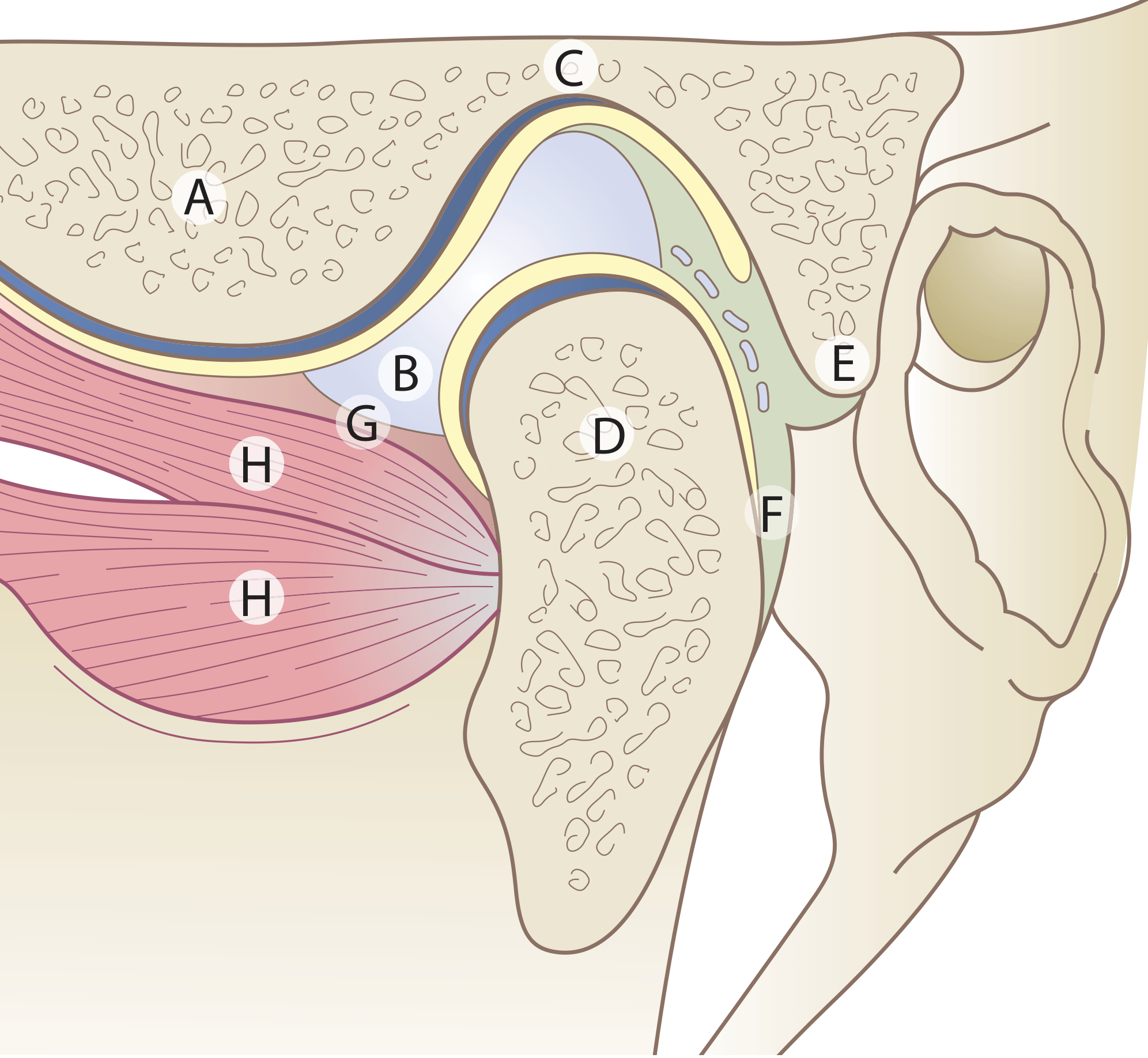
Anterior Relations
- Lateral pterygoid muscle - its superior head attaches to the articular disc and the pterygoid fovea on the anterior surface of the condylar neck. This is the most important anterior relation.
- The articular eminence (tubercle of the temporal bone) lies anteriorly within the joint itself and is the surface over which the condyle glides.
- Masseteric nerve and deep temporal nerves (branches of V3) approach the joint anteriorly.
Posterior Relations
- External acoustic meatus (EAM) - the TMJ is intimately related to the EAM posteriorly. This is why TMJ disorders can produce earache, and why EAM infections or tumors can involve the joint.
- Squamotympanic and petrotympanic fissures - the posterior capsule attaches here. The superior lamina of the retrodiscal tissue also attaches to the anterior margin of the squamotympanic fissure.
- Tympanic plate of the temporal bone separates the joint from the middle ear.
- Superficial temporal artery runs just behind and lateral to the joint.
Lateral Relations
- Skin and fascia (the joint is subcutaneous laterally, making it accessible for injection/aspiration)
- Lateral ligament (temporomandibular ligament) - the main supporting ligament, running from the articular tubercle to the lateral surface of the condylar neck
- Auriculotemporal nerve - a branch of V3 that runs just deep to the superficial temporal vessels, passes within the capsule adjacent to the neck of the mandible and behind the TMJ before winding round to supply the joint and scalp
- Superficial temporal vessels (artery and vein) cross the lateral aspect
Medial Relations
- Maxillary artery (and its branches: anterior tympanic and deep auricular) - lies medial to the joint. The deep auricular branch arises in the parotid gland behind the TMJ and pierces the cartilage of the external meatus
- Auriculotemporal nerve loops medially around the condylar neck before running upward
- Middle meningeal artery passes upward medially
- Chorda tympani (from CN VII) passes close medially
- There is no medial ligament - medial stability relies on the lateral ligament of the opposite side
Superior Relations
- Middle cranial fossa - the roof of the mandibular fossa is a thin bony plate separating the joint from the middle cranial fossa. Severe condylar fractures or tumors can breach this
- Temporalis muscle and temporal fascia lie superolaterally
- Middle meningeal vessels in the extradural space above
Inferior Relations
- Neck of mandible - the capsule is attached below to the neck of the condyle
- Medial pterygoid muscle lies inferiorly and medially
- Parotid gland - its deepest (retromandibular) part wraps around the condylar neck. Parotid tumors, especially deep-lobe tumors, can directly involve the TMJ
Nerve Supply (and its clinical significance)
| Nerve | Origin | Significance |
|---|---|---|
| Auriculotemporal nerve | V3 (post-ganglionic from otic ganglion) | Main sensory supply; referred otalgia in TMJ disorders |
| Masseteric nerve | V3 | Supplies joint and masseter |
| Deep temporal nerves | V3 | Contribute proprioceptive fibers |
The auriculotemporal nerve also carries postganglionic parasympathetic fibers from the otic ganglion to the parotid gland. In Frey syndrome, aberrant regeneration of these fibers after parotid surgery causes gustatory sweating over the skin of the TMJ region.
Blood Supply
| Vessel | Source |
|---|---|
| Superficial temporal artery | External carotid |
| Anterior tympanic artery | First part of maxillary artery |
| Deep auricular artery | First part of maxillary artery (arises in parotid behind TMJ) |
| Deep temporal and transverse facial arteries | Contribute minor branches |
Key Clinical Correlations
- Referred otalgia: The auriculotemporal nerve explains why TMJ disorders commonly cause earache - the joint and external ear share sensory supply via V3.
- Parotid tumors: Deep-lobe parotid tumors grow into the parapharyngeal space and can directly involve the TMJ capsule.
- Condylar fractures: The thin neck of the condyle is a weak point; fractures can drive the condyle through the roof of the fossa into the middle cranial fossa.
- External acoustic meatus: Posterior wall of EAM is directly adjacent to the TMJ - erosion by TMJ pathology or vice versa is possible.
- Frey syndrome: Due to the proximity of the auriculotemporal nerve, parotidectomy can result in inappropriate sweating over the TMJ/temporal area during eating.
Sources: Scott-Brown's Otorhinolaryngology Head & Neck Surgery, Ch. 41; Gray's Anatomy for Students; Bailey and Love's Short Practice of Surgery, 28th Ed.
This is a shared conversation. Sign in to Orris to start your own chat.