
Benign prostatic hyperplasia
"Prostatic Hyperplasia"[MeSH Terms] AND "therapy"
benign prostatic hyperplasia
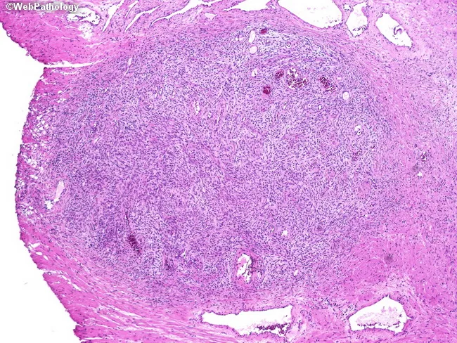
This is a histopathology slide of prostatic tissue obtained from transurethral resection of the prostate (TURP). Imaging modality: light microscopy; staining: hematoxylin and eosin. The specimen demonstrates nodular stromal hyperplasia composed of irregularly sized acini intimately admixed with dense fibromuscular stroma. The nodules display papillary infolding and variable gland size, a hallmark of nodular prostatic hyperplasia (benign prostatic hyperplasia, BPH). The glandular epithelium is typically cuboidal to columnar without significant cytologic atypia; stromal cells are fibromuscular and nonmalignant. Overall architecture is nodular with coarse borders and occasional dilated acini, consistent with benign prostatic tissue arranged in nodules rather than a uniform adenocarcinoma. No malignant features are identified: mitotic activity is not increased, and there is no prominent necrosis or invasion. In TURP specimens, the diagnostic focus rests on obstructive voiding symptoms and clinical presentation rather than morphologic abnormalities. The findings correlate with lower urinary tract symptoms due to prostatic enlargement and bladder outlet obstruction; they support a benign etiology for LUTS and guide management toward medical therapy or TURP as required. Important differentials include prostatic adenocarcinoma, prostatitis, and atrophy; however, absence of invasion or significant atypia favors benign nodular hyperplasia. Correlation with clinical history and imaging enhances diagnostic confidence overall.
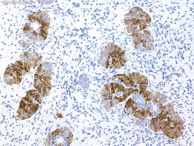
This histopathology slide depicts a formalin-fixed, paraffin-embedded prostatic needle biopsy specimen subjected to immunohistochemical staining. The tissue showcases prostatic glandular units with a preserved, continuous basal cell layer; gland lumina are variably sized and lined by two cell layers in many foci. The basal cell population exhibits diffuse, strong positivity for high molecular weight cytokeratin (HMWCK) with a brown chromogen, outlining the acinar architecture. Adjacent stromal and epithelial cells show non-specific blue hematoxylin counterstain. The staining pattern contrasts with adenocarcinoma, where the basal layer is typically disrupted or absent; here, the intact basal cell layer argues against neoplastic invasion and favors basal cell hyperplasia. p63 immunoreactivity is present in basal cells, reinforcing benign histology. In differential, prostatic hyperplasia with epithelial budding may mimic carcinoma on H&E, but the IHC demonstrates basal cells per gland. Clinically relevant use: helps distinguish benign prostatic hyperplasia with basal cell hyperplasia from prostatic adenocarcinoma in suspicious needle biopsy cores, reducing overtreatment and guiding management. The image demonstrates an educational example of diagnostic IHC utility, where basal cell markers are essential in challenging cases. The technique is commonly employed in uropathology practice to confirm benign versus malignant prostatic lesions. Correlation with morphology improves diagnostic confidence significantly.
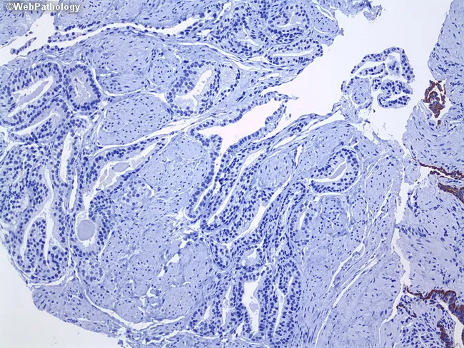
This is a formalin-fixed paraffin-embedded prostatic biopsy tissue section, analyzed by light microscopy with immunohistochemical staining for basal cell markers. The primary histology in the image demonstrates a pseudohyperplastic pattern of prostatic adenocarcinoma, characterized by gland-forming proliferations with cribriform and back-to-back glands and mild to moderate cytologic atypia. The malignant glands completely lack a discernible basal cell layer, a key feature distinguishing cancer from benign prostatic hyperplasia. By contrast, an internal positive control of benign prostatic glands along the right edge shows intact basal cells positive for high molecular weight cytokeratin (34βE12) and p63, validating the stain and serving as an internal control. The tumor glands display absence of basal cells while the luminal epithelium remains columnar to cuboidal with small nucleoli, resulting in a histologic appearance that can mimic hyperplasia. Immunostaining thus supports diagnosis of prostatic adenocarcinoma of the pseudohyperplastic type. Clinically this diagnosis is relevant in PSA interpretation, prognosis, and management decisions; differential diagnoses include benign prostatic hyperplasia and other mimickers such as high-grade PIN or intraductal carcinoma when interpreted in full. This image is suitable for educational demonstration of basal cell–deficient glands and internal control use in IHC diagnostics.
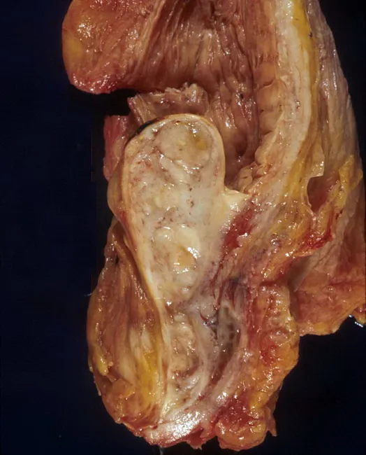
Gross pathology photograph of an autopsy prostatic specimen demonstrating benign prostatic hyperplasia with median lobe enlargement projecting into the bladder neck. The tissue appears tan‑brown and nodular, with a prominent midline lobe extending anteriorly toward the bladder outlet and creating a ball‑valve type obstruction at the bladder neck. The surrounding capsule and adjacent prostatic zones show variable, fibromuscular hyperplasia consistent with nodular hyperplasia rather than malignant invasion. This gross appearance reproduces the classic mechanism of lower urinary tract symptoms in aging men: progressive prostatic enlargement compresses the urethral channel, especially at the bladder outlet, producing obstructive and irritative voiding disorders. The autopsy context confirms a non‑malignant process, though histology would be required to definitively exclude focal carcinoma. Notable features include bilateral symmetry of nodular growth and conspicuous midline protrusion, which can be associated with reduced urinary flow, incomplete emptying, and increased postvoid residuals. Clinically, these findings underpin diagnostic considerations such as benign prostatic hyperplasia and differential diagnoses including prostatic adenocarcinoma or prostatitis; radiologic or endoscopic correlation may help stage obstruction. Educationally, this image serves as a reference for urology trainees and pathology learners to understand gross anatomy, angioarchitecture, and the anatomic basis of bladder outlet obstruction in BPH.
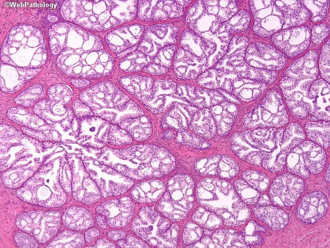
Prostate histopathology slide prepared from either transurethral resection (TURP) specimen or needle core biopsy, evaluated by light microscopy following Hematoxylin and Eosin staining. The architectural pattern consists of small to medium-sized glandular units arranged in lobules with sieve-like, cribriform cradles, creating an irregular lattice within the prostatic parenchyma. Glands display variable cytoplasmic clearing in some acini, leading to a pale, vacuolated appearance; however, not all glands show cytoplasmic clearing. The epithelial cells typically exhibit low to absent cytologic atypia: nuclei are uniform, with minimal pleomorphism, rare nucleoli, and lack of conspicuous mitotic activity. A prominent basal cell layer is preserved and readily evident around many glands, which supports a benign process and helps distinguish from prostatic adenocarcinoma. A variant of epithelial hyperplasia, commonly termed clear cell cribriform hyperplasia, frequently seen in TURP specimens but can be encountered in needle biopsy samples. The overall impression is benign cribriform prostatic epithelial proliferation without invasion. Diagnostic significance includes exclusion of high-grade prostatic intraepithelial neoplasia (PIN) and prostate adenocarcinoma. Clinically, recognizing this entity prevents overtreatment; correlated with imaging and PSA history in differential diagnoses such as benign prostatic hyperplasia and other prostatic lesions. This entity does not typically require urgent management changes beyond standard monitoring and routine clinical correlation.
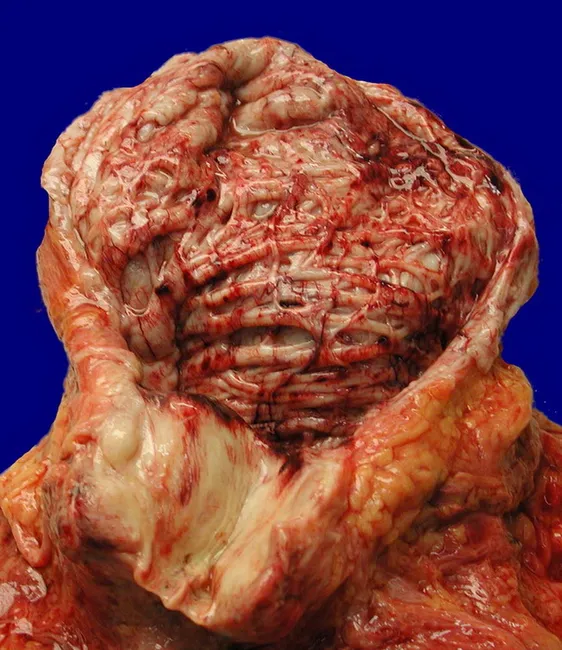
Gross pathology photograph of a pelvic prostatic specimen showing marked enlargement of the middle and posterior prostatic lobes, producing bladder outlet obstruction. The image captures exposed tissue with thickened detrusor muscle and subepithelial fibrosis consistent with chronic obstruction. Prostatic hyperplasia results in nodular enlargement of the periurethral transitional zone, with irregular surface architecture and vascularity typical of benign prostatic hyperplasia (BPH). The bladder wall and detrusor are hypertrophied as a compensatory response, reflecting long-standing outlet resistance and increased bladder workload. This specimen illustrates the pathophysiology of aging-related prostatic enlargement: progressive glandular proliferation, stromal expansion, and smooth muscle hypertrophy that impede urine flow, elevate intravesical pressure, and predispose to symptoms such as hesitancy, weak stream, and nocturia. Clinically relevant terms include BPH, benign prostatic hyperplasia, LUTS, urinary retention risk, detrusor hypertrophy, chronic obstruction, and pelvic genitourinary anatomy. Pathological significance lies in correlating anatomic changes with functional impairment and guiding management decisions in urology. Differential considerations encompass prostate adenocarcinoma in enlargement, prostatitis with nodularity, and secondary bladder remodeling. Educationally, this image supports discussions of prostatic architecture, disease progression, and the gross features used in diagnosis and teaching within pathology and urology. Education.
AUA guideline management of benign prostatic hyperplasia lower urinary tract symptoms 2023 amendment recommendations
Benign Prostatic Hyperplasia (BPH)
Definition and pathology
Clinical features
- Obstructive (voiding) symptoms: hesitancy, straining, weak stream, terminal dribbling, prolonged voiding, incomplete emptying.
- Irritative (storage) symptoms: frequency, urgency, urge incontinence, nocturia, small voided volumes.
- Complications: acute urinary retention, recurrent UTI, bladder stones, gross hematuria, and chronic retention with hydronephrosis and renal impairment. Chronic obstruction also thickens the detrusor.
Evaluation
- History and symptom score: use the AUA Symptom Index (AUASI), which is the same as the IPSS. Repeat it to follow response to treatment. - Harrison's, p. 747
- Examination: digital rectal exam for size, nodules and induration.
- Urinalysis: to exclude infection and hematuria.
- Creatinine: if renal impairment is suspected.
- PSA: after shared decision-making, mainly when it would change management.
- Optional tests:
- Uroflowmetry identifies men with normal flow who are unlikely to benefit from treatment.
- Bladder ultrasound measures post-void residual.
- Pressure-flow urodynamics detects primary bladder dysfunction.
- Cystoscopy is advised for hematuria and before surgery.
- Upper tract imaging is advised for hematuria, stones or prior urinary tract problems. - Harrison's, p. 747
- Frequency-volume chart: useful for nocturia, to look for nocturnal polyuria.
- Differentials: prostate cancer, prostatitis, urethral stricture, neurogenic bladder, overactive bladder, UTI, and drug causes such as anticholinergics and decongestants.
Treatment
| Option | Mechanism and notes |
|---|---|
| Watchful waiting and lifestyle | For mild symptoms. Reduce evening fluids, caffeine and alcohol, and review offending drugs. |
| α-blockers (tamsulosin, alfuzosin, doxazosin, terazosin, silodosin) | Treat the dynamic component by relaxing smooth muscle at the bladder outlet. Onset is fast. Adverse effects are orthostatic hypotension, retrograde ejaculation and intraoperative floppy iris syndrome. |
| 5α-reductase inhibitors (finasteride, dutasteride) | Treat the static component by shrinking the gland, with a delayed effect. They reduce acute urinary retention and the need for surgery. They suit larger prostates (about 30 g or more, or PSA above about 1.5 ng/mL). Adverse effects are decreased libido, erectile dysfunction and ejaculatory problems. They lower PSA by about 50% after 6 to 12 months. |
| Combination (α-blocker plus 5ARI) | Gives symptom relief and prevents progression. Evidence is from MTOPS and CombAT. - Campbell-Walsh Urology |
| PDE5 inhibitors (tadalafil) | Improve LUTS, especially with coexisting erectile dysfunction. Short-acting agents like sildenafil need separate dosing from α-blockers because of hypotension. - Harrison's, p. 747 |
| Anticholinergics or β3 agonists | For predominant storage symptoms, with a post-void residual check first. |
| Surgery | Second-line after medical therapy, or first-line for complications. Options are TURP (the standard), transurethral incision of the prostate for small glands, laser enucleation or vaporization (HoLEP, GreenLight), and open simple prostatectomy for very large glands. - Harrison's, p. 747 |
| Minimally invasive options | Prostatic urethral lift, Rezūm water vapor therapy, and prostate artery embolization. Embolization gives AUA-SI improvements comparable to TURP, but TURP gives better Qmax and post-void residual. Long-term data are limited. - Smith and Tanagho's General Urology, 19th ed., p. 121 |
- retrograde ejaculation (very common)
- bleeding
- TUR syndrome (hyponatremia from absorbed irrigant)
- urethral stricture
- incontinence
- erectile dysfunction
Recent evidence and guidelines
- The AUA published an updated BPH guideline in 2026. It replaces the 2021 guideline as amended in 2023. Full access now requires a free login, so I could not review its specific recommendations. Check it for current drug and procedure recommendations.
- Recent systematic reviews cover minimally invasive therapies. One found reintervention rates after minimally invasive BPH therapies need careful counselling, and industry involvement is common in these studies (PMID 40817983).
- Another review covered minimally invasive options in older men (PMID 38656693).
- A 2024 European Urology review compared relief of bladder outlet obstruction and symptom improvement across medical and surgical therapies (PMID 38749852). I have not read the full texts of these reviews. None seems to overturn the textbook approach above.