
Can you explain general anatomy?
Reading File
Now let me search the library for general anatomy content and look for relevant images in parallel.
Finding Sources
Finding Sources
Excellent! Let me read the main general anatomy content from the Color Atlas and the THIEME Atlas in parallel.
Reading File
Reading File
Let me now read more of both textbooks for richer content on tissues, skeletal, muscular, and other key anatomy topics.
Reading File
Reading File
Now let me fetch a couple of key diagrams - the anatomical planes diagram and the bone structure diagram - to include in the answer, while also reading more content on muscles and vessels.
Loading Image
Loading Image
Reading File
Let me fetch the bone types diagram and muscle types diagram, plus read more content on muscles, vessels, and the nervous system.
Loading Image
Loading Image
Reading File
Let me fetch one more key image - the cell structure diagram - and also read content from the THIEME atlas on the overview of the human body.
Loading Image
Reading File
I now have a strong foundation to write a thorough, well-illustrated answer on general anatomy. Let me compose the full response.
General Anatomy
General anatomy is the study of the structure of the human body - its organization from cells up to organ systems. Here is a systematic overview of all the major components.
1. Anatomical Terminology and Body Planes
Before studying any region of the body, you need a common language to describe positions and directions.
Anatomical position is the reference stance: standing upright, facing forward, arms at the sides with palms facing anteriorly.
Anatomical planes divide the body for description:
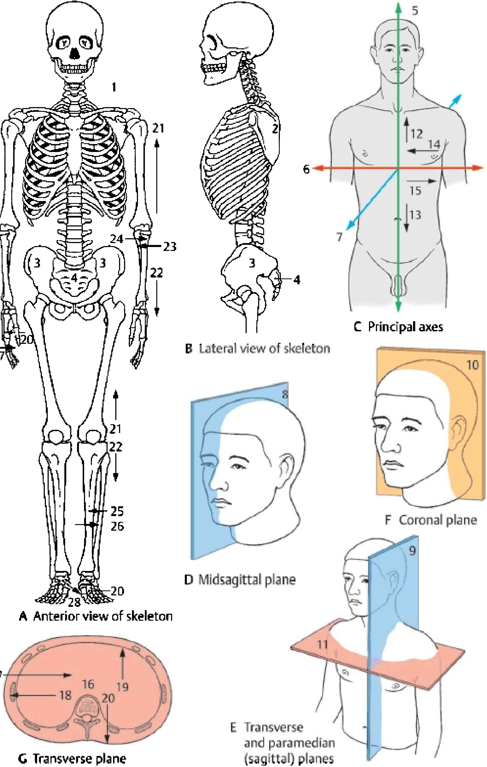
| Plane | Description |
|---|---|
| Sagittal (median) | Divides the body into left and right halves |
| Coronal (frontal) | Divides the body into anterior (front) and posterior (back) |
| Transverse (axial) | Divides the body into superior (upper) and inferior (lower) |
Key directional terms:
- Superior/Inferior - toward the head / toward the feet
- Anterior/Posterior - toward the front / toward the back
- Medial/Lateral - toward the midline / away from the midline
- Proximal/Distal - closer to the origin / farther from the origin
- Superficial/Deep - toward the surface / toward the interior
(Color Atlas of Human Anatomy 1, p. 21)
2. The Cell - The Basic Unit of Life
All body structures are built from cells. The human cell contains a number of key organelles:
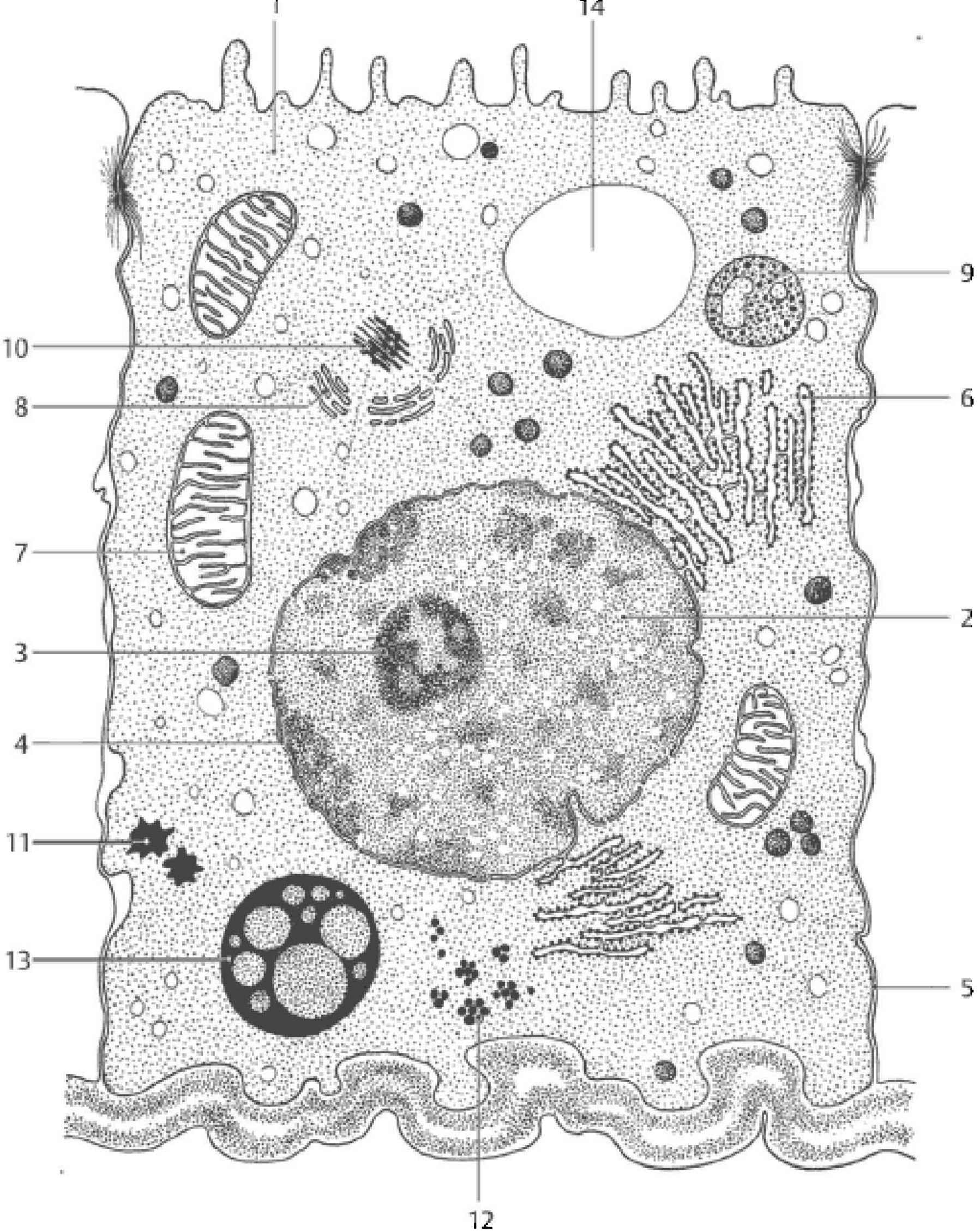
Key organelles and their functions:
- Nucleus (3) - contains DNA; controls cell activity
- Mitochondria (7) - site of ATP production ("powerhouse of the cell")
- Rough ER (6) - protein synthesis and processing
- Golgi apparatus - sorts and packages proteins for export
- Ribosomes (5) - translate mRNA into protein
- Lysosomes (13) - intracellular digestion
- Cell membrane (1) - selectively permeable barrier with microvilli (14)
(Color Atlas of Human Anatomy 1, p. 24)
3. Tissues
Four fundamental tissue types make up all organs:
a. Epithelial Tissue
- Covers body surfaces, lines cavities, and forms glands
- Classified by cell shape (squamous, cuboidal, columnar) and layers (simple vs. stratified)
- Functions: protection, secretion, absorption, filtration
b. Connective Tissue
- The most abundant tissue type; binds and supports other tissues
- Includes loose connective tissue, dense connective tissue, cartilage, bone, blood, and adipose tissue
- Characterized by cells embedded in an extracellular matrix (fibers + ground substance)
c. Muscle Tissue
Three types:
- Skeletal muscle - striated, voluntary, attached to bones
- Cardiac muscle - striated, involuntary, found only in the heart
- Smooth muscle - non-striated, involuntary, found in walls of hollow organs
d. Nervous Tissue
- Composed of neurons (excitable cells that transmit impulses) and glial cells (supporting cells)
- Found in the brain, spinal cord, and peripheral nerves
(Color Atlas of Human Anatomy 1, p. 27-36)
4. The Skeletal System
The adult skeleton contains 206 bones and serves as the framework of the body.
Functions of bone:
- Support and protection
- Movement (via joints and muscle attachments)
- Mineral storage (calcium and phosphate)
- Blood cell production (hematopoiesis in red marrow)
Bone classification by shape:
| Type | Example |
|---|---|
| Long bones (Ossa longa) | Femur, humerus |
| Short bones (Ossa brevia) | Carpals, tarsals |
| Flat bones (Ossa plana) | Skull, sternum, ribs |
| Irregular bones | Vertebrae, facial bones |
| Sesamoid bones | Patella |
Bone composition: Bone is a living tissue made of ~1/3 organic matrix (mainly collagen fibers) and ~2/3 inorganic mineral salts (calcium phosphate/hydroxyapatite). The salts confer hardness and strength; a salt-free ("decalcified") bone is pliable. Deficiency in calcification leads to conditions like rickets (in children) or osteomalacia (in adults). (Color Atlas of Human Anatomy 1, p. 37)
Divisions of the skeleton:
- Axial skeleton - skull, vertebral column, thoracic cage (80 bones)
- Appendicular skeleton - limbs and girdles (126 bones)
5. Joints (Arthrology)
Joints connect bones and allow varying degrees of movement.
Classification by structure:
| Type | Tissue connecting bones | Mobility | Example |
|---|---|---|---|
| Fibrous (Junctura fibrosa) | Dense fibrous tissue | Immovable (synarthrosis) | Skull sutures |
| Cartilaginous (Junctura cartilaginea) | Cartilage | Slightly movable (amphiarthrosis) | Intervertebral discs |
| Synovial (Junctura synovialis) | Joint capsule + synovial fluid | Freely movable (diarthrosis) | Knee, hip, shoulder |
Types of synovial joints:
- Hinge (Ginglymus) - elbow, knee: flexion/extension only
- Ball-and-socket (Articulatio sphaeroidea) - hip, shoulder: multiaxial movement
- Saddle (Articulatio sellaris) - carpometacarpal joint of thumb
- Plane/pivot - gliding or rotational movement
- Ellipsoidal/condylar - wrist joint
Articular cartilage is avascular and loses elasticity with aging, predisposing to degenerative joint disease (osteoarthritis). (Color Atlas of Human Anatomy 1, p. 50-56)
6. The Muscular System
The human body has over 600 skeletal muscles, classified by their fiber arrangement:
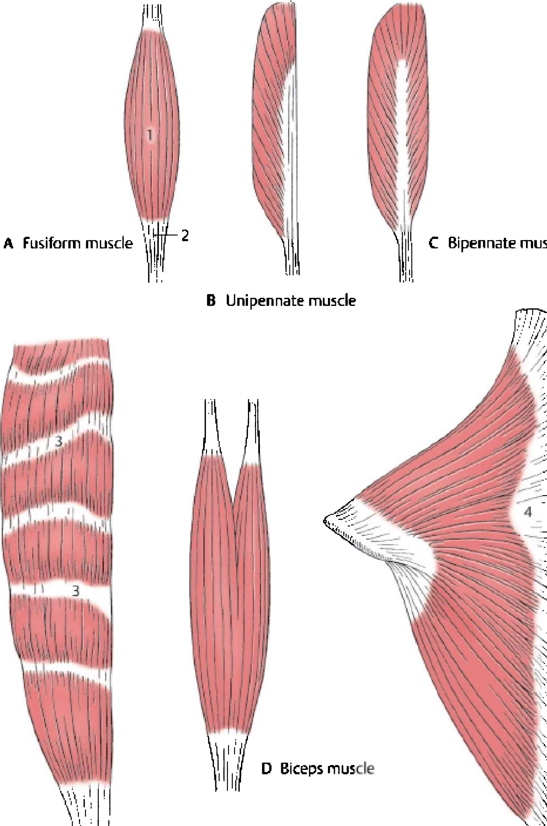
| Shape | Description | Example |
|---|---|---|
| Fusiform | Spindle-shaped, parallel fibers | Biceps brachii |
| Unipennate | Fibers angled on one side of tendon | Extensor digitorum longus |
| Bipennate | Fibers angled on both sides | Rectus femoris |
| Multi-belly | Two or more bellies in series | Digastric |
| Fan-shaped | Broad origin, converge to tendon | Pectoralis major |
Muscle attachments:
- Origin - the more fixed, proximal attachment
- Insertion - the more mobile, distal attachment
- Muscles attach via tendons (cord-like) or aponeuroses (flat, sheet-like)
(Color Atlas of Human Anatomy 1, p. 63)
7. The Vascular System
Arteries carry oxygenated blood away from the heart (except pulmonary arteries). They have thick, elastic walls. The aorta is the largest artery.
Veins return deoxygenated blood to the heart. They have thinner walls and contain valves to prevent backflow.
Capillaries are microscopic vessels where gas, nutrient, and waste exchange occurs between blood and tissues.
The two circulations:
- Systemic circulation: Left heart → aorta → body tissues → venae cavae → right heart
- Pulmonary circulation: Right heart → pulmonary arteries → lungs → pulmonary veins → left heart
8. The Lymphatic System
- A network of vessels, nodes, and lymphoid organs that:
- Returns excess interstitial fluid to the bloodstream
- Filters lymph through lymph nodes (containing lymphocytes)
- Transports dietary fats (chylomicrons) from the gut
- Primary lymphoid organs: thymus (T-cell maturation), bone marrow (B-cell maturation)
- Secondary lymphoid organs: lymph nodes, spleen, tonsils, Peyer's patches
(THIEME Atlas of Anatomy - General Anatomy, Ch. 7)
9. The Nervous System
Divisions:
Nervous System
├── Central Nervous System (CNS)
│ ├── Brain (cerebrum, cerebellum, brainstem)
│ └── Spinal cord
└── Peripheral Nervous System (PNS)
├── Somatic NS (voluntary - skeletal muscle control, sensory)
└── Autonomic NS (involuntary - visceral control)
├── Sympathetic ("fight or flight")
└── Parasympathetic ("rest and digest")
The functional unit is the neuron, which consists of:
- Cell body (soma) - contains nucleus and organelles
- Dendrites - receive incoming signals
- Axon - transmits signals away from cell body; can be myelinated for faster conduction
(THIEME Atlas of Anatomy - General Anatomy, Ch. 8)
10. Body Cavities and Organ Systems
The body is organized into cavities that contain and protect organs:
| Cavity | Contents |
|---|---|
| Cranial | Brain |
| Vertebral | Spinal cord |
| Thoracic | Heart, lungs, great vessels, esophagus |
| Abdominal | Stomach, intestines, liver, pancreas, spleen, kidneys |
| Pelvic | Bladder, rectum, reproductive organs |
The 11 organ systems of the body:
| System | Primary Function |
|---|---|
| Integumentary | Protection, thermoregulation |
| Skeletal | Support, protection, movement, hematopoiesis |
| Muscular | Movement, posture, heat production |
| Nervous | Control, coordination, sensation |
| Endocrine | Hormonal regulation of homeostasis |
| Cardiovascular | Transport of O2, nutrients, hormones, wastes |
| Lymphatic/Immune | Immunity, fluid balance |
| Respiratory | Gas exchange (O2/CO2) |
| Digestive | Nutrient absorption |
| Urinary | Excretion of metabolic wastes, fluid balance |
| Reproductive | Perpetuation of the species |
11. Human Development (Ontogeny)
Ontogeny is the study of the individual's development from fertilization to birth. It encompasses:
- Histogenesis - formation of tissues
- Organogenesis - formation of organs
- Morphogenesis - shaping of the body
During gastrulation (week 3), the embryonic disc differentiates into three germ layers:
| Germ Layer | Derivatives |
|---|---|
| Ectoderm | Skin, nervous system, sensory organs |
| Mesoderm | Muscle, bone, connective tissue, cardiovascular system, kidneys |
| Endoderm | Gut lining, respiratory epithelium, liver, pancreas |
(THIEME Atlas of Anatomy - General Anatomy, p. 22-26)
Sources:
- Color Atlas of Human Anatomy 1 (Thieme, 9783132424432)
- General Anatomy and Musculoskeletal System - THIEME Atlas of Anatomy (9781626237186)
- Gray's Anatomy for Students (9780323934237)
This is a shared conversation. Sign in to Orris to start your own chat.