
Pilonidal sinus surgery steps
pilonidal sinus surgery excision procedure
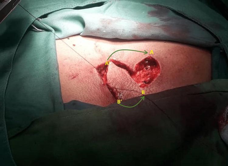
This clinical photograph captures an intraoperative view of a surgical procedure for pilonidal sinus disease (PSD) in the sacrococcygeal region. The image demonstrates the creation of a rotational 'swing flap' to reconstruct a surgical defect. A primary elliptical wound is visible where the pilonidal sinus was excised, exposing deep subcutaneous tissue and underlying fascia. Adjacent to this, a linear incision has been made to mobilize a vascularized skin flap. Annotated green arrows with labels A' to A and B' to B indicate the planned vector of transposition, showing how the mobilized tissue will be rotated to cover the primary excision site. The surgical field is surrounded by green sterile drapes. This visual highlights the technique of using local flap transposition rather than primary midline closure to reduce tension and move the scar away from the midline, which are key principles in preventing PSD recurrence. This material is intended for surgical residents and medical students studying plastic and reconstructive surgery techniques or colorectal management of sinus tracts.
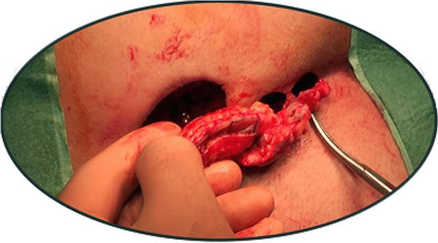
This clinical photograph captures an intraoperative view of a pilonidal sinus excision utilizing a modified Lord-Millar technique. The image demonstrates multiple distinct, circular surgical openings in the sacrococcygeal region, where a skin bridge has been intentionally preserved between the excision sites to facilitate healing by secondary intention. A gloved hand is visible holding a segment of excised, erythematous tissue, likely containing the pilonidal cyst or sinus tract. A metallic surgical probe or dissector is inserted into one of the smaller openings, assisting in the exploration or dissection of the tract. The surrounding skin shows post-surgical erythema and inflammation. This visual illustrates the surgical principle of limited excision and the preservation of healthy skin bridges to reduce the overall wound surface area while ensuring the complete removal of the pathological sinus, a key concept in proctological and dermatological surgery for pilonidal disease.
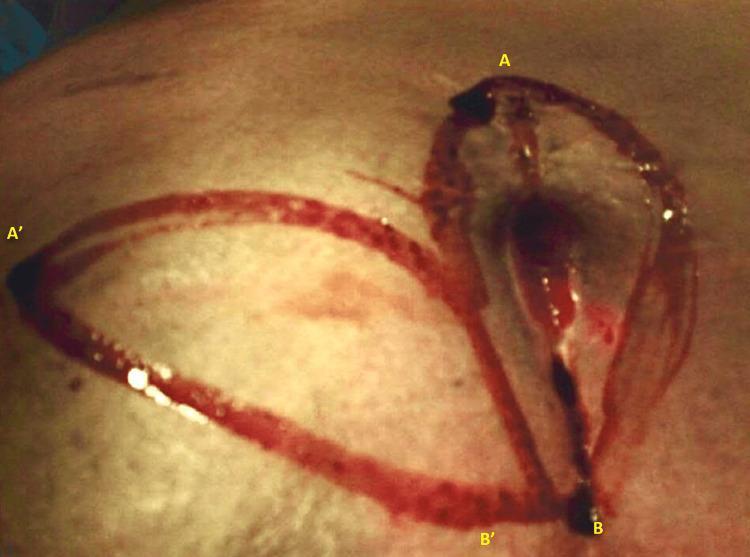
This clinical photograph displays a surgical field during the initial stages of a slide-swing skin flap procedure for pilonidal sinus disease (PSD). The image shows the sacrococcygeal region with fresh surgical incisions. There are two primary curvilinear, off-midline elliptical incisions designed to encompass the pilonidal pits. The incision lines are marked with yellow anatomical labels: A to B identifies a vertical, teardrop-shaped elliptical incision over the primary pathology, while A' to B' indicates the complementary arc-shaped incision used for the rotational flap. The surgical borders demonstrate sharp edges with visible dermal and subcutaneous tissue disruption, accompanied by moderate serosanguinous fluid and blood pooling within the deeper aspects of the teardrop-shaped defect. This visual represents the flap design phase, essential for surgeons to visualize the geometric relationship between the excision site and the donor flap area to ensure tension-free closure and off-midline scarring, which are critical for reducing recurrence in pilonidal sinus surgery.
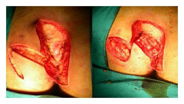
This clinical photograph shows two perspectives of an intraoperative site during pilonidal sinus surgery. The images demonstrate a wide surgical excision in the sacrococcygeal region with the patient in the prone position. An S-shaped oblique incision has been utilized for the initial excision, extending down to the postsacral fascia. Visible in the operative field is a full-thickness, broad-pedicled transposition flap, resembling a Dufourmentel flap, which includes skin, subcutaneous fat, and the fascia over the gluteus maximus. The flap is dissected and reflected to reveal the underlying deep tissue defect and anatomical landmarks, including the reddish gluteal muscle fibers and yellowish lobulated subcutaneous adipose tissue. The incision edges are sharp and clean-cut, indicating precise surgical margins for reconstruction. This visual serves to illustrate the surgical technique for managing complex or wide pilonidal disease by flattening the intergluteal sulcus and moving the eventual scar away from the midline to reduce recurrence risk.
Pilonidal Sinus Surgery - Steps and Techniques
Overview of Operative Approaches
- Minimal surgery (pit picking / Bascom procedure)
- Lay open of sinus tracks (unroofing / marsupialization)
- Wide excision with open healing, primary closure, or flap reconstruction
Preoperative Preparation
- Position: Prone (jack-knife) position with buttocks taped apart to expose the natal cleft
- Anaesthesia: General, spinal, or local (for minor procedures)
- Antibiotics: Bacterial colonization is present in 50-70% (S. aureus, Bacteroides); perioperative antibiotics are recommended
- Hair removal: Pre-op shaving/depilation of the area
- Bowel prep: Not always required, but some surgeons prefer it
A. Management of Acute Pilonidal Abscess (Emergency)
- Incision and drainage - make a small longitudinal incision over the most fluctuant point, placed off the midline (lateral, not through the natal cleft)
- Thorough curettage of the abscess cavity - remove all embedded hair and devitalized tissue
- No primary closure at this stage; pack open to allow drainage
- Definitive elective surgery is deferred until inflammation settles
B. Minimal Surgery / Bascom (Pit Excision) Procedure
- Mark and identify all midline pits
- Excise each midline pit with a fine scalpel blade - small circular/elliptical excisions down to (but not obliterating) the presacral cavity; leave these pit wounds open
- Make a separate lateral incision ~1 cm off the midline to access the presacral cavity beneath the pits
- Open and scoop out the cavity contents (hair, granulation tissue, debris) through this lateral incision
- Close the lateral incision primarily (the midline pit wounds are left open to granulate)
- Instruct patient on strict local hair removal and hygiene
C. Lay Open (Unroofing) of Sinus Tracks
- Pass a probe through each pilonidal sinus tract
- Divide the overlying skin and roof of the track with scalpel or diathermy along the probe
- Curette the base - remove all granulation tissue, hair and debris from the floor
- Excise the skin edges of the laid-open wound to convert it from a slit to a flat, open granulating wound
- Leave wound open to heal by secondary intention with careful wound packing
- Meticulous wound care: keep clean, free of hair by regular shaving/depilation
- If wound fails to heal: secondary skin graft or advancement flap
D. Wide Excision of Sinus Tracks
- Mark the excision margins to encompass all midline pits, lateral openings, and sinus tracks
- Elliptical/midline incision around the entire disease complex, down to the presacral fascia (the deep margin)
- En-bloc excision of all affected skin, subcutaneous fat, sinuses, and hair-bearing tissue
- The defect is then closed by one of the methods below:
D1 - Open / Secondary Intention Healing
- Wound is left open, packed with dressings
- Long healing time (weeks to months) but low recurrence
- Can use VAC (negative-pressure) dressing to accelerate healing
D2 - Marsupialization
- The wound edges (skin) are sutured down to the presacral fascia, creating a flat, shallow open wound
- Reduces wound size, shortens healing time compared to fully open wound
D3 - Primary Midline Closure (least preferred)
- Subcutaneous tissue closed in layers to eliminate dead space; skin closed with interrupted sutures
- Suction drains placed to prevent haematoma/seroma
- High breakdown rate due to shearing forces at the natal cleft - not recommended
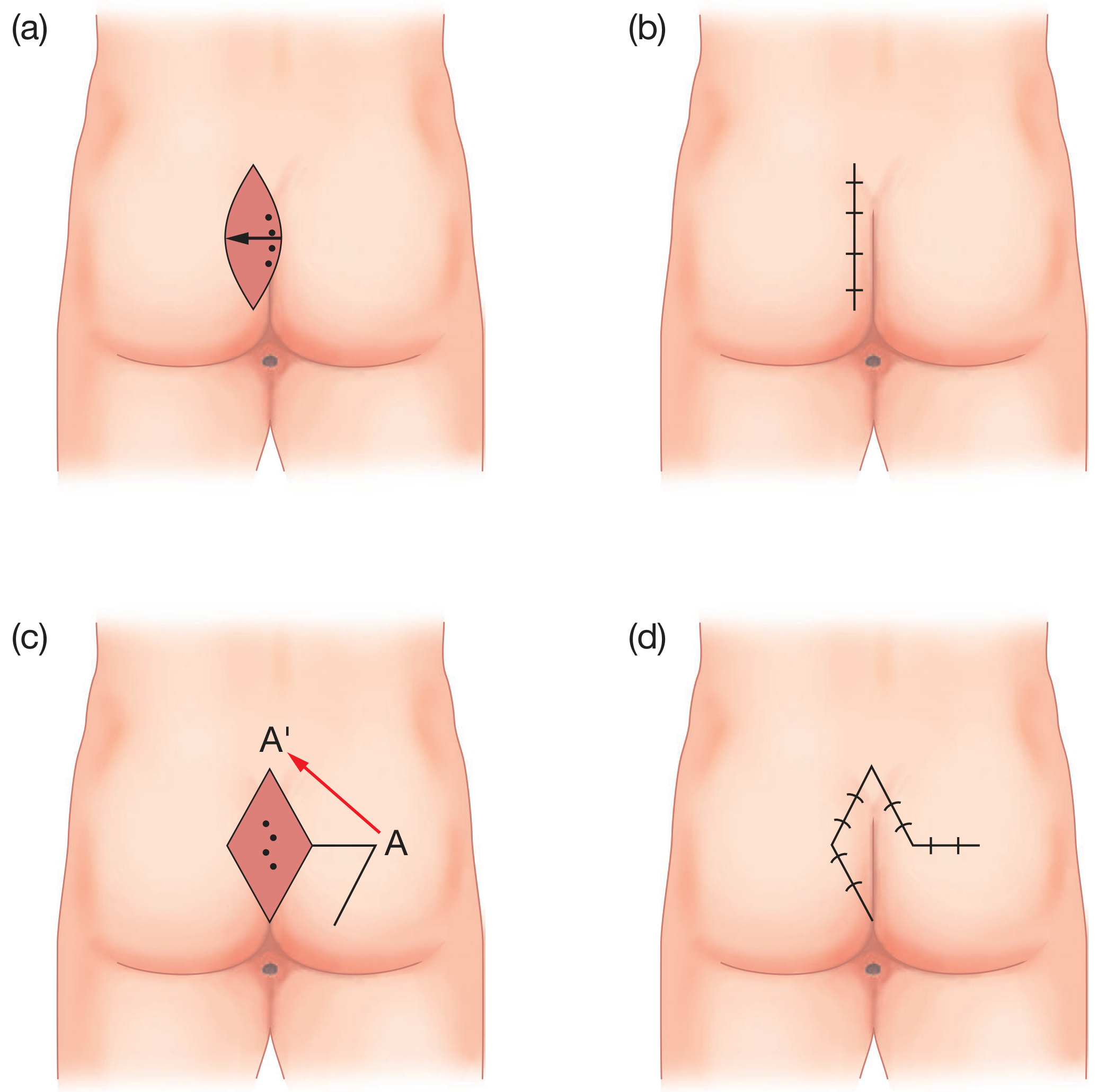
D4 - Off-Midline Closure: Karydakis Procedure (preferred)
- Elliptical incision made off the midline to encompass the disease
- A contralateral flap of skin and subcutaneous tissue is mobilised and advanced to achieve tension-free off-midline closure
- The scar lies to one side of the cleft, away from the midline - this is the key to reducing recurrence
D5 - Limberg (Rhomboid) Flap
- Disease excised using a rhomboid-shaped incision
- A measured, vascularized flap (point A) is rotated to fill the defect (point A') - see diagram above
- Achieves off-midline scar, good tissue bulk, low tension
D6 - Other Flap Options
- Z-plasty - transposes triangular skin flaps to break up scar line and relieve tension
- Bascom cleft lift - excises deep cleft and recreates a flattened natal cleft, reducing hair accumulation
- V-to-Y advancement flap - local tissue advancement
- Dufourmentel / S-flap - for wide defects with extensive disease
- Myocutaneous flap (superior gluteal artery based) - reserved for complex non-healing wounds, often with plastic surgery input
Intraoperative Images
Postoperative Care
- Wound care: Daily cleaning, packing (for open wounds), district nurse input
- Hair removal: Shaving/laser depilation of surrounding skin continued for months - critical to prevent recurrence
- Antibiotics: Continued if bacterial contamination confirmed
- Activity: Avoid prolonged sitting in early recovery; gentle walking encouraged
- Suture removal: Typically day 10-14 for closed wounds
- Follow-up: Monitor for wound breakdown, seroma, haematoma, or recurrence
Key Principles (Summary)
| Principle | Rationale |
|---|---|
| Avoid midline closure | Midline wounds are under shearing forces and break down frequently |
| Off-midline incision preferred | Reduces recurrence and non-healing (Karydakis, Limberg) |
| Drain dead space | Prevents seroma/haematoma that becomes infected |
| Thorough curettage | Remove all hair - foreign body reaction drives recurrence |
| Ongoing depilation | Recurrence is high if surrounding hair not controlled |
| No primary closure if infected | Wait for inflammation to settle |