
At the end of session, the students will be able to describe the anatomy of oesophagus, discuss the investigations and principles of management of achalasia cardia. Compare and contrast the treatment options — pneumatic dilation, Heller's myotomy (laparoscopic), POEM (peroral endoscopic myotomy), and botulinum toxin injection — including indications, outcomes, and complications.(15 marks)
Reading File
Now let me run parallel searches across the medical library for all relevant topics.
Finding Sources
Finding Sources
Finding Sources
Finding Sources
Excellent results. Now let me read the key sections in parallel.
Reading File
Reading File
Reading File
Finding Sources
Excellent content retrieved. Let me now read more detailed sections and fetch the key images.
Reading File
Reading File
Reading File
Loading Image
Excellent - I now have comprehensive content. Let me read a few more critical sections covering POEM, Heller's myotomy details, and oesophagus anatomy continuation, plus fetch the oesophagus muscle image.
Reading File
Reading File
Finding Sources
Loading Image
Now let me get the POEM and Heller's myotomy details plus achalasia investigation content.
Reading File
Finding Sources
Finding Sources
I now have comprehensive content from multiple authoritative textbooks. Let me also get the POEM and investigations content from Harrison's and Sabiston's.
Reading File
Reading File
I now have all the content needed for a comprehensive, well-structured answer. Let me also fetch the HRM subtypes image which is highly relevant.
Loading Image
Loading Image
I now have all the material needed to write a comprehensive, exam-quality answer. Let me compose the full response.
Anatomy of the Oesophagus, Achalasia Cardia: Investigations, Management Principles, and Comparative Treatment Options
PART I: ANATOMY OF THE OESOPHAGUS
Overview
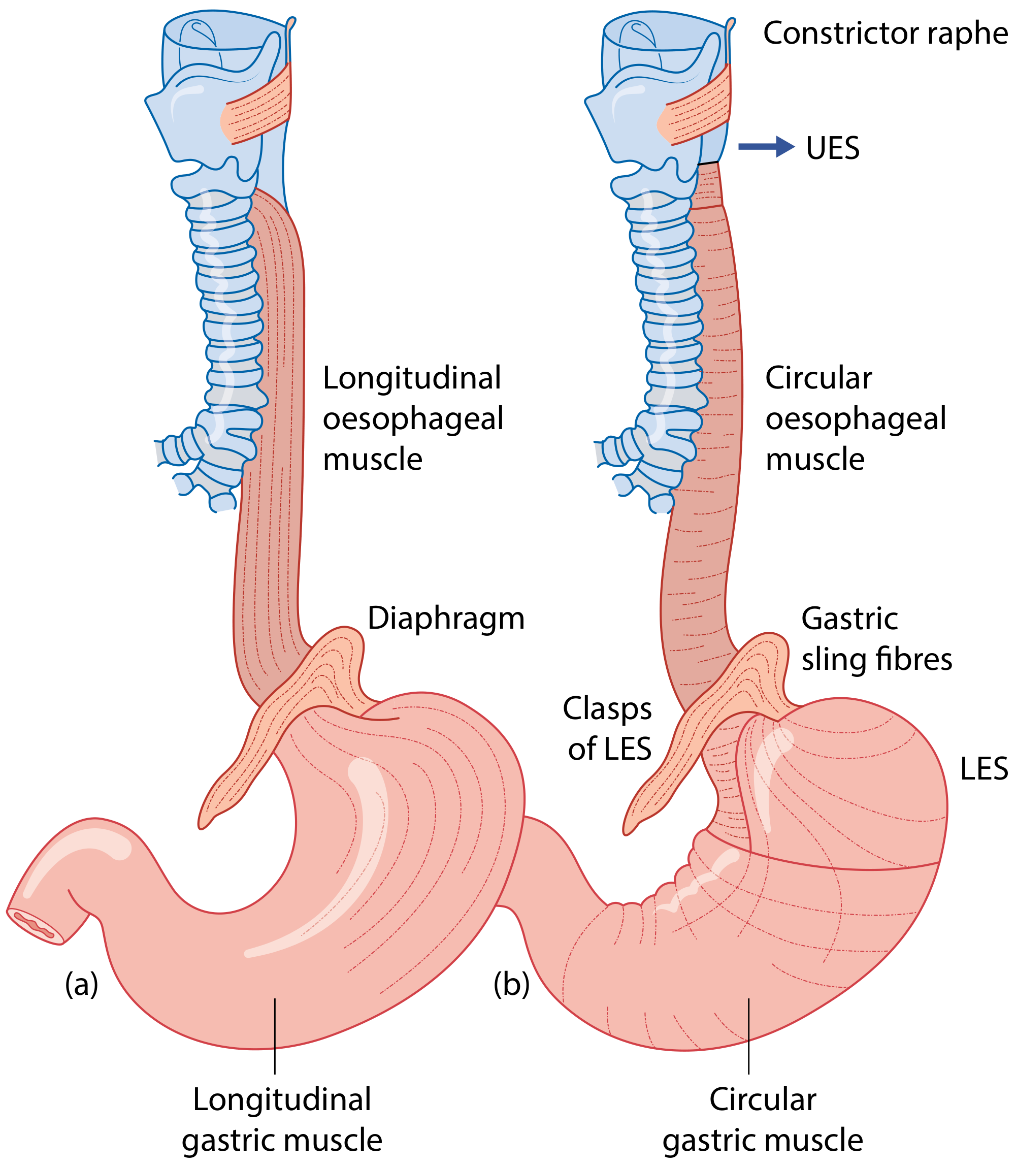
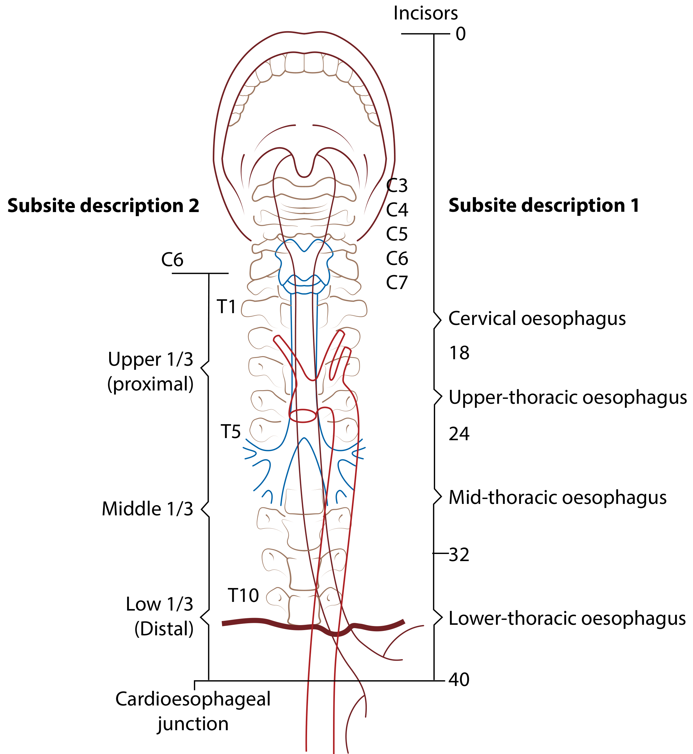
The oesophagus is a 25 cm long flattened muscular tube extending from the inferior border of the cricoid cartilage (C6) to the cardiac orifice of the stomach (T11). It is the narrowest part of the digestive tract (excluding the appendix) and passes through the oesophageal hiatus of the diaphragm at T10. It has three segments: cervical, thoracic, and abdominal. - Scott-Brown's Otorhinolaryngology Head & Neck Surgery
Segments
| Segment | Extent | Key Relations |
|---|---|---|
| Cervical | C6 to T1 (~5 cm) | Anterior: trachea; Posterior: vertebral column/longus colli; Lateral: carotid sheaths, thyroid lobes; Left: thoracic duct |
| Thoracic | T1 to T10 (~20 cm) | Superior mediastinum: anterior = trachea + left RLN; Posterior mediastinum: anterior = left main bronchus, pericardium, left atrium; Posterior = vertebrae T1-4, thoracic duct, azygos vein, descending aorta |
| Abdominal | T10-T11 (~1.5-2.5 cm) | Passes through oesophageal hiatus; anterior = left lobe of liver; gastro-oesophageal junction |
Three Anatomical Constrictions
Visible at rigid oesophagoscopy and clinically important for foreign body lodgement:
- Upper (cricopharyngeal/pharyngo-oesophageal junction) - at 15 cm from incisors - site of Killian's triangle, most common site for foreign body impaction
- Middle (aorto-bronchial) - at ~25 cm from incisors - where aortic arch and left main bronchus cross
- Lower (diaphragmatic) - at ~38 cm from incisors - at oesophageal hiatus
Wall Structure (Layers)
- Mucosa: Stratified squamous epithelium (changes to columnar at Z-line/squamocolumnar junction)
- Submucosa: Contains mucous glands
- Muscularis externa: Two layers - inner circular + outer longitudinal
- Upper 1/3: striated (voluntary) muscle
- Middle 1/3: mixed striated and smooth muscle
- Lower 1/3: smooth muscle
- No serosa - this has important surgical implications (spread of malignancy, anastomotic leak risk)
Blood Supply
- Cervical: Inferior thyroid artery
- Thoracic: Oesophageal branches of descending thoracic aorta, bronchial arteries
- Abdominal: Left gastric artery, left inferior phrenic artery
Venous Drainage
- Cervical → inferior thyroid veins → brachiocephalic vein
- Thoracic → azygos and hemiazygos veins → superior vena cava
- Abdominal → left gastric (coronary) vein → portal vein (portosystemic anastomosis - clinically important in portal hypertension causing oesophageal varices)
Lymphatic Drainage
- Cervical → deep cervical nodes
- Thoracic → posterior mediastinal nodes, tracheobronchial nodes
- Abdominal → coeliac nodes
- Note: submucosal lymphatics run longitudinally, allowing spread of malignancy over long distances before transmural invasion
Nerve Supply
- Parasympathetic: Vagus nerve (CN X) - forms oesophageal plexus around the lower thoracic oesophagus
- Sympathetic: From T5-T10 sympathetic ganglia via splanchnic nerves
- Intrinsic plexus: Auerbach's (myenteric) plexus between muscle layers; Meissner's (submucosal) plexus - loss of Auerbach's plexus neurons (particularly inhibitory nitrergic neurons) is the pathological basis of achalasia
Lower Oesophageal Sphincter (LES)
A functional, not anatomical, sphincter (~3-4 cm long, resting pressure 10-45 mmHg). Maintained by:
- Intrinsic circular smooth muscle tone
- Extrinsic crural diaphragm compression
- Angle of His (acute angle between oesophagus and stomach)
- Intra-abdominal segment of oesophagus
PART II: ACHALASIA CARDIA - INVESTIGATIONS AND PRINCIPLES OF MANAGEMENT
Definition and Pathophysiology
Achalasia cardia is a primary oesophageal motility disorder characterised by:
- Failure of LES relaxation (impaired LES relaxation on swallowing)
- Absent peristalsis in the oesophageal body
The underlying pathology is progressive degeneration and loss of inhibitory ganglion cells (nitric oxide-releasing and VIP neurons) in Auerbach's plexus. This destroys the inhibitory neurotransmission that normally triggers LES relaxation. The excitatory cholinergic neurons remain relatively intact, producing sustained high LES pressure. - Sleisenger and Fordtran's Gastrointestinal and Liver Disease
Epidemiology: Incidence ~1/100,000/year; affects both sexes equally; peak age 25-60 years. The aetiology remains poorly understood; viral triggers, autoimmune mechanisms, and genetic factors have been proposed.
Investigations
1. Barium Swallow (Upper GI Contrast Study)
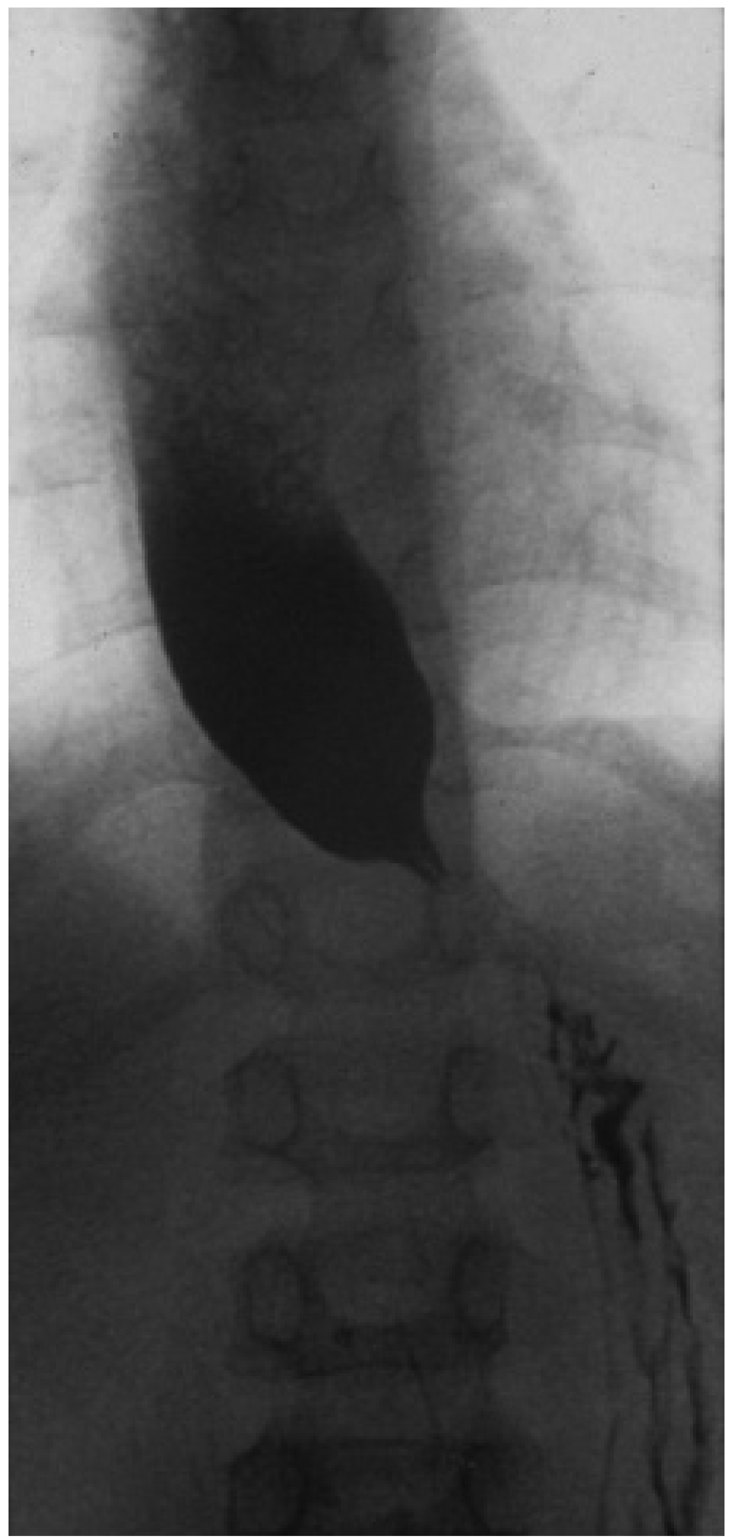
- Classic finding: "Bird's beak" or "rat-tail" appearance at the gastro-oesophageal junction
- Dilated, baggy oesophagus with tapered narrowing distally
- Absent primary peristalsis
- Food debris visible in dilated oesophagus
- In advanced disease: massively dilated, sigmoid-shaped oesophagus ("megaoesophagus")
2. High-Resolution Manometry (HRM) - Gold Standard
- Most sensitive diagnostic test - identifies disease before dilatation occurs
- Mandatory findings: (1) impaired LES relaxation (elevated integrated relaxation pressure, IRP > 15 mmHg) + (2) absent peristalsis
- Subclassifies achalasia into 3 types per the Chicago Classification:
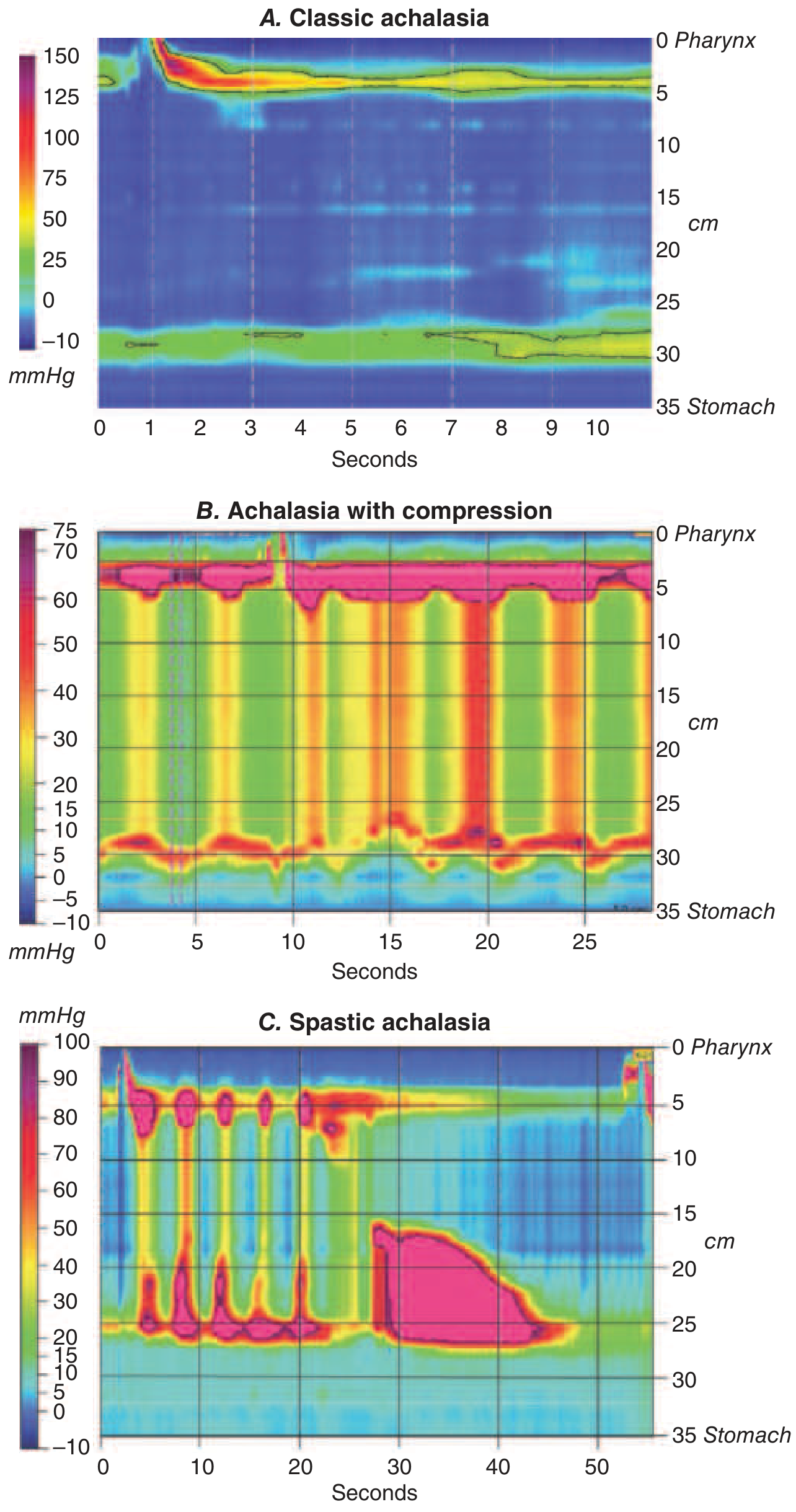
| Type | HRM Pattern | Treatment Response |
|---|---|---|
| Type I (Classic) | Absent peristalsis, minimal pressurisation | Good response to myotomy/PD |
| Type II (with compression) | Pan-oesophageal pressurisation >20 mmHg | Best response to all therapies (100% to PD; 93% to LHM) |
| Type III (Spastic) | Premature/spastic contractions | Poorest response to PD (40%); better with LHM (86%) or POEM |
3. Upper GI Endoscopy (OGD)
- Mandatory to exclude pseudoachalasia (secondary achalasia due to carcinoma at cardia - especially if age >60, rapid weight loss, short symptom duration)
- Findings: dilated oesophagus, food/fluid retention, "popping" sensation as endoscope passes through LES (pathognomonic), inflamed mucosa from stasis
- Biopsies from GEJ to rule out malignancy
4. CT Chest/Abdomen
- When pseudoachalasia is suspected (malignancy, external compression, e.g., lymphoma, lung cancer)
- Assess degree of dilatation and any mediastinal involvement
5. Timed Barium Oesophagram
- Column height of barium measured at 1, 2, and 5 minutes
- Used to monitor treatment response objectively
6. FLIP (Functional Lumen Imaging Probe)
- Newer modality assessing distensibility of the EGJ
- Useful when HRM results are inconclusive
Principles of Management
Since the underlying neuropathology cannot be reversed, all treatments are palliative - aimed at reducing LES pressure to allow gravity-assisted oesophageal emptying. - Harrison's Principles of Internal Medicine 22E
Principles:
- Reduce LES pressure (pharmacological, endoscopic, or surgical)
- Prevent complications (aspiration, oesophageal dilatation, carcinoma)
- Preserve oesophageal mucosa and avoid iatrogenic reflux
- Select treatment modality based on patient fitness, achalasia subtype, centre expertise
Pharmacological therapy (temporising only):
- Sublingual nifedipine 30-40 mg before meals (calcium channel blocker - reduces LES pressure)
- Isosorbide dinitrate sublingually (nitrate - smooth muscle relaxant)
- Sildenafil (PDE-5 inhibitor)
- Significant side effects (headache, hypotension, flushing), poor long-term efficacy; used as bridge to definitive therapy or in frail patients
PART III: COMPARATIVE ANALYSIS OF TREATMENT OPTIONS (15 Marks)
Overview Summary Table
| Feature | Pneumatic Dilation | Laparoscopic Heller's Myotomy | POEM | Botulinum Toxin Injection |
|---|---|---|---|---|
| Nature | Endoscopic/non-surgical | Laparoscopic surgery | Endoscopic submucosal tunnel surgery | Endoscopic injection |
| Mechanism | Forceful disruption of LES circular muscle | Surgical division of circular (and longitudinal) muscle fibres | Submucosal endoscopic division of inner circular muscle | Inhibits acetylcholine release at cholinergic nerve terminals |
| Efficacy (1-year) | 60-90% | 84-93% | ~90% | ~66% at 6 months |
| Durability | Moderate (requires repeat sessions) | High (durable at 5+ years) | High | Short (9 months average) |
| Risk of perforation/serious AE | ~1-5% perforation | ~1% perforation | ~1% mucosal injury | Minimal |
| GERD risk post-procedure | Low (10-20%) | Reduced with partial fundoplication (~8.8%) | High (up to 40-50%) | Negligible |
| Invasiveness | Outpatient, conscious sedation | General anaesthesia, 3-5 laparoscopic ports | General anaesthesia, endoscopic | Outpatient, conscious sedation |
| Repeatability | Yes (graded approach) | Revision possible but complex | Revision possible | Yes (but decreasing efficacy) |
| Preferred in | Good surgical risk, types I/II | Good surgical risk, type III, failed PD | Type III, failed LHM/PD, surgical high risk | Elderly/frail, diagnostic uncertainty, bridge therapy |
1. Pneumatic Dilation (PD)
Mechanism: A non-compliant cylindrical balloon (Rigiflex, 3.0-4.0 cm diameter) is passed over a guidewire and positioned fluoroscopically across the LES. The balloon is inflated until the radiological "waist" disappears, forcefully tearing the circular muscle fibres of the LES.
Technique:
- Patient fasted (liquid diet 1-2 days pre-procedure)
- Savary guidewire placed endoscopically into stomach
- Rigiflex balloon (3.0 cm first; 3.5 cm, then 4.0 cm in graded approach) inflated to 7-15 psi for 60 seconds under fluoroscopy
- Patient monitored for 2-6 hours post-procedure; gastrografin swallow if perforation suspected
Indications:
- First-line definitive therapy (especially type II achalasia - 100% efficacy in European trial)
- Good fit for most achalasia patients as initial treatment
- Preferred when surgical risk is high
- Type I and II achalasia
Outcomes:
- Initial success rates: 60-90%
- European Achalasia Trial (multicenter RCT): ~90% efficacy at 5 years when graded dilation used - equivalent to laparoscopic Heller myotomy
- Type II: 100% efficacy with PD vs 93% with LHM
- Type III: only ~40% efficacy - NOT preferred
- Repeat dilation improves cumulative success rates
Complications:
- Perforation: ~1-5% (most serious; identified by pain, subcutaneous emphysema, gastrografin leak - managed surgically or endoscopically)
- GERD: ~10-20% (less than after POEM, more than after LHM with fundoplication)
- Mucosal haematoma, post-procedure chest pain
- Recurrence requiring repeat dilation (~25-30% need repeat within 5 years)
Advantages: Outpatient procedure, low cost, repeatable, no surgery required, equivalent long-term efficacy to LHM with graded approach.
Disadvantages: Requires fluoroscopy, multiple sessions often needed, risk of perforation, not ideal for type III achalasia.
2. Laparoscopic Heller's Myotomy (LHM)
Mechanism: Surgical division of the circular (and longitudinal) muscle fibres of the distal oesophagus (6-8 cm above GEJ) extended 2.5-3 cm onto the gastric cardia, eliminating the mechanical LES obstruction. The first minimally invasive approach was thoracoscopic (Pellegrini, 1992); laparoscopic became the standard.
Technique:
- General anaesthesia, 5 laparoscopic ports
- Circumferential dissection of distal oesophagus
- Myotomy extended 6-8 cm proximally on oesophagus and 2.5-3 cm onto stomach
- Partial fundoplication added: Dor (anterior, 180°) or Toupet (posterior, 270°) fundoplication to prevent GERD without obstructing the myotomised segment. Complete Nissen fundoplication is NOT recommended as it recreates functional obstruction.
Indications:
- Gold standard surgical treatment for achalasia
- Preferred for type III (spastic) achalasia (86% success vs 40% with PD)
- Failed or repeated pneumatic dilation
- Young patients (more durable than PD)
- Patients with tortuous/sigmoid oesophagus
- Preference for single definitive procedure
Outcomes:
- Systematic review of >3000 LHMs: ~90% clinical success
- Boeckxstaens et al review (2264 patients, 42-month follow-up): 84% efficacy
- European Achalasia Trial: 84% success at 5 years
- With partial fundoplication: GERD rate 8.8% vs 31.5% with myotomy alone
- Equivalent to PD when graded dilation approach used
Complications:
- Oesophageal perforation/mucosal tear: ~1% (managed intraoperatively by direct suture repair)
- GERD if no fundoplication added (~30-47% without fundoplication)
- Dysphagia (incomplete myotomy, too tight fundoplication)
- Pneumothorax, mediastinal emphysema
- Splenic injury, bleeding (~1-2%)
- Conversion to open: rare
Advantages: Most durable single definitive treatment; fundoplication added to protect against GERD; ideal for type III; long-term data available.
Disadvantages: Requires general anaesthesia and laparoscopic surgery expertise; hospital admission; failed prior botulinum toxin injections may cause perilesional fibrosis making myotomy more difficult.
3. Peroral Endoscopic Myotomy (POEM)
Mechanism: Endoscopic third-space surgery (submucosal tunnel technique). The inner circular muscle fibres of the distal oesophagus and LES are divided from within a submucosal tunnel, without external incision. Pioneered by Inoue et al. (2010, Endoscopy).
Technique:
- General anaesthesia, patient supine
- Mucosal incision made ~12 cm proximal to GEJ on anterior wall
- Submucosal tunnel created distally to extend 2-3 cm onto gastric cardia
- Selective inner circular muscle layer divided using electrocautery knife (~10-12 cm myotomy)
- Mucosal entry site closed with endoscopic clips
- No external incision; hospital stay 1-2 days
Indications:
- Type III (spastic) achalasia - POEM allows longer myotomy proximally compared to LHM, ideal for spastic type
- Failed pneumatic dilation or LHM
- High surgical risk patients (avoids laparotomy)
- Younger patients desiring minimal invasiveness
- Prior surgery making laparoscopic approach difficult
- Re-do procedures
Outcomes:
- Short-term success: ~90%
- RCT (JAMA 2019): POEM vs pneumatic dilation - POEM was superior in treatment-naive patients (83% vs 72% success at 2 years)
- RCT (NEJM 2019): POEM vs LHM - POEM was non-inferior to LHM + Dor fundoplication for dysphagia control at 2 years; however, GERD rates significantly higher with POEM
- Particularly effective for type III: myotomy can be extended more proximally than LHM
- Long-term data (>5 years) still accumulating
Complications:
- GERD: Major drawback - 40-50% develop pathological reflux (vs ~10% with LHM + fundoplication). No fundoplication is added during POEM. Requires long-term PPI therapy.
- Capnoperitoneum/capnomediastinum (CO2 insufflation during tunnelling): usually self-limiting
- Mucosal perforation: ~1%
- Bleeding: rare
- Mediastinitis: rare
- Barrett's oesophagus risk with chronic reflux (long-term surveillance needed)
Advantages: Endoscopic, no external incision, flexible myotomy length (excellent for type III), can be used in failed surgical cases, shorter recovery, day 1-2 discharge.
Disadvantages: High GERD rate requiring PPI; no fundoplication possible in same session; operator-dependent technically demanding procedure; more reinterventions vs LHM reported in some series; higher cost than PD.
4. Botulinum Toxin Injection (BTX)
Mechanism: Botulinum toxin A (100 units) is injected endoscopically into 4 quadrants of the LES (1 cm proximal to Z-line), 1 cc per quadrant. It irreversibly blocks the release of acetylcholine from excitatory cholinergic nerve terminals, thereby reducing LES tone. Effect eventually reverses with axonal regeneration (typically 6-9 months). First described by Pasricha et al., Johns Hopkins, 1994. - Clinical Gastrointestinal Endoscopy, Expert Consult 3e
Indications:
- Elderly or frail patients who are high surgical risk and cannot tolerate PD, LHM, or POEM
- Diagnostic uncertainty - if positive response confirms functional LES obstruction and identifies candidates for definitive therapy
- Bridge therapy before definitive treatment
- Patients with recurrent symptoms after LHM or POEM (residual short sphincter segment)
- Chest pain-predominant achalasia with spasm features
- Patients refusing invasive procedures
NOT recommended as first-line therapy in young, fit patients.
Outcomes:
- Initial response: ~90% at 1 week (double-blind RCT data)
- Sustained response: ~66% at 6 months; only ~35% at 1 year
- Repeat injections can work but effectiveness diminishes with time
- Dose >100 units does not improve efficacy
Complications:
- Chest pain/discomfort (transient, several days)
- Occasional skin rash at injection site
- Generally very safe - no perforation risk
- Important: Repeated injections cause local fibrosis and inflammatory reaction, which can increase technical difficulty and complication rates if subsequent LHM or POEM is needed
- Rare: mediastinitis, pleural effusion
Advantages: Safest modality, outpatient procedure, no perforation risk, can be diagnostic/therapeutic simultaneously.
Disadvantages: Short duration of benefit (~9 months), repeated injections needed, decreasing efficacy with each injection, causes fibrosis that complicates future definitive procedures, not curative.
Head-to-Head Comparative Points
Efficacy Hierarchy (long-term, single session): LHM ≈ POEM > PD > BTX
GERD Risk Post-Procedure: POEM (40-50%) > PD (~15%) > LHM without fundoplication (31%) > LHM + Dor fundoplication (~8.8%) >> BTX (negligible)
Safety Profile: BTX > PD ≈ LHM ≈ POEM (perforation ~1% for last three)
Achalasia Subtype Guidance:
- Type I & II: PD is preferred first-line (low cost, 90-100% efficacy, equivalent to LHM at 5 years with graded approach); LHM equally valid
- Type III: POEM or LHM preferred (PD only 40% success; POEM allows longer proximal myotomy)
The European Achalasia Trial (Boeckxstaens et al., NEJM 2011): landmark multicenter RCT showed no statistically significant difference between PD and LHM at 5 years (~90% each). However, graded PD approach was used; single PD session is inferior.
The JAMA 2019 RCT (POEM vs PD): POEM showed superior symptom control (83% vs 72%) but significantly higher GERD.
The NEJM 2019 RCT (POEM vs LHM): POEM was non-inferior to LHM for dysphagia control but had substantially higher GERD (44% vs 10%).
When to Escalate / End-Stage Disease
~5% of patients eventually require oesophagectomy for:
- Massively dilated sigmoid oesophagus (megaoesophagus)
- Severe dysphagia unresponsive to all LES-directed therapy
- Peptic stricture or carcinoma
- Procedure: subtotal oesophagectomy with gastric pull-up; mortality ~2.7%; significant morbidity. Last resort. - Clinical Gastrointestinal Endoscopy 3e
Key Takeaway Points (Exam Summary)
- Anatomy: Oesophagus is 25 cm, no serosa, three constrictions (15/25/38 cm from incisors), dual nerve supply via vagus and sympathetic chain; submucosal venous plexus forms portosystemic anastomosis.
- Achalasia pathology: Loss of inhibitory (nitrergic) ganglion cells in Auerbach's plexus → absent LES relaxation + aperistalsis.
- Investigations: HRM (gold standard), barium swallow (bird's beak), OGD (exclude pseudoachalasia), CT (if malignancy suspected).
- Achalasia subtypes (HRM): Type I (classic), II (compression, best prognosis), III (spastic, hardest to treat).
- Treatment choice:
- Type I/II + fit patient: PD (first-line, graded) or LHM
- Type III: POEM or LHM (long myotomy)
- Frail/elderly: BTX injection
- Failed LHM or PD: POEM or re-do LHM
- End-stage megaoesophagus: oesophagectomy
- POEM vs LHM: Non-inferior efficacy, but POEM has higher GERD (requires PPI); LHM adds fundoplication for reflux control.
- BTX: Safe, short-acting, non-curative; fibrosis from repeated use complicates future surgery.
Sources: Scott-Brown's Otorhinolaryngology Head & Neck Surgery; Sleisenger and Fordtran's Gastrointestinal and Liver Disease; Harrison's Principles of Internal Medicine 22E; Clinical Gastrointestinal Endoscopy Expert Consult 3e; Sabiston Textbook of Surgery; Schwartz's Principles of Surgery 11E; Fischer's Mastery of Surgery 8E
This is a shared conversation. Sign in to Orris to start your own chat.