
teach me full concept of atherosclerosis pathology mbbs
Atherosclerosis - Complete Pathology for MBBS
1. Definition & Overview
- A soft, friable lipid core (mainly cholesterol, cholesterol esters, necrotic debris)
- Covered by a fibrous cap
2. Risk Factors
Non-Modifiable (Constitutional)
| Factor | Details |
|---|---|
| Genetics | Most important independent risk factor. Familial hypercholesterolemia (LDL receptor mutations) causes MI before age 20 in homozygotes |
| Age | Progressive process; symptomatic disease typically emerges after age 40 in men |
| Sex | Males at higher risk; females gain equal risk post-menopause (estrogen is atheroprotective) |
Modifiable (Major)
| Factor | Mechanism / Impact |
|---|---|
| Hyperlipidemia | Elevated LDL is the dominant lipid risk. Lowering cholesterol slows progression and causes partial plaque regression |
| Hypertension | Increases IHD risk ~60% vs. normotensives; mechanical stress causes endothelial injury |
| Cigarette smoking | Prolonged use doubles IHD death rate; cessation substantially reduces risk |
| Diabetes mellitus | 2x MI risk; 100x increased risk of atherosclerotic gangrene in lower extremities; induces hypercholesterolemia |
Additional Risk Factors
- Inflammation / CRP - CRP is a strong independent marker of risk for MI, stroke, and sudden cardiac death (even in apparently healthy people). CRP levels fall with statins, exercise, and weight loss.
- Hyperhomocysteinemia - Correlates with coronary, peripheral, and cerebrovascular disease
- Metabolic syndrome - Central obesity + insulin resistance + hypertension + dyslipidemia + hypercoagulability
- Lipoprotein(a) [Lp(a)] - Elevated levels independently raise coronary and cerebrovascular risk
- Hemostatic factors - Elevated plasminogen activator inhibitor-1 (PAI-1) predicts MI and stroke
Note: ~20% of cardiovascular events occur in people with none of the classic major risk factors. More than 75% of cardiovascular events in previously healthy females occur with LDL < 160 mg/dL.
3. Pathogenesis - "Response to Injury" Hypothesis
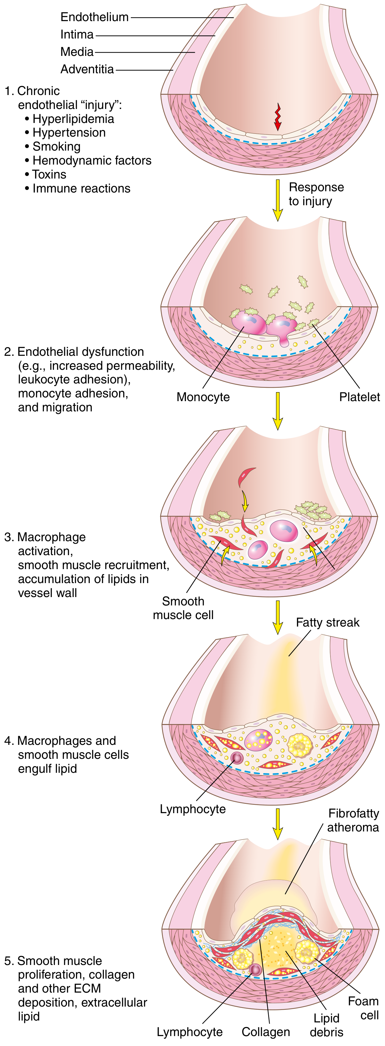
Step-by-Step Sequence:
Step 1 - Endothelial Injury & Dysfunction
- Endothelial cell (EC) injury is the cornerstone of atherosclerosis
- Early lesions begin at sites of intact but dysfunctional endothelium
- "Dysfunction" = defective vascular tone, impaired barrier function, reduced hemostatic regulation, and enhanced inflammation
- Two most important causes of EC dysfunction:
- Hemodynamic disturbances - Turbulent blood flow at ostia, branch points, and posterior abdominal aorta preferentially induces EC injury. Non-turbulent laminar flow induces "atheroprotective" genes - explaining why plaques form at specific anatomical sites
- Hypercholesterolemia - Chronic hyperlipidemia directly impairs ECs by increasing reactive oxygen species (ROS), which accelerate nitric oxide (NO) decay, dampening vasodilator activity
- Other triggers: inflammatory cytokines (TNF-α), cigarette smoke toxins, hypertension, immune reactions
Step 2 - Lipoprotein Accumulation & Oxidation
- With chronic hyperlipidemia, LDL enters and accumulates in the intima
- Oxidized LDL (ox-LDL) forms when LDL is oxidized by free radicals from inflammatory cells - this is particularly dangerous:
- Ox-LDL is toxic to ECs, SMCs, and macrophages
- It is taken up by macrophages via scavenger receptors (not the normal LDL receptor) - leading to foam cell formation since it cannot be degraded
- Stimulates release of growth factors, cytokines, and chemokines
- Recruits more monocytes - creating a vicious cycle
Step 3 - Monocyte Recruitment & Macrophage Activation
- Dysfunctional ECs express adhesion molecules (VCAM-1, ICAM-1, E-selectin) that capture monocytes and T lymphocytes from blood
- Monocytes migrate into the intima via chemokines (e.g., MCP-1) and differentiate into macrophages
- Macrophages engulf oxidized LDL via scavenger receptors → transform into foam cells (lipid-laden macrophages)
- Macrophages also sense cholesterol crystals and free fatty acids via the inflammasome → produce IL-1 → activates ECs, recruits more mononuclear cells
- Activated macrophages produce: ROS (enhance LDL oxidation), growth factors (drive SMC proliferation), and proteases (break down ECM)
Step 4 - Platelet Adhesion
- Exposed intimal components trigger platelet adhesion and activation
- Activated platelets release platelet-derived growth factor (PDGF) and other cytokines
Step 5 - T-Cell Recruitment & Inflammatory Amplification
- T lymphocytes enter the intima and release interferon-γ (IFN-γ) which:
- Activates macrophages further
- Activates ECs and SMCs
- Promotes inflammation (contributing to plaque instability)
Step 6 - Smooth Muscle Cell (SMC) Proliferation & Matrix Synthesis
- Growth factors (PDGF from platelets/macrophages, FGF, TGF-β) recruit SMCs from the media into the intima (phenotypic switch: contractile → synthetic)
- Intimal SMCs proliferate and synthesize ECM (especially collagen) - converting the fatty streak into a mature atheroma
- Collagen deposition forms and stabilizes the fibrous cap
- However, activated macrophages can also induce SMC apoptosis and ECM breakdown → unstable plaques
Step 7 - Lipid Accumulation (Extracellular)
- As foam cells die, lipids spill into the extracellular space → necrotic lipid core forms
- Cholesterol crystals become incorporated into the plaque
4. Morphology (Lesion Types)
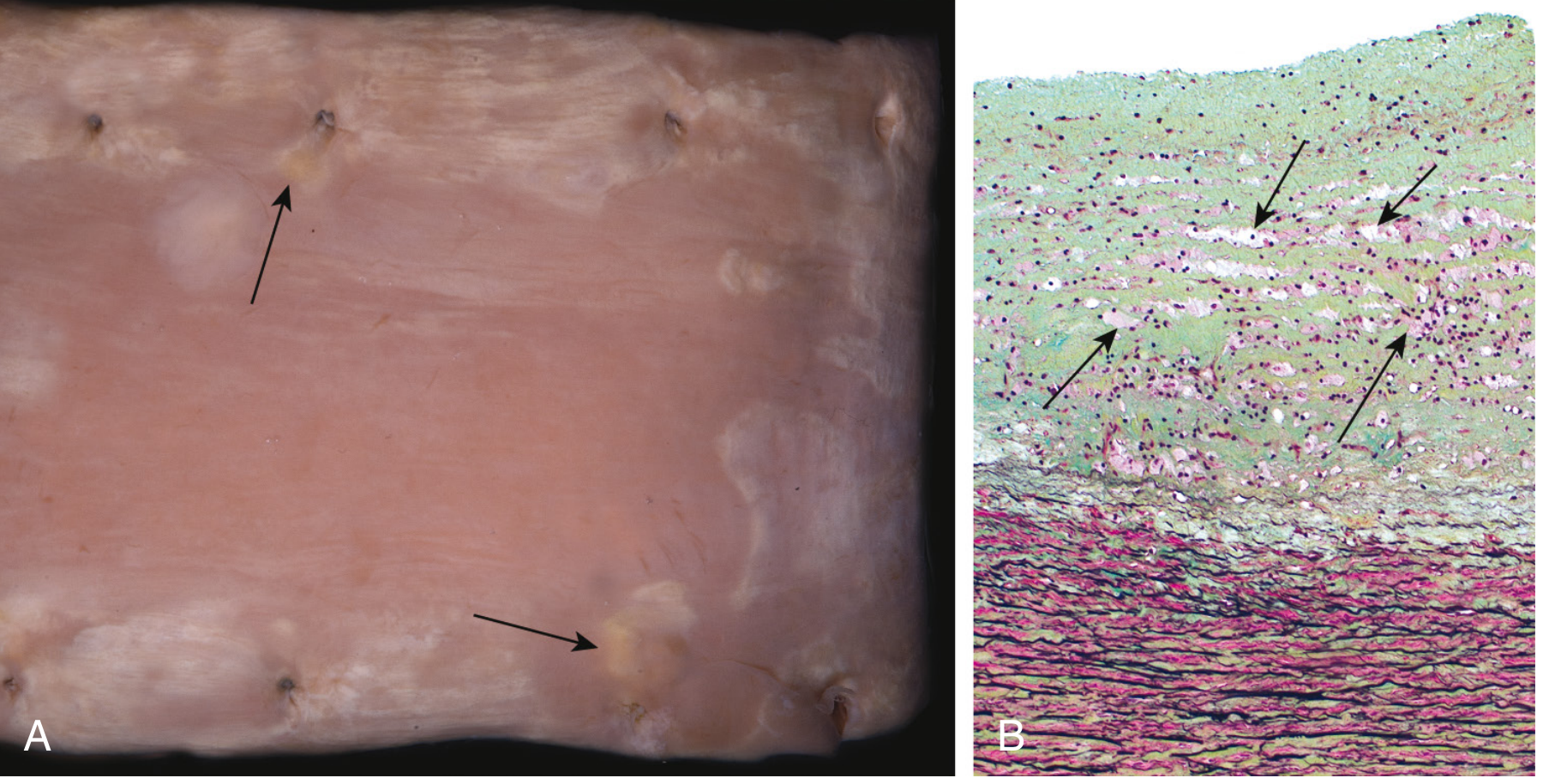
Stage 1 - Fatty Streak (Earliest Lesion)
- Gross: Minute, flat, yellow macules that coalesce into elongated lesions ≥1 cm long
- Microscopy: Intimal lipid-filled foam cells (macrophage-derived) - no significant flow disturbance
- Age: Can appear in the aorta of infants <1 year old; present in virtually ALL children >10 years, regardless of risk factors
- Significance: Not all progress to plaques - but coronary fatty streaks form during adolescence at the same sites that develop plaques later in life
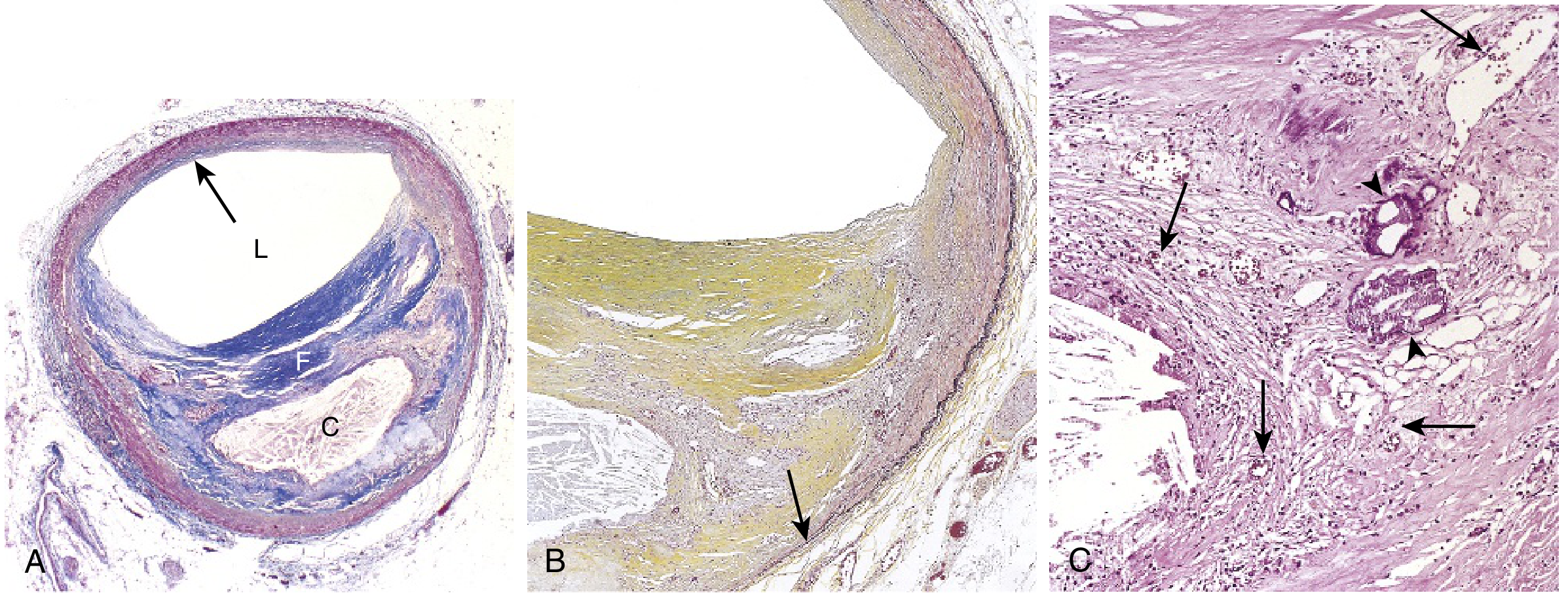
Stage 2 - Atherosclerotic Plaque (Atheromatous Plaque / Fibrous Plaque)
- Gross: White to yellow raised lesions, 0.3-1.5 cm in diameter (may coalesce); eccentric (involves only part of the vessel wall)
- Microscopy: Three key components:
- Fibrous cap - Smooth muscle cells, collagen (dense), proteoglycans, elastin; relatively few inflammatory cells
- Cellular zone (shoulder region) - Active area with macrophages, foam cells, T lymphocytes, SMCs; most prone to rupture
- Necrotic lipid core - Extracellular lipids, cholesterol crystals, necrotic debris, calcification; no collagen
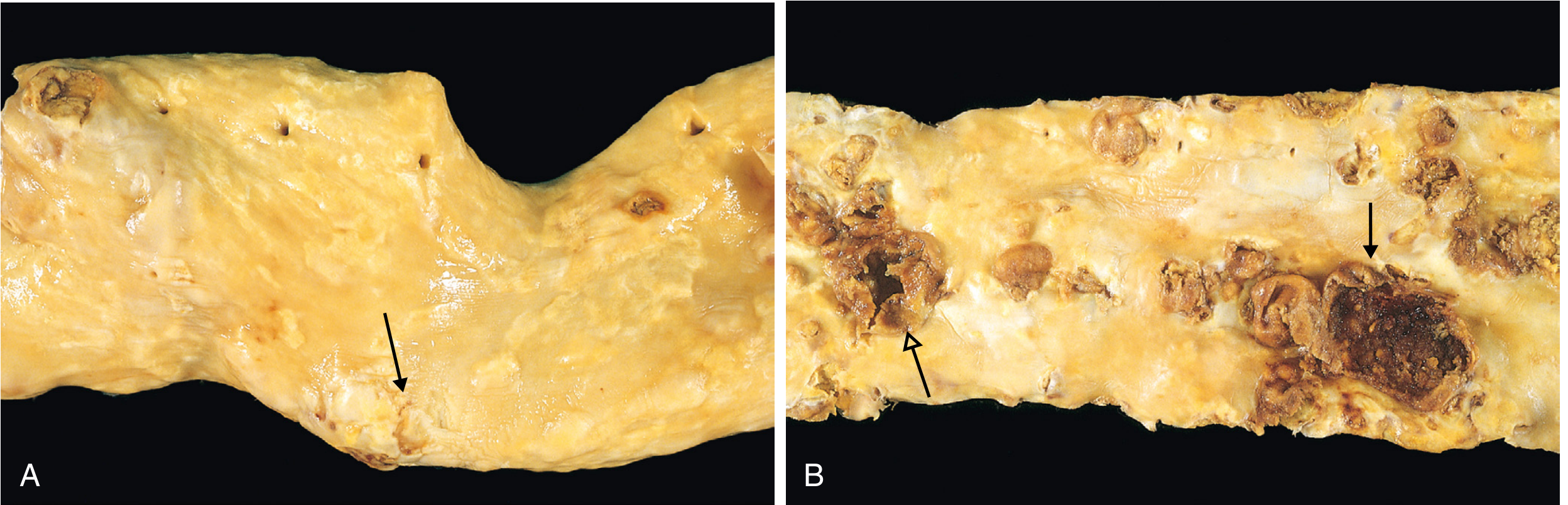
Distribution of Plaques (Descending Frequency):
- Infrarenal abdominal aorta (most common/severe)
- Coronary arteries
- Popliteal arteries
- Internal carotid arteries
- Circle of Willis
5. Stable vs. Vulnerable (Unstable) Plaque
| Feature | Stable Plaque | Vulnerable (Unstable) Plaque |
|---|---|---|
| Fibrous cap | Thick, rich in collagen | Thin, poorly supported |
| Lipid core | Small | Large (>40% of plaque volume) |
| Inflammatory cells | Few | Abundant macrophages and T cells |
| SMCs | Abundant | Reduced (apoptotic) |
| Clinical behavior | Stable angina | ACS: unstable angina, STEMI, NSTEMI |
| Risk | Gradual stenosis | Rupture → thrombosis |
- Macrophage-derived metalloproteinases (MMPs) that digest collagen in the fibrous cap
- T cell IFN-γ inhibiting collagen synthesis by SMCs
- SMC apoptosis from inflammatory signals
6. Consequences of Atherosclerosis / Acute Plaque Change
1. Plaque Rupture / Erosion / Ulceration
- Exposes highly thrombogenic subendothelial components and lipid core
- Triggers thrombus formation - can cause acute coronary syndrome (unstable angina, STEMI, NSTEMI)
- Most ruptures occur at the shoulder region of the plaque (highest inflammatory cell density, thinnest fibrous cap)
2. Hemorrhage into Plaque
- Rupture of overlying fibrous cap or thin-walled neovessels within the plaque
- Sudden expansion of plaque volume → acute luminal narrowing
3. Atheroembolism
- Plaque rupture can discharge atherosclerotic debris (cholesterol crystals, etc.) into the bloodstream
- Causes microemboli downstream (e.g., "blue toe syndrome")
4. Aneurysm Formation
- Atherosclerosis-induced ischemia of the underlying media with loss of elastic tissue → wall weakening → aneurysmal dilation and rupture
- Most common: infrarenal abdominal aortic aneurysm (AAA)
Atherosclerotic Stenosis (Chronic)
- Critical stenosis: ~70% occlusion is the tipping point in coronary arteries
- At rest: adequate perfusion; with exertion: demand exceeds supply → stable angina
- Chronic arterial hypoperfusion causes: bowel ischemia, chronic IHD, ischemic encephalopathy, intermittent claudication
7. Major Clinical Consequences
| Artery Involved | Clinical Consequence |
|---|---|
| Coronary arteries | Myocardial infarction, stable angina, sudden cardiac death |
| Cerebral / carotid arteries | Ischemic stroke, TIA |
| Aorta | Aortic aneurysm (AAA), aortic dissection |
| Peripheral arteries (popliteal, iliac) | Peripheral arterial disease, intermittent claudication, gangrene |
| Renal arteries | Renovascular hypertension, renal failure |
| Mesenteric arteries | Bowel ischemia / infarction |
8. Key High-Yield Points for MBBS Exams
- Response to injury hypothesis (Ross) = the core concept - endothelial injury is the initiating event
- Foam cells = lipid-laden macrophages (and SMCs) formed via scavenger receptor uptake of ox-LDL
- Fatty streak = earliest visible lesion; present in virtually all children >10 years; does NOT obstruct flow
- Fibrous cap = collagen + SMCs; protects against plaque rupture
- Vulnerable plaque = thin cap, large lipid core, heavy inflammation - responsible for ACS
- Most severe atherosclerosis = infrarenal abdominal aorta > coronary > popliteal > internal carotid
- Critical stenosis = 70% in coronary arteries = stable angina threshold
- CRP = strong independent inflammatory marker of cardiovascular risk (not causative)
- Hyperhomocysteinemia - additional risk factor (caused by vit B12/B6/folate deficiency or inborn errors)
- Diabetes = 100-fold increased risk of lower limb gangrene
Summary Diagram of Pathogenesis (Memorization Framework)
Endothelial Injury (hyperlipidemia, HTN, smoking, turbulence)
↓
Endothelial Dysfunction (↑ permeability, ↑ leukocyte adhesion)
↓
LDL enters intima → Oxidized LDL (ox-LDL)
↓
Monocytes adhere (VCAM-1) → migrate → Macrophages
↓
Macrophages engulf ox-LDL via scavenger receptors → FOAM CELLS → FATTY STREAK
↓
Platelet adhesion → PDGF released
Macrophages → IL-1, TNF-α, ROS, MMPs
T cells → IFN-γ
↓
SMC migration (media → intima) + proliferation + collagen synthesis
↓
Fibrous cap forms over lipid core → ATHEROSCLEROTIC PLAQUE
↓
Plaque growth → stenosis OR plaque rupture → THROMBOSIS → ACS/Stroke
teach me full concept of antihypertensive drugs pharmacology for mbbs in a understandable way
Antihypertensive Drugs - Complete Pharmacology for MBBS
Understanding Blood Pressure First
| Category | Systolic | Diastolic |
|---|---|---|
| Normal | 90-120 | 60-80 |
| Elevated | 120-129 | <80 |
| Stage I HTN | 130-139 | 80-89 |
| Stage II HTN | ≥140 | ≥90 |
Sites of Drug Action - The Big Picture
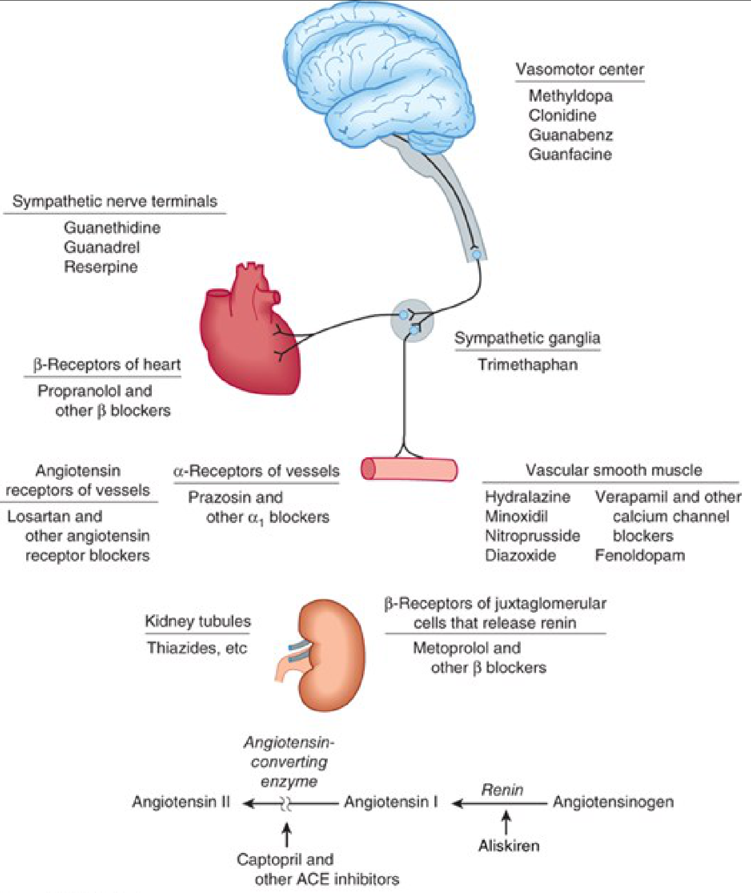
- Brain (vasomotor center) - centrally acting drugs
- Heart - beta blockers
- Blood vessels - CCBs, alpha blockers, vasodilators, ARBs
- Kidney - diuretics, beta blockers (↓renin), ACE inhibitors, ARBs
Classification of Antihypertensive Drugs
ANTIHYPERTENSIVE DRUGS
│
├── 1. DIURETICS
│ ├── Thiazides: Hydrochlorothiazide, Chlorthalidone, Indapamide
│ ├── Loop: Furosemide, Bumetanide, Torsemide
│ └── K⁺-sparing: Spironolactone, Eplerenone, Amiloride, Triamterene
│
├── 2. RENIN-ANGIOTENSIN SYSTEM (RAS) BLOCKERS
│ ├── ACE Inhibitors: Captopril, Enalapril, Lisinopril, Ramipril
│ ├── ARBs: Losartan, Valsartan, Olmesartan, Telmisartan
│ └── Direct Renin Inhibitor: Aliskiren
│
├── 3. CALCIUM CHANNEL BLOCKERS (CCBs)
│ ├── Dihydropyridines (vasodilators): Amlodipine, Nifedipine, Felodipine
│ └── Non-dihydropyridines (cardiac): Verapamil, Diltiazem
│
├── 4. SYMPATHOPLEGIC AGENTS
│ ├── Beta-blockers: Propranolol, Atenolol, Metoprolol, Carvedilol
│ ├── Alpha-1 blockers: Prazosin, Doxazosin, Terazosin
│ ├── Alpha + Beta blockers: Carvedilol, Labetalol
│ └── Central acting: Methyldopa, Clonidine
│
└── 5. DIRECT VASODILATORS
├── Oral: Hydralazine, Minoxidil
└── Parenteral: Sodium Nitroprusside, Diazoxide, Fenoldopam
CLASS 1: DIURETICS
How They Work
- Initial (first 6-8 weeks): Reduce blood volume and cardiac output → ↓ BP. Peripheral vascular resistance may actually go up initially.
- Long-term: Cardiac output returns toward normal but peripheral vascular resistance falls - this is the sustained mechanism.
A. Thiazide Diuretics
| Side Effect | Mechanism |
|---|---|
| Hypokalemia | ↑ Na delivery to collecting duct → K⁺ loss |
| Hyperuricemia | Compete with uric acid excretion → gout |
| Hyperglycemia | ↓ insulin secretion, ↑ insulin resistance |
| Hyperlipidemia | ↑ LDL, triglycerides |
| Hypercalcemia | Thiazides RETAIN calcium (unlike loop diuretics) |
| Hyponatremia | Can cause severe rapid hyponatremia |
Memory trick: Thiazides cause "GLUD" - Glucose up, Lipids up, Uric acid up, potassium Down. But calcium goes UP (opposite of loop diuretics).
B. Loop Diuretics
- Renal insufficiency (GFR < 30-40 mL/min) - thiazides don't work well
- Severe hypertension with multiple sodium-retaining drugs
- Pulmonary edema, cardiac failure, cirrhosis
- Short duration → only once-daily → rebound sodium retention (baroreceptor activation of RAS offsets the diuresis)
- Must be given twice daily; then become too aggressive
C. Potassium-Sparing Diuretics
- Mechanism: Block mineralocorticoid receptors in the collecting duct → block aldosterone-driven Na⁺ reabsorption / K⁺ secretion
- Use: Resistant hypertension (add-on), heart failure, primary hyperaldosteronism
- Side effects: Hyperkalemia; spironolactone causes gynecomastia (steroidal structure); eplerenone is more selective and avoids this
- Mechanism: Directly block epithelial Na⁺ channels (ENaC) in collecting duct
- Use: Usually combined with thiazides to prevent K⁺ loss; rarely used alone
- Liddle syndrome (genetic ENaC overactivity) responds specifically to amiloride
CLASS 2: RAS BLOCKERS
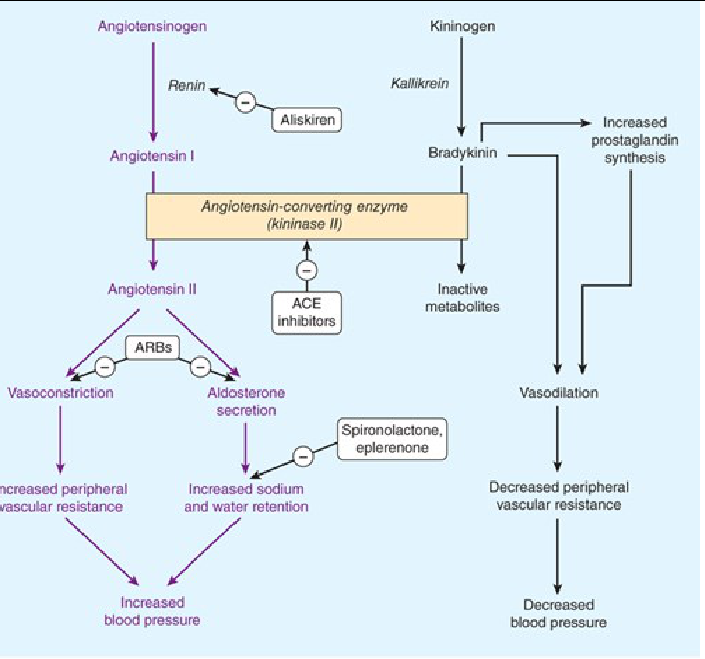
Angiotensinogen
↓ (Renin - from kidney JG cells)
Angiotensin I (inactive decapeptide)
↓ (ACE / Kininase II - in lung endothelium)
Angiotensin II (active octapeptide)
↓ acts on AT₁ receptors
→ Vasoconstriction (↑ TPR)
→ Aldosterone release (↑ Na retention → ↑ volume → ↑ BP)
→ ADH release
→ Sympathetic activation
A. ACE Inhibitors (ACEIs)
- Blocks conversion of Ang I → Ang II → ↓ vasoconstriction + ↓ aldosterone → ↓ BP
- Prevents breakdown of bradykinin → bradykinin accumulates → vasodilation (via NO, prostacyclin)
- Diabetic nephropathy: Reduce intraglomerular pressure (dilate efferent arteriole) → reduce proteinuria → protect kidneys
- Heart failure + post-MI: Reduce afterload, prevent cardiac remodeling
- Reduce incidence of new-onset diabetes
| Side Effect | Mechanism |
|---|---|
| Dry cough (10-15%) | Bradykinin + substance P accumulation → irritates bronchial mucosa; most common reason for discontinuation |
| Angioedema (rare, 0.1-0.2%) | Bradykinin accumulation; life-threatening; switch to ARB |
| Hyperkalemia | ↓ aldosterone → ↓ K⁺ excretion |
| First-dose hypotension | Especially in volume-depleted patients or high-renin states |
| Acute renal failure | In bilateral renal artery stenosis (RAS critically dependent on Ang II to maintain GFR) |
| Teratogenic | CONTRAINDICATED in pregnancy - causes fetal renal agenesis, oligohydramnios, skull defects (especially 2nd/3rd trimester) |
B. Angiotensin Receptor Blockers (ARBs)
| Feature | ACEIs | ARBs |
|---|---|---|
| Block Ang II formation | Yes | No (Ang II forms but can't act) |
| Bradykinin effect | ↑ bradykinin | No effect on bradykinin |
| Dry cough | Yes (common) | NO cough - major advantage |
| Angioedema | Yes (rare) | Very rare (bradykinin-independent) |
| Efficacy in HTN | Similar | Similar |
| Kidney protection | Yes | Yes |
Do NOT combine ACEIs + ARBs (dual RAS blockade) - dangerous: risk of acute renal failure, hyperkalemia, hypotension; no additional BP benefit.
C. Direct Renin Inhibitor
CLASS 3: CALCIUM CHANNEL BLOCKERS (CCBs)
Two Subgroups - Very Different Clinical Profiles:
| Feature | Dihydropyridines (DHPs) | Non-DHPs |
|---|---|---|
| Drugs | Amlodipine, Nifedipine, Felodipine, Nimodipine, Nicardipine | Verapamil, Diltiazem |
| Main action | Vascular selective vasodilation | Cardiac (heart rate + contractility) + mild vasodilation |
| Effect on HR | Reflex tachycardia (especially nifedipine) | ↓ Heart rate (rate-limiting) |
| Use in HTN | Yes - preferred | Yes, but with caution |
| Use in angina | Vasospastic (Prinzmetal) angina | Stable angina, SVT, AF rate control |
| Cardiac effects | Minimal depression | Significant depression |
- Amlodipine is the most widely used DHP - very long half-life (35-50 hrs), smooth BP control, once daily, well tolerated
- Nifedipine short-acting - should NOT be used for chronic HTN (rapid BP fall → reflex tachycardia → ischemia); only sustained-release forms acceptable
- Verapamil - greatest cardiac depressant; can cause AV block, bradycardia, constipation
- Verapamil/Diltiazem: Heart failure with reduced EF, 2nd/3rd degree AV block, sick sinus syndrome
- Verapamil + beta-blockers: Dangerous combination (additive cardiac depression → heart block)
CLASS 4: SYMPATHOPLEGIC AGENTS
A. Beta-Blockers (β-Blockers)
- Non-selective: Propranolol (β₁ + β₂), Carvedilol (α₁ + β₁ + β₂), Labetalol (α₁ + β)
- Cardioselective (β₁ preferential): Atenolol, Metoprolol, Bisoprolol
- ↓ Cardiac output - block β₁ receptors in heart → ↓ heart rate + ↓ contractility
- ↓ Renin secretion - block β₁ receptors on juxtaglomerular cells → ↓ renin → ↓ Ang II → ↓ aldosterone
- Central effect - reduce sympathetic outflow from vasomotor center
- Presynaptic β₂ block - reduce norepinephrine release
- HTN with post-MI: Excellent (reduce mortality)
- HTN with heart failure: Carvedilol, Bisoprolol, Metoprolol
- HTN with angina: Beta-blockers are first-line
- HTN with atrial fibrillation (rate control): Yes
- HTN with aortic aneurysm: Beta-blockers specifically
| Side Effect | Reason |
|---|---|
| Bradycardia | β₁ block on heart |
| AV block | β₁ block on conduction |
| Bronchospasm | β₂ block in lungs (use cardioselective in asthmatics with caution) |
| Fatigue, depression | CNS effects |
| Cold extremities | β₂ block → peripheral vasoconstriction |
| Masking of hypoglycemia | β₂ block → impair glucagon response; mask tachycardia of hypoglycemia |
| Dyslipidemia | ↑ triglycerides, ↓ HDL |
| Rebound hypertension | Never stop abruptly - taper slowly |
Current guidelines suggest restricting beta-blockers to patients with compelling indications (heart failure, post-MI, angina, AF) rather than using them as first-line for uncomplicated HTN.
B. Alpha-1 Blockers
- First-dose phenomenon: Severe postural hypotension after first dose (especially prazosin) - give at bedtime, start low
- Reflex tachycardia
- Sodium and water retention
- Dizziness, nasal stuffiness
C. Centrally Acting Agents
- Clonidine: Stimulates α₂ receptors in the brainstem vasomotor center → ↓ sympathetic outflow → ↓ heart rate + ↓ BP
- Methyldopa: Converted to α-methylnorepinephrine in the brain → acts as α₂ agonist (same mechanism)
- Sedation, dry mouth (clonidine - most common)
- Rebound hypertension with abrupt withdrawal of clonidine (catecholamine surge) - must taper slowly
- Methyldopa: Drug of choice in pregnancy (proven safety); also causes hemolytic anemia, positive Coombs test, hepatotoxicity, lupus-like syndrome
- HTN emergencies (oral/transdermal)
- Opioid/nicotine/alcohol withdrawal
- ADHD, menopausal flushing
CLASS 5: DIRECT VASODILATORS
A. Hydralazine
- Severe/resistant hypertension (combined with beta-blocker + diuretic)
- Hypertensive urgency in pregnancy (IV hydralazine is preferred for preeclampsia)
- Heart failure combined with nitrates (BiDil) in patients who cannot tolerate ACEIs/ARBs (especially in Black patients)
- Headache, nausea, flushing, palpitations, tachycardia
- Drug-induced lupus syndrome: At doses ≥400 mg/day; arthralgia, myalgia, rash, fever. No renal damage (unlike true lupus); reverses on stopping drug. More common in slow acetylators.
- Peripheral neuropathy (treat with pyridoxine)
- Reflex tachycardia + angina in patients with coronary artery disease
B. Minoxidil
- Severe reflex tachycardia, palpitations, angina
- Severe fluid retention, edema
- Hypertrichosis (hair growth on face/body) - problematic especially in women; paradoxically used topically as "Rogaine" for baldness treatment
C. Sodium Nitroprusside (Parenteral)
Drug Selection Based on Compelling Indications
| Clinical Condition | Preferred Drug(s) | Avoid |
|---|---|---|
| Uncomplicated HTN | Any first-line: ACEI/ARB, CCB, Thiazide | - |
| Diabetes + HTN | ACEI or ARB (protect kidneys) | - |
| Diabetic nephropathy/proteinuria | ACEI or ARB | - |
| Heart failure | ACEI/ARB + Beta-blocker + Diuretic + MRA | Non-DHP CCBs, alpha-blockers |
| Post-MI | Beta-blocker + ACEI/ARB | - |
| Coronary artery disease/Angina | Beta-blocker, CCB, ACEI | - |
| AF - rate control | Beta-blocker or Verapamil/Diltiazem | Dihydropyridine CCBs alone |
| Aortic aneurysm | Beta-blocker | - |
| BPH + HTN | Alpha-1 blocker (Doxazosin) | - |
| Pregnancy | Methyldopa (1st line), Hydralazine, Labetalol | ACEIs, ARBs (teratogenic!) |
| Renal artery stenosis (bilateral) | CCB, Beta-blocker | ACEIs, ARBs |
| Isolated systolic HTN (elderly) | Thiazide, DHP-CCB, ACEI/ARB | - |
| Resistant HTN | Add Spironolactone (4th drug) | - |
| Left ventricular hypertrophy | ACEI, ARB, CCB | - |
| Black patients | CCB + Thiazide (ACEIs less effective alone) | - |
| Gout | CCB, ARB (losartan is uricosuric) | Thiazides |
| Asthma/COPD | CCB, ACEI/ARB, Thiazide | Beta-blockers, ACEIs (cough worsens) |
| Hypertensive emergency | IV Nitroprusside, IV Labetalol, IV Nicardipine | - |
| Pheochromocytoma | Alpha-blocker first, then add beta-blocker | Never give beta-blocker alone first (unmasked alpha → BP crisis) |
Combination Therapy - Why and What Works Together
- Vasodilators → reflex tachycardia + sodium retention
- Adding a beta-blocker blocks tachycardia; adding a diuretic blocks sodium retention
- All three drugs then work synergistically
- ACEI/ARB + CCB - Excellent (NICE guidelines first choice in most patients): additive vasodilation, CCB-induced edema reduced by ACEI
- ACEI/ARB + Thiazide - Very effective; diuretic activates RAS → more substrate for ACEI to block
- CCB + Thiazide - Complementary mechanisms
- ACE + ARB - Avoid (dual RAS blockade: renal failure, hyperkalemia)
- Beta-blocker + Verapamil/Diltiazem - Avoid (severe bradycardia/AV block)
- Beta-blocker + Alpha-blocker - Avoid (Labetalol/Carvedilol already combine both)
Hypertensive Emergencies (Quick Reference)
| Drug | Route | Best For |
|---|---|---|
| Sodium nitroprusside | IV infusion | Most emergencies |
| Labetalol | IV bolus/infusion | Aortic dissection, eclampsia |
| IV Nicardipine | IV infusion | Most emergencies, stroke |
| IV Hydralazine | IV | Eclampsia/preeclampsia |
| IV Esmolol | IV | Aortic dissection, tachycardia |
| Fenoldopam | IV | Renal impairment (↑ renal blood flow) |
Summary Memory Framework
BP = CO × TPR
DIURETICS → ↓ volume → ↓ CO (initial), then ↓ TPR
BETA-BLOCKERS → ↓ HR and contractility → ↓ CO; + ↓ renin → ↓ Ang II → ↓ TPR
CCBs → ↓ TPR (DHPs) or ↓ CO + ↓ TPR (non-DHPs)
ACEIs/ARBs → ↓ Ang II effects → ↓ TPR + ↓ volume (↓ aldosterone)
ALPHA-BLOCKERS → block NE → ↓ TPR
CENTRAL DRUGS → ↓ sympathetic outflow → ↓ CO + ↓ TPR
VASODILATORS → direct ↓ TPR (but cause reflex ↑ CO - need combination)
High-Yield Exam Points
- ACEIs cause dry cough due to bradykinin; switch to ARB if intolerable
- ACEIs/ARBs are teratogenic - absolutely contraindicated in pregnancy
- Methyldopa = drug of choice in pregnancy
- Thiazides retain calcium (opposite of loop diuretics which waste calcium)
- Clonidine rebound - never stop abruptly; causes sympathetic storm
- Hydralazine slow acetylators → lupus-like syndrome at lower doses
- Minoxidil must be given with beta-blocker + diuretic
- Nitroprusside → cyanide toxicity → treat with sodium thiosulfate
- Beta-blocker first in pheochromocytoma = contraindicated (give alpha-blocker first, always)
- Losartan is uricosuric (useful when patient has both HTN and gout)
- Spironolactone causes gynecomastia (steroid structure) → use eplerenone if this is an issue
- Verapamil + beta-blocker = dangerous combination (severe AV block)
- Bilateral RAS = ACEIs/ARBs contraindicated (GFR depends on Ang II to maintain efferent arteriolar tone)
- Amlodipine = longest acting DHP CCB; preferred in most stable hypertensives
make it shorter with most knowing points
Antihypertensive Drugs - High-Yield MBBS Summary
The Core Formula
Classification at a Glance
| Class | Key Drugs |
|---|---|
| Diuretics | HCTZ, Furosemide, Spironolactone |
| ACE Inhibitors | Captopril, Enalapril, Lisinopril, Ramipril |
| ARBs | Losartan, Valsartan, Telmisartan |
| CCBs | Amlodipine, Nifedipine, Verapamil, Diltiazem |
| Beta-Blockers | Propranolol, Atenolol, Metoprolol, Carvedilol |
| Alpha-1 Blockers | Prazosin, Doxazosin |
| Central Acting | Methyldopa, Clonidine |
| Direct Vasodilators | Hydralazine, Minoxidil, Nitroprusside |
Drug-by-Drug: Mechanism + Key Points
1. Thiazide Diuretics (HCTZ, Chlorthalidone)
- MOA: Block Na⁺/Cl⁻ transporter in distal convoluted tubule → ↓ volume → ↓ BP
- Side effects (remember "GLUD + HH"):
- Glucose↑ (hyperglycemia)
- Lipids↑ (hyperlipidemia)
- Uric acid↑ (hyperuricemia → gout)
- K (potassium) ↓ (hypokalemia)
- Hypercalcemia (thiazides RETAIN calcium - opposite of loop diuretics)
- Hyponatremia (can be severe)
- Use: First-line for mild-moderate uncomplicated HTN
2. Loop Diuretics (Furosemide)
- MOA: Block Na⁺/K⁺/2Cl⁻ in loop of Henle
- Use: HTN with renal failure (GFR < 30), heart failure, pulmonary edema
- Side effects: Hypokalemia, hypocalcemia (LOSES calcium - opposite of thiazides), ototoxicity
- Less preferred for HTN alone (short acting → rebound Na retention)
3. Spironolactone / Eplerenone
- MOA: Aldosterone receptor antagonist → blocks Na retention, K loss
- Use: Resistant HTN (4th drug add-on), heart failure, primary hyperaldosteronism
- Side effect: Hyperkalemia; spironolactone → gynecomastia (use eplerenone to avoid this)
4. ACE Inhibitors
- MOA: Block ACE → ↓ Ang II (↓ vasoconstriction, ↓ aldosterone) + ↑ bradykinin (vasodilation)
- Benefits beyond BP:
- Renal protection in diabetics (↓ proteinuria)
- Heart failure, post-MI (↓ remodeling)
- Side effects - HIGH YIELD:
| Side Effect | Why |
|---|---|
| Dry cough (10-15%) | Bradykinin accumulation - most common reason to stop |
| Angioedema (rare) | Bradykinin; life-threatening; switch to ARB |
| Hyperkalemia | ↓ aldosterone → ↓ K⁺ excretion |
| Teratogenic | Fetal renal agenesis - absolutely banned in pregnancy |
| Acute renal failure | In bilateral renal artery stenosis |
5. ARBs (Losartan, Valsartan)
- MOA: Block AT₁ receptors directly; Ang II forms but can't act
- Key advantage over ACEIs: No cough, no angioedema (no bradykinin effect)
- Same indications and contraindications as ACEIs
- Do NOT combine ACEI + ARB - dual RAS blockade causes renal failure + hyperkalemia
- Losartan bonus: uricosuric → useful in HTN + gout
6. Calcium Channel Blockers (CCBs)
| Feature | Dihydropyridines (DHPs) | Non-DHPs |
|---|---|---|
| Drugs | Amlodipine, Nifedipine | Verapamil, Diltiazem |
| Main action | Vasodilation (↓ TPR) | Cardiac depression (↓ HR, ↓ contractility) |
| Heart rate | ↑ (reflex tachycardia) | ↓ |
| Use in AF | No | Yes (rate control) |
| Key side effect | Peripheral edema, flushing | Bradycardia, AV block, constipation (verapamil) |
- Amlodipine = most preferred DHP (long half-life, once daily, smooth BP control)
- Short-acting nifedipine = AVOID in chronic HTN (reflex tachycardia → ischemia)
- Verapamil + Beta-blocker = DANGEROUS (severe bradycardia/AV block - never combine)
7. Beta-Blockers
- Block β₁ in heart → ↓ HR + ↓ contractility → ↓ CO
- Block β₁ in kidney JG cells → ↓ renin → ↓ Ang II
- ↓ Central sympathetic outflow
| Effect | Mechanism |
|---|---|
| Bronchospasm | β₂ block in lungs |
| Masking hypoglycemia | β₂ block (also masks tachycardia of hypoglycemia) |
| Cold extremities | β₂ block → peripheral vasoconstriction |
| Bradycardia/AV block | β₁ block |
| Rebound HTN on abrupt stop | Receptor upregulation → always taper |
8. Alpha-1 Blockers (Prazosin, Doxazosin)
- MOA: Block α₁ on vessels → prevent NE-mediated vasoconstriction → ↓ TPR
- Unique use: HTN + BPH (also relaxes urethral smooth muscle)
- First-dose phenomenon: Severe postural hypotension after first dose - give at bedtime, start low
9. Methyldopa + Clonidine (Central Acting)
- MOA: α₂ agonists in brainstem → ↓ sympathetic outflow → ↓ BP
- Methyldopa: Drug of choice in pregnancy (proven safety record). Also causes hemolytic anemia, positive Coombs test
- Clonidine: Rebound hypertension if stopped abruptly (always taper). Also used for opioid withdrawal, ADHD
10. Direct Vasodilators
| Drug | MOA | Key Points |
|---|---|---|
| Hydralazine | Dilates arterioles (↓ TPR) | Drug of choice for HTN in pregnancy (IV); lupus-like syndrome at high doses (>400 mg/day); slow acetylators at higher risk; must combine with beta-blocker + diuretic |
| Minoxidil | Opens K⁺ channels → hyperpolarization → vasodilation | Most potent oral vasodilator; causes hypertrichosis (hair growth); must combine with beta-blocker + diuretic; topical form = Rogaine |
| Sodium Nitroprusside | Releases NO → dilates arteries + veins | IV only; hypertensive emergencies; risk of cyanide toxicity (treat with sodium thiosulfate) |
Drug Selection - Must Know Table
| Condition | Use | Avoid |
|---|---|---|
| Uncomplicated HTN | ACEI/ARB, CCB, Thiazide | - |
| Diabetes/Proteinuria | ACEI or ARB | - |
| Heart Failure | ACEI + Beta-blocker + Diuretic + MRA | Non-DHP CCBs |
| Post-MI | Beta-blocker + ACEI | - |
| Angina | Beta-blocker, CCB | - |
| Pregnancy | Methyldopa, Labetalol, Hydralazine | ACEIs, ARBs |
| Asthma | CCB, ACEI, Thiazide | Beta-blockers |
| BPH + HTN | Alpha-1 blocker | - |
| Gout + HTN | Losartan (uricosuric), CCB | Thiazides |
| AF rate control | Verapamil, Diltiazem, Beta-blocker | - |
| Bilateral renal artery stenosis | CCB, Beta-blocker | ACEIs, ARBs |
| Pheochromocytoma | Alpha-blocker FIRST, then beta-blocker | Beta-blocker alone first (→ BP crisis) |
| Resistant HTN (4th drug) | Spironolactone | - |
| Hypertensive emergency | IV Nitroprusside, IV Labetalol | - |
| Black patients | CCB + Thiazide | ACEIs less effective alone |
Top 15 Exam One-Liners
- ACEIs cause dry cough → bradykinin accumulation → switch to ARB
- ACEIs/ARBs = teratogenic → absolutely contraindicated in pregnancy
- Methyldopa = drug of choice in pregnancy
- Thiazides retain Ca²⁺; Loop diuretics lose Ca²⁺ - opposite effects
- Spironolactone causes gynecomastia → use eplerenone instead
- Clonidine rebound → never stop abruptly (taper always)
- Hydralazine + slow acetylators → lupus-like syndrome at lower doses
- Minoxidil causes hypertrichosis → topical form used for baldness (Rogaine)
- Nitroprusside → cyanide toxicity → treat with sodium thiosulfate
- Verapamil + beta-blocker = dangerous → severe AV block
- First-dose hypotension → Prazosin (give at bedtime)
- Pheochromocytoma: Always give alpha-blocker before beta-blocker
- Losartan is uricosuric → preferred in HTN with gout
- Short-acting nifedipine → avoid in chronic HTN (reflex tachycardia)
- ACEI + ARB together → never combine (renal failure + hyperkalemia)
teach me mbbs final exam concepts of cholinergic drugs
-
Autonomic basis
- Acetylcholine synthesis, release, degradation
- Muscarinic vs nicotinic receptors
- Parasympathetic organ effects
-
Direct-acting cholinomimetics
- Bethanechol, pilocarpine, carbachol, methacholine
-
Indirect-acting cholinomimetics
- Reversible anticholinesterases: neostigmine, pyridostigmine, physostigmine, donepezil
- Irreversible anticholinesterases: organophosphates
-
Clinical uses
- Myasthenia gravis
- Postoperative ileus and urinary retention
- Glaucoma and xerostomia
- Alzheimer disease
- Reversal of nondepolarizing neuromuscular block
-
Adverse effects and contraindications
- Muscarinic toxicity, nicotinic toxicity, CNS toxicity
- Why asthma, peptic ulcer disease, bradycardia, and urinary obstruction matter
-
Organophosphate poisoning
- Cholinergic toxidrome
- Diagnosis
- Treatment with atropine, oxime therapy, decontamination, and airway support
-
High-yield comparisons
- Neostigmine vs physostigmine
- Atropine vs pralidoxime
- Muscarinic vs nicotinic manifestations
- Cholinergic crisis vs myasthenic crisis
Explain cholinergic drug classification and mechanisms
cholinergic drugs pharmacology
cholinergic agonists acetylcholinesterase inhibitors pharmacology mechanism Merck Manual
Cholinergic Drugs: Classification and Mechanisms
1. What are cholinergic drugs?
- Parasympathetic postganglionic nerves
- Autonomic ganglia
- Neuromuscular junction
- Some CNS pathways
- Sympathetic nerves supplying sweat glands
- Directly stimulating cholinergic receptors, or
- Indirectly increasing ACh by inhibiting acetylcholinesterase (AChE).
2. Receptors to know first
A. Muscarinic receptors
| Receptor | G protein | Main location | Major effect |
|---|---|---|---|
| M1 | Gq | CNS, autonomic ganglia, gastric glands | Neural excitation, gastric acid secretion |
| M2 | Gi | Heart | ↓ Heart rate, ↓ AV conduction, ↓ atrial contractility |
| M3 | Gq | Glands, smooth muscle, eye, endothelium | Secretion, smooth muscle contraction, miosis, accommodation |
| M4, M5 | CNS | Less important clinically | CNS modulation |
Important signaling
- M1, M3, M5 = Gq → IP3/DAG → ↑ intracellular Ca²⁺
- M2, M4 = Gi → ↓ cAMP; M2 also opens K⁺ channels in the heart
M3 on endothelium
Blood vessels have little direct parasympathetic innervation, but they can dilate when a muscarinic agonist is administered.
B. Nicotinic receptors
| Receptor | Location | Effect |
|---|---|---|
| Nn | Autonomic ganglia, adrenal medulla, CNS | Ganglionic transmission, catecholamine release |
| Nm | Neuromuscular junction | Skeletal muscle depolarization and contraction |
3. Classification of Cholinergic Drugs
CHOLINERGIC DRUGS
│
├── I. Direct-acting cholinomimetics
│ ├── Choline esters
│ │ ├── Acetylcholine
│ │ ├── Methacholine
│ │ ├── Carbachol
│ │ └── Bethanechol
│ │
│ └── Alkaloids / related agents
│ ├── Pilocarpine
│ ├── Muscarine
│ └── Nicotine
│
└── II. Indirect-acting cholinomimetics
└── Anticholinesterases
├── Reversible
│ ├── Edrophonium
│ ├── Neostigmine
│ ├── Pyridostigmine
│ ├── Physostigmine
│ ├── Donepezil
│ ├── Rivastigmine
│ └── Galantamine
│
└── Irreversible
├── Echothiophate
└── Organophosphates:
Malathion, parathion, sarin
4. Direct-Acting Cholinomimetics
Basic mechanism
- Most clinically useful direct-acting drugs act mainly on muscarinic receptors
- Nicotine acts at nicotinic receptors
- Their effects do not depend on endogenous ACh release
A. Choline esters
| Drug | Receptor preference | Key mechanism / property |
|---|---|---|
| Acetylcholine | Muscarinic + nicotinic | Rapidly destroyed by AChE; no systemic therapeutic use |
| Methacholine | Mainly muscarinic | Resistant to AChE; used in bronchial challenge test |
| Carbachol | Muscarinic + nicotinic | Resistant to AChE; topical ophthalmic use |
| Bethanechol | Mainly muscarinic | Resistant to AChE; increases bladder and gut motility |
Why are bethanechol and carbachol longer acting than ACh?
Why do choline esters not enter the CNS?
B. Alkaloids
Pilocarpine
- Direct muscarinic agonist
- Tertiary amine, so it can enter the CNS
- Causes miosis, ciliary muscle contraction, salivation, sweating
- Used in xerostomia and sometimes glaucoma
Nicotine
- Agonist at Nn and Nm nicotinic receptors
- Low dose: stimulates autonomic ganglia and adrenal medulla
- High/prolonged dose: persistent depolarization followed by ganglionic blockade
- Used in nicotine replacement therapy
5. Indirect-Acting Cholinomimetics: Anticholinesterases
Basic mechanism
ACh released → binds receptor → acetylcholinesterase breaks down ACh
Acetylcholine → choline + acetate
↓ ACh breakdown
↓
↑ ACh at synapses
↓
Increased muscarinic, nicotinic, and sometimes CNS effects
- Parasympathetic neuroeffector junctions: muscarinic effects
- Autonomic ganglia: nicotinic effects
- Neuromuscular junction: improved skeletal muscle contraction
- CNS, only if the drug crosses the BBB
A. Reversible anticholinesterases
1. Edrophonium
- Very short acting
- Binds AChE noncovalently
- Reversible inhibition lasts only minutes
- Historically used in diagnosis of myasthenia gravis, now largely replaced by safer diagnostic approaches
2. Carbamate inhibitors
Mechanism
AChE + carbamate drug → carbamylated AChE
Important comparison
| Drug | Charge | Crosses BBB? | Important use |
|---|---|---|---|
| Neostigmine | Quaternary | No | Myasthenia gravis, reversal of nondepolarizing neuromuscular blockade, ileus |
| Pyridostigmine | Quaternary | No | Long-term treatment of myasthenia gravis |
| Physostigmine | Tertiary | Yes | Antimuscarinic, especially atropine, poisoning |
| Donepezil | Tertiary | Yes | Alzheimer disease |
| Rivastigmine | Tertiary | Yes | Alzheimer disease, Parkinson disease dementia |
| Galantamine | Tertiary | Yes | Alzheimer disease |
Neostigmine special point
- Indirect action: inhibits AChE
- Some direct stimulatory action at Nm receptors at the neuromuscular junction
B. Irreversible anticholinesterases: Organophosphates
Mechanism
AChE + organophosphate → phosphorylated AChE
- Muscarinic receptors
- Nicotinic receptors
- CNS synapses
Aging
Phosphorylated AChE → aged AChE
Therefore, pralidoxime should be given early in organophosphate poisoning.
6. Direct vs Indirect Cholinergic Drugs
| Feature | Direct agonists | Indirect agonists |
|---|---|---|
| Primary action | Direct receptor stimulation | AChE inhibition |
| Need endogenous ACh release? | No | Yes |
| Receptors affected | Depends on drug, often muscarinic | Muscarinic + nicotinic + CNS if BBB penetration |
| Example | Bethanechol, pilocarpine | Neostigmine, physostigmine |
| NMJ effect | Usually limited | Strong, due to increased ACh at Nm receptor |
7. Organ Effects of Excess Cholinergic Stimulation
| Organ | Predominant receptor | Effect |
|---|---|---|
| Eye | M3 | Miosis, accommodation for near vision, increased aqueous drainage |
| Heart | M2 | Bradycardia, reduced AV conduction |
| Bronchi | M3 | Bronchoconstriction, increased secretions |
| GIT | M3 | Increased motility, secretions, cramps, diarrhea |
| Bladder | M3 | Detrusor contraction, sphincter relaxation, urination |
| Salivary/lacrimal glands | M3 | Salivation, lacrimation |
| Sweat glands | M3 | Sweating |
| Skeletal muscle | Nm | Fasciculations, weakness, then paralysis in toxicity |
| CNS | Muscarinic/nicotinic | Agitation, confusion, seizures, coma in severe poisoning |
8. Exam Memory Points
- Direct-acting drugs activate cholinergic receptors directly.
- Indirect-acting drugs inhibit AChE and increase ACh concentration.
- Muscarinic receptors are GPCRs; nicotinic receptors are ligand-gated ion channels.
- M1/M3/M5 = Gq; M2/M4 = Gi.
- Nn = autonomic ganglia; Nm = neuromuscular junction.
- Bethanechol is mainly muscarinic and resistant to AChE.
- Pilocarpine is a direct muscarinic agonist.
- Neostigmine and pyridostigmine do not cross the BBB.
- Physostigmine crosses the BBB and is used in atropine toxicity.
- Organophosphates irreversibly inhibit AChE by phosphorylation.
- Pralidoxime works before enzyme aging.
explain its clinicals uses
cholinergic drugs clinical use
cholinergic agonists acetylcholinesterase inhibitors clinical uses FDA label bethanechol pilocarpine neostigmine
Clinical Uses of Cholinergic Drugs
- Contracts bladder and gut
- Increases secretions
- Causes miosis
- Improves neuromuscular transmission
- Enhances central cholinergic activity
1. Urinary Retention and Paralytic Ileus
Drug: Bethanechol
- Direct muscarinic agonist
- Stimulates M3 receptors in bladder and gut
Uses
- Non-obstructive urinary retention, especially postoperative or postpartum
- Neurogenic bladder
- Paralytic ileus or postoperative intestinal atony
Why it works
M3 stimulation
→ detrusor muscle contracts
→ bladder sphincter relaxes
→ urination
M3 stimulation in gut
→ ↑ peristalsis
→ improves bowel movement
Do not use in
If urine cannot pass because of a prostate obstruction or urethral blockage, do not give bethanechol. Contraction against a blockage can be harmful.
2. Xerostomia: Dry Mouth
Drug: Pilocarpine
Uses
- Dry mouth due to Sjögren syndrome
- Dry mouth after head and neck radiotherapy
Why it works
M3 stimulation → ↑ salivary secretion
3. Glaucoma and Ophthalmic Use
Drugs
- Pilocarpine
- Carbachol
- Rarely, echothiophate as a topical AChE inhibitor
Mechanism
M3 activation
→ contraction of sphincter pupillae → miosis
→ contraction of ciliary muscle
→ opens trabecular meshwork
→ ↑ aqueous humor outflow
→ ↓ intraocular pressure
Uses
- Selected cases of glaucoma, especially to induce miosis
- To reverse drug-induced mydriasis in selected situations
- Acute angle-closure glaucoma: pilocarpine can be used once initial reduction of intraocular pressure allows iris perfusion and sphincter response
In modern chronic glaucoma management, prostaglandin analogues, beta-blockers, alpha-agonists, and carbonic anhydrase inhibitors are more commonly preferred. Pilocarpine remains important pharmacologically and for certain clinical settings.
4. Myasthenia Gravis
Drugs
- Pyridostigmine: preferred for long-term symptomatic treatment
- Neostigmine: alternative, especially when parenteral administration is needed
Mechanism
AChE inhibition
→ ACh is not broken down
→ ↑ ACh concentration at NMJ
→ more ACh reaches remaining Nm receptors
→ improved skeletal muscle contraction
Important comparison
| Drug | Role |
|---|---|
| Pyridostigmine | Main drug for chronic symptomatic MG |
| Neostigmine | MG and reversal of nondepolarizing neuromuscular block |
| Edrophonium | Historically used diagnostically; now rarely used |
| Physostigmine | Not used for MG because it enters CNS and has more central adverse effects |
5. Reversal of Nondepolarizing Neuromuscular Block After Surgery
Drug: Neostigmine
Mechanism
Neostigmine inhibits AChE
→ ↑ ACh at NMJ
→ ACh competes with nondepolarizing blocker at Nm receptor
→ skeletal muscle function recovers
- Glycopyrrolate, or
- Atropine
6. Atropine and Other Antimuscarinic Poisoning
Drug: Physostigmine
Use
Why physostigmine specifically?
AChE inhibition in CNS
→ ↑ ACh
→ overcomes central muscarinic blockade
→ improves delirium and agitation
Important caution
7. Alzheimer Disease and Related Dementias
Drugs
- Donepezil
- Rivastigmine
- Galantamine
Use
- Mild to moderate Alzheimer disease
- Donepezil may also be used in severe Alzheimer disease
- Rivastigmine is also used in Parkinson disease dementia
Mechanism
CNS AChE inhibition
→ ↑ synaptic acetylcholine in brain
→ modest symptomatic improvement in memory/cognition or daily function
8. Organophosphate Poisoning: A Special Role of Cholinergic Drugs
Drug: Pralidoxime is not a cholinergic drug
Drug: Pyridostigmine
High-Yield Drug-to-Use Table
| Drug | Main clinical use | Key reason |
|---|---|---|
| Bethanechol | Non-obstructive urinary retention, paralytic ileus | Direct M3 stimulation contracts bladder and gut |
| Pilocarpine | Xerostomia, selected glaucoma uses | Direct muscarinic agonist, increases secretions and miosis |
| Carbachol | Ophthalmic miosis/glaucoma | Direct muscarinic and nicotinic agonist |
| Neostigmine | MG, ileus, reversal of nondepolarizing block | Peripheral AChE inhibitor |
| Pyridostigmine | Chronic myasthenia gravis | Longer-acting peripheral AChE inhibitor |
| Physostigmine | Severe atropine/antimuscarinic toxicity | Crosses BBB |
| Donepezil | Alzheimer disease | CNS AChE inhibitor |
| Rivastigmine | Alzheimer disease, Parkinson dementia | CNS AChE inhibitor |
| Galantamine | Alzheimer disease | CNS AChE inhibitor |
| Echothiophate | Rarely glaucoma | Irreversible topical AChE inhibitor |
One-line exam answers
- Drug of choice for chronic myasthenia gravis: Pyridostigmine
- Drug for reversal of nondepolarizing neuromuscular block: Neostigmine with glycopyrrolate or atropine
- Drug for atropine poisoning: Physostigmine
- Drug for non-obstructive postoperative urinary retention: Bethanechol
- Drug for xerostomia in Sjögren syndrome: Pilocarpine
- Cholinesterase inhibitors in Alzheimer disease: Donepezil, rivastigmine, galantamine
Teach organophosphate poisoning and its treatment
organophosphate poisoning treatment atropine pralidoxime
organophosphate poisoning clinical management atropine pralidoxime WHO
https://www.merckmanuals.com/professional/injuries-poisoning…