
I want to know everything related to limbic system and its applied aspect give in utmost details as they are 30 marks question each
Reading File
Now let me search the medical library comprehensively for the limbic system.
Finding Sources
Finding Sources
Excellent finds. Now let me read the most comprehensive sections in parallel - from Kaplan Psychiatry, Guyton, Adams & Victor, Gray's Anatomy, and Neuroanatomy through Clinical Cases.
Reading File
Reading File
Reading File
Excellent content. Now let me read more - the Guyton physiology section (functions/circuits), the Kaplan psychiatry applied section, and the Adams & Victor physiology section.
Reading File
Reading File
Reading File
Great material. Let me now read the amygdala and applied clinical sections from Kaplan, and the Neuroanatomy clinical cases section.
Reading File
Reading File
Finding Sources
Now let me read the clinical/applied section from Neuroanatomy through Clinical Cases and the Papez circuit details.
Reading File
Finding Sources
Finding Sources
Let me now get the Klüver-Bucy syndrome details, hippocampus memory details, and the fornix/connections section.
Reading File
Reading File
Finding Sources
Reading File
Reading File
I now have comprehensive information from multiple authoritative textbooks. Let me fetch the key anatomical diagram and compile the full answer.
Loading Image
Loading Image
I now have all the information needed for a comprehensive 30-mark answer. Let me compile it.---
THE LIMBIC SYSTEM - Complete 30-Mark Answer
1. INTRODUCTION AND ETYMOLOGY
The word "limbic" is derived from the Latin limbus, meaning "border" or "margin." The term was originally introduced to neurology by Paul Broca (1878), who used it to describe the ring of gray matter - primarily the cingulate and parahippocampal gyri - that flanks the corpus callosum and sits at the junction of the diencephalon and cerebral hemispheres (Broca's "grand lobe limbique"). In 1952, Paul MacLean coined the term limbic system to describe this cortical lobe plus associated subcortical nuclei as the neural substrate for emotion. The concept was underpinned by James Papez (1937), who postulated a circuit linking these structures to emotional behavior, and by Klüver and Bucy, who showed that temporal lobe lesions alter affective responses in primates.
Key Mnemonic: HOME - the four major functions of the limbic system: Homeostasis, Olfaction, Memory, Emotion (Neuroanatomy through Clinical Cases, 3rd Ed.)
2. ANATOMY OF THE LIMBIC SYSTEM
Diagram - Limbic System Anatomy (Guyton & Hall):
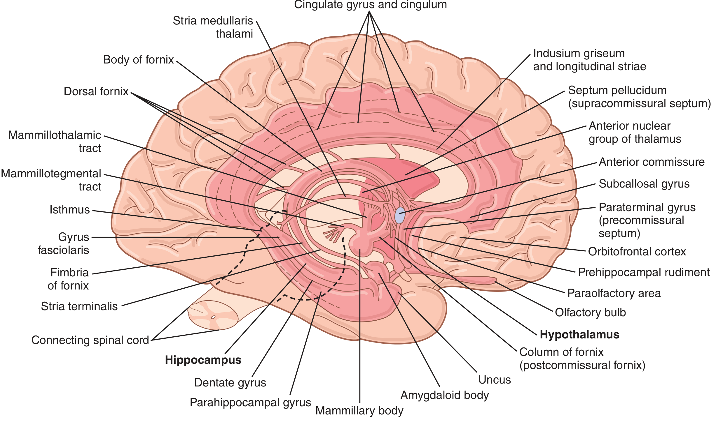
Anatomy of the limbic system (highlighted in dark pink). From Guyton & Hall Textbook of Medical Physiology.
Schematic showing the key central position of the hypothalamus:
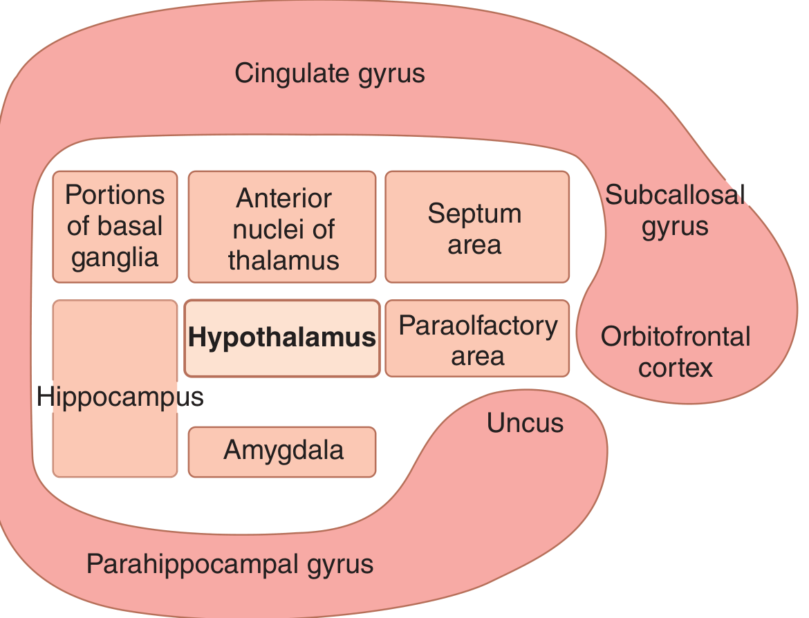
The limbic system schematic showing the hypothalamus at its center, surrounded by hippocampus, amygdala, septal area, basal ganglia portions, anterior thalamic nuclei, and paraolfactory area - with the outer ring of limbic cortex (cingulate gyrus, parahippocampal gyrus, orbitofrontal cortex, subcallosal gyrus) surrounding them.
2.1 Components - Cortical Structures (Limbic Cortex / Limbic Lobe)
The limbic cortex forms a ring of mostly paleocortex on the medial and ventral surfaces of each cerebral hemisphere:
- Orbitofrontal area (ventral surface of frontal lobe) - starting point of the ring
- Subcallosal gyrus (below the corpus callosum anteriorly)
- Cingulate gyrus - C-shaped, lies dorsal to and follows the corpus callosum along the medial hemisphere. Subdivided into:
- Subgenual anterior cingulate cortex (sACC) - Brodmann area 25, connected to amygdala and hypothalamus; overactive in depression; target for deep brain stimulation (DBS)
- Pregenual anterior cingulate cortex (pACC) - Brodmann area 32/24
- Midcingulate cortex (MCC)
- Posterior cingulate cortex (PCC)
- Retrosplenial cortex (RSC)
- Parahippocampal gyrus - medial temporal lobe; includes the entorhinal cortex (Brodmann area 28, the principal gateway to the hippocampus)
- Uncus - anteromedial knob of the parahippocampal gyrus
- Temporal poles and anterior insular cortex are also included in some classifications
Cytoarchitecture: Unlike the six-layered neocortex, limbic cortex is trilaminate. The inner hippocampus is archicortex (allocortex), the cingulate is mesocortex (transitional), and the entorhinal cortex is also transitional mesocortex.
(Guyton & Hall; Adams & Victor; Neuroanatomy through Clinical Cases)
2.2 Subcortical Components
| Structure | Location | Key features |
|---|---|---|
| Hippocampal formation | Medial temporal lobe, floor of temporal horn of lateral ventricle | Dentate gyrus + hippocampus (CA1-CA3) + subiculum; archicortex; C-shaped; 3-layered |
| Amygdala (amygdaloid complex) | Anterior temporal lobe, abuts the hippocampus | Basolateral (lateral + basal nuclei), centromedial, cortical nuclei; generates fear, emotional memory |
| Septal nuclei | Above anterior commissure | Reciprocal with hippocampus, amygdala, hypothalamus; "septohippocampal circuit" for anxiety |
| Hypothalamus | Diencephalon; central position in limbic system | Supraoptic, infundibular, mammillary zones; autonomic/endocrine control |
| Anterior thalamic nuclei | Thalamus | Part of Papez circuit; relay from mammillary bodies to cingulate gyrus |
| Mediodorsal thalamic nucleus | Thalamus | Connected to prefrontal and amygdala |
| Habenula (lateral subdivision) | Epithalamus | Hub connecting limbic, basal ganglia, brainstem circuits; projects to raphe nuclei (serotonin) and VTA (dopamine); implicated in mood disorders |
| Mammillary bodies | Posterior hypothalamus | Relay from fornix (hippocampus) to anterior thalamus |
| Nucleus accumbens | Ventral striatum | Reward, addiction |
| Ventral striatum / Basal forebrain | Subcortical | Including ventral pallidum, septal nuclei, nucleus basalis of Meynert (cholinergic) |
| Olfactory bulb and olfactory cortex | Anterior temporal, piriform cortex | Gateway for olfactory input to limbic system |
3. KEY CIRCUITS AND CONNECTIONS
3.1 The Papez Circuit (1937) - Circuit of Emotion and Memory
This is the most important limbic circuit, described by James Papez as the neural substrate of emotion. It is the internal core of the limbic system.
HIPPOCAMPUS
    ↓ (via Fornix)
MAMMILLARY BODIES (hypothalamus)
    ↓ (via Mammillothalamic tract / Bundle of Vicq d'Azyr)
ANTERIOR NUCLEUS OF THALAMUS
    ↓ (thalamocortical radiation)
CINGULATE GYRUS
    ↓ (via Cingulum)
PARAHIPPOCAMPAL GYRUS → ENTORHINAL CORTEX
    ↓ (perforant path / Schaffer collaterals)
HIPPOCAMPUS (circuit complete)
Key white matter tracts in the Papez circuit:
- Fornix - the principal output of the hippocampus; carries information from hippocampal subiculum → mammillary bodies + septal nuclei
- Mammillothalamic tract (bundle of Vicq d'Azyr) - from mammillary nuclei → anterior thalamic nuclei
- Cingulum - runs concentric to the corpus callosum; connects cingulate gyrus → parahippocampal gyrus; receives input from inferior parietal and temporal association cortex
- Perforant path - from entorhinal cortex → dentate gyrus (main input to hippocampus)
(Adams & Victor's Principles of Neurology; Neuroanatomy through Clinical Cases)
3.2 Amygdala Circuits
The amygdala has three nuclear groups with distinct connections:
Basolateral complex (BLA) - lateral, basal, and accessory basal nuclei:
- Receives sensory input from all modalities via thalamus ("low road" - fast) and sensory cortex ("high road" - slow/more processed)
- Projects to: hypothalamus, prefrontal cortex, hippocampus, nucleus accumbens
- Critical for fear conditioning and emotional memory
Centromedial complex - central and medial nuclei:
- Central nucleus: outputs to brainstem and hypothalamus → autonomic fear responses (heart rate, freezing behavior)
- Part of the extended amygdala - continuous through the substantia innominata with the bed nucleus of the stria terminalis (BNST)
- Central extended amygdala: connected to brainstem viscerosensory/visceromotor regions
- Medial extended amygdala: connected to medial (endocrine) hypothalamus
Efferent pathways from amygdala:
- Stria terminalis - main efferent tract; curves around fornix; projects to hypothalamus and septal nuclei
- Ventral amygdalofugal pathway - passes through substantia innominata; more direct route to hypothalamus and brainstem
3.3 Hypothalamus as the Central Hub
The hypothalamus is at the anatomical and functional center of the limbic system. It:
- Receives input from all limbic structures
- Controls the autonomic nervous system (sympathetic + parasympathetic)
- Regulates the pituitary (neuroendocrine control)
- Contains sensory receptors for hunger, thirst, temperature, osmolality
- Controls the fight-or-flight response (sympathoadrenal axis)
Subdivisions of the hypothalamus:
| Zone | Nuclei | Functions |
|---|---|---|
| Supraoptic | Suprachiasmatic, supraoptic, anterior, preoptic | Circadian rhythms; vasopressin synthesis; temperature regulation |
| Infundibular (tuberal) | Ventromedial, dorsomedial, arcuate, lateral hypothalamic area | Feeding/satiety; releasing hormones (CRH, TRH, GnRH, GHRH, somatostatin); lateral area = "reward"; ventromedial = "satiety center" |
| Mammillary | Posterior nucleus, mammillary nuclei | Fight/flight (posterior); Papez circuit relay (mammillary) |
3.4 Medial Forebrain Bundle (MFB)
A complex bidirectional fiber system at the core of the limbic system connecting:
- Rostral: Orbitofrontal cortex → septal nuclei → amygdala → hippocampus
- Caudal: Brainstem nuclei (VTA, raphe, locus coeruleus) → hypothalamus → limbic forebrain
The MFB carries dopaminergic fibers from VTA → nucleus accumbens (reward pathway) and noradrenergic fibers from locus coeruleus to widespread cortex.
3.5 Hippocampal Internal Circuitry (Tri-synaptic Circuit)
Entorhinal cortex
    ↓ Perforant path
Dentate gyrus (granule cells → mossy fibers)
    ↓ Mossy fiber pathway
CA3 pyramidal cells (→ Schaffer collaterals)
    ↓ Schaffer collateral pathway
CA1 pyramidal cells
    ↓
Subiculum
    ↓
Entorhinal cortex / Fornix (outputs)
CA1 and subiculum are the main output regions of the hippocampus. The subiculum projects via the fornix to the mammillary bodies, as well as directly to the amygdala, orbitofrontal cortex, and ventral striatum.
(Kaplan & Sadock's Comprehensive Textbook of Psychiatry)
4. FUNCTIONS OF THE LIMBIC SYSTEM
4.1 Olfaction
- The olfactory bulb → olfactory tract → primary olfactory (piriform) cortex (anterior temporal and deep sylvian fissure)
- Unique among sensory modalities: olfactory input reaches limbic structures (amygdala, entorhinal cortex) without thalamic relay
- Explains the powerful emotional and memory associations of smells (Proust phenomenon)
- The hippocampus originated as olfactory cortex phylogenetically; in many lower animals it decides what is food, danger, or a mate
4.2 Emotion
Hypothalamic control: Bard (1928) produced "sham rage" (pseudo-anger: intense aggression, autonomic overactivity) in decorticate cats, showing the hypothalamus + brainstem was sufficient for emotional expression. Removal of amygdala on both sides suppressed sham rage, producing placidity.
Amygdala in Fear:
- Best-studied emotional function of any brain structure
- The amygdala evaluates the emotional significance of stimuli - especially threat
- Fear conditioning: pairing a neutral stimulus (tone) with an aversive event (shock) → amygdala encodes this association
- Bilateral amygdala damage → inability to recognize fearful facial expressions, failure of fear conditioning
- LeDoux's "two roads to the amygdala": fast thalamo-amygdala route (pre-conscious fear) vs. slower thalamo-cortical-amygdala route (conscious appraisal)
Reward and Punishment:
- Stimulation of lateral hypothalamus and septal area → intense pleasure/reward; animals self-stimulate up to 5,000 times/hour
- Stimulation of periventricular areas and parts of amygdala → punishment/aversion
- These reward-punishment circuits select what information gets consolidated into memory (> 99% of sensory input is discarded)
(Guyton & Hall; Adams & Victor)
4.3 Memory
The limbic system is central to declarative (explicit) memory - both episodic (events) and semantic (facts).
Medial temporal lobe memory system:
- Entorhinal cortex receives convergent input from association cortex of frontal, parietal, temporal lobes
- → Perirhinal cortex and parahippocampal cortex relay it further → entorhinal cortex → hippocampus
- Hippocampus consolidates new memories and transfers them to neocortex for long-term storage
Case of H.M. (Henry Molaison): Bilateral medial temporal lobectomy (removal of hippocampus, amygdala, parahippocampal gyrus) for epilepsy → profound anterograde amnesia - could not form any new long-term declarative memories; was capable of short-term memory (seconds to minutes) but could not consolidate beyond that. Pre-existing remote memories were intact. This proved the hippocampus is necessary for memory consolidation, not for memory storage per se.
(Guyton & Hall; Neuroanatomy through Clinical Cases)
Medial diencephalic memory system:
- Damage to mammillary bodies (Korsakoff syndrome) or mediodorsal/anterior thalamic nuclei also produces severe anterograde amnesia
- The fornix is critical: interruption → amnesia (though less severe than hippocampal removal)
Role of the basal forebrain:
- Nucleus basalis of Meynert provides cholinergic projections to the entire cortex, modulating neuronal excitability and synaptic plasticity
- Destruction in Alzheimer's disease explains the profound memory impairment
4.4 Homeostasis and Vegetative Functions
Via the hypothalamus, the limbic system regulates:
- Body temperature (anterior hypothalamus = heat dissipation; posterior = heat conservation)
- Hunger and satiety (lateral hypothalamus = feeding center; ventromedial = satiety center)
- Thirst and water balance (osmoreceptors)
- Sexual drives and behavior (medial preoptic area)
- Circadian rhythms (suprachiasmatic nucleus)
- Autonomic regulation (heart rate, blood pressure, gastrointestinal motility)
- Neuroendocrine control of pituitary (HPA axis, HPT axis, HPG axis)
5. APPLIED / CLINICAL ASPECTS
5.1 Temporal Lobe / Limbic Epilepsy (Complex Partial Seizures)
The hippocampus has three-layered archicortex instead of six layers, making it intrinsically hyperexcitable and the most common focus for epilepsy.
Clinical features of a limbic seizure:
- Aura: Often the initial symptom - olfactory hallucinations (uncinate fits), rising epigastric sensation, déjà vu/jamais vu, fear, autonomic symptoms
- Stereotyped unpleasant odor + nausea + panicky sensation (as in the opening case in Neuroanatomy through Clinical Cases)
- Ictal phase: Automatisms (lip-smacking, hand fumbling), altered consciousness, slow inappropriate speech
- Postictal confusion
- Psychomotor symptoms: olfactory, visual, auditory, tactile hallucinations that the patient knows are unreal but cannot suppress (due to hippocampal hyperexcitability)
Causes of TLE: Mesial temporal sclerosis (most common), tumors, herpes simplex encephalitis (which has a tropism for limbic cortex), cavernous malformations, cortical dysplasia
Treatment: AEDs; surgical temporal lobectomy (anterior temporal resection or selective amygdalohippocampectomy) can be curative; vagus nerve stimulation, responsive neurostimulation (RNS)
(Guyton & Hall; Neuroanatomy through Clinical Cases; Kaplan & Sadock)
5.2 Klüver-Bucy Syndrome
First described in 1939 in monkeys after bilateral temporal lobectomy. Human cases occur from herpes simplex encephalitis, bilateral temporal surgery, Pick disease, TBI, or Alzheimer disease.
Classic features (mnemonic: "HOVE" or note the components):
| Feature | Mechanism |
|---|---|
| Psychic blindness / visual agnosia | Inability to recognize objects visually; due to disruption of ventral temporal visual streams |
| Oral-exploratory / hyperorality | Compulsive oral examination of objects; amygdala lesion |
| Hypersexuality | Bilateral lateral amygdala resection especially; disinhibition of sexual behavior |
| Tameness / placidity | Loss of aggression and fear; amygdala lesion |
| Hypermetamorphosis | Compulsive attention and response to every visual stimulus |
| Dietary changes | Hyperphagia, change in food preferences |
| Memory loss | Due to concurrent hippocampal damage; amnesic component |
In humans: Usually partial syndrome; often includes aphasia and amnesia that are not typically seen in experimental animals. Treatment is "difficult and often unsatisfactory."
(Bradley & Daroff's Neurology; Localization in Clinical Neurology; Neuroanatomy through Clinical Cases)
5.3 Alzheimer's Disease
The hippocampus, entorhinal cortex, and basal forebrain cholinergic neurons (nucleus basalis of Meynert) are preferentially and earliest affected in Alzheimer's disease.
- Neurofibrillary tangles (tau) and amyloid plaques first deposit in the entorhinal cortex (transentorhinal stage), then spread to hippocampus (Braak stages I-IV), then association cortex
- Loss of cholinergic neurons in nucleus basalis → depleted ACh in cortex → impaired synaptic plasticity and memory consolidation
- Amnesia (especially episodic anterograde amnesia) is the cardinal early symptom
- Hippocampal atrophy on MRI is a key imaging biomarker
(Kaplan & Sadock's Comprehensive Textbook of Psychiatry)
5.4 Korsakoff Syndrome (Wernicke-Korsakoff)
Cause: Thiamine (vitamin B1) deficiency → bilateral destruction of mammillary bodies and mediodorsal thalamic nuclei (Papez circuit disruption).
Clinical features:
- Anterograde amnesia (cannot form new memories)
- Retrograde amnesia (loss of recent past memories)
- Confabulation (fabricating plausible but false memories - characteristic)
- Relatively preserved immediate recall and procedural memory
- Associated with chronic alcoholism, malnutrition, hyperemesis gravidarum
Pathology: The mammillary body atrophy visible on MRI is classic. The mediodorsal thalamic nucleus damage contributes to the amnesic syndrome by disconnecting the Papez circuit.
5.5 Depression and Limbic Dysfunction
Multiple limbic structures show abnormalities in major depressive disorder:
- Subgenual anterior cingulate cortex (Brodmann area 25 / sACC): Overactive in depression; normalizes with antidepressants, ECT, rTMS, and DBS
- DBS of the sACC (and its white matter connections - cingulum bundle, uncinate fasciculus, subcortical fascicle) was pioneered by Mayberg et al. as a treatment for treatment-resistant depression
- Amygdala: Hyperactive; enhanced threat-sensitivity; prolonged activation to negative stimuli in depression
- Habenula: The lateral habenula projects to dopaminergic VTA and serotonergic raphe nuclei; inhibits these reward/mood systems. It is considered the brain's "punishment center." DBS of the lateral habenula has shown early success in treatment-resistant depression
(Kaplan & Sadock's Comprehensive Textbook of Psychiatry)
5.6 Anxiety Disorders (PTSD, GAD, Phobias)
- PTSD: Hyper-responsive amygdala (exaggerated fear response), failure of prefrontal cortex (especially ventromedial PFC and anterior cingulate) to inhibit amygdala activity
- GAD: Disruptions in functional (ACC-amygdala) and structural (uncinate fasciculus) connectivity; weaker frontoamygdala connectivity underlies emotion dysregulation
- Fear extinction (the basis of exposure therapy) depends on the medial prefrontal cortex signaling to the amygdala to suppress fear responses
- Septohippocampal pathway (septal nuclei ↔ hippocampus) forms a neurobiologic circuit for anxiety regulation
(Kaplan & Sadock's Comprehensive Textbook of Psychiatry)
5.7 Schizophrenia and Psychosis
- Reduced hippocampal volume is one of the most replicated neuroimaging findings in schizophrenia
- Hyperactivity of the hippocampus (due to reduced GABAergic interneuron inhibition) → excessive prediction errors → delusions and hallucinations via the dopaminergic VTA
- Temporal lobe epilepsy (limbic epilepsy) is associated with interictal psychosis, particularly with left-sided foci; this links limbic dysfunction to psychotic symptoms
(Kaplan & Sadock)
5.8 Herpes Simplex Encephalitis (HSE)
- HSV-1 has a specific tropism for limbic cortex (temporal poles, parahippocampal gyrus, cingulate gyrus, insula, orbitofrontal cortex)
- Presents with fever, temporal lobe seizures, behavioral change, olfactory hallucinations, aphasia, memory disturbance
- Untreated: severe bilateral limbic destruction → persistent Klüver-Bucy syndrome, dense amnesia, personality change
- Treatment: IV Acyclovir (urgent); MRI shows T2/FLAIR hyperintensity in bilateral medial temporal lobes
(Neuroanatomy through Clinical Cases)
5.9 Bilateral Hippocampal Damage / Amnesia Syndromes
Causes: HSE, ischemia (CA1 sector most vulnerable to hypoxia - "Sommer's sector"), bilateral posterior cerebral artery infarction, limbic encephalitis (anti-NMDA receptor, anti-LGI1 antibodies), TBI
Result: Profound anterograde amnesia with relatively preserved:
- Immediate memory (seconds)
- Procedural memory (motor skills, habits - via basal ganglia)
- Remote memory (long-term memories already consolidated)
5.10 Uncal Herniation (Transtentorial Herniation)
Clinical relevance: The uncus (amygdaloid area) is the anteromedial portion of the parahippocampal gyrus. When there is supratentorial mass effect (e.g., extradural hematoma, large MCA infarct), the uncus herniates over the tentorium cerebelli.
Consequences:
- Compression of CN III (runs between PCA and SCA) → ipsilateral fixed dilated pupil ("blown pupil") - earliest sign
- Compression of midbrain → contralateral hemiplegia (ipsilateral may occur due to Kernohan's notch)
- Compression of posterior cerebral artery → occipital infarction, contralateral visual field defect
- Coma (midbrain ARAS compression)
This is a neurosurgical emergency.
5.11 Reward, Addiction, and the Limbic System
Mesolimbic dopamine system:
- VTA (ventral tegmental area) → nucleus accumbens (ventral striatum) via the medial forebrain bundle
- Drugs of abuse (cocaine, amphetamines, opioids, alcohol, nicotine) all converge on this pathway, flooding the nucleus accumbens with dopamine
- This creates pathological reward learning - the drug stimulus gets encoded as the most important survival signal
- Repeated stimulation → downregulation of dopamine receptors → tolerance and dependence
- Withdrawal → underactivation of nucleus accumbens → dysphoria, anhedonia
(Guyton & Hall; Neuroanatomy through Clinical Cases)
5.12 Rage and Aggression
- Stimulation of amygdala → rage and aggressive behavior
- Stimulation of lateral hypothalamus → pleasure/reward
- Bilateral amygdaloidectomy in a normally aggressive macaque → placidity (Klüver-Bucy)
- "Sham rage" in decorticate animals is abolished by additional amygdala removal (Bard and Mountcastle)
- Psychosurgical relevance: Bilateral amygdalotomy has been used (controversially) in cases of intractable pathological aggression
5.13 Sexual Behavior and the Limbic System
- Stimulation of certain limbic areas → hypersexuality (also seen in Klüver-Bucy syndrome)
- The limbic system is directly involved in self-preservation and species-survival behaviors - sexual functioning
- Medial preoptic area of the hypothalamus: critical for male sexual behavior
- The extended amygdala (medial nucleus) is reciprocally connected with the endocrine hypothalamus and governs hormonally-mediated sexual behavior
(Kaplan & Sadock's Comprehensive Textbook of Psychiatry)
5.14 Depersonalization/Derealization
- Limbic temporal lobe involvement is implicated in depersonalization disorder and in temporal lobe epilepsy seizures
- Left-sided temporal lobe seizure foci are particularly associated with depersonalization/derealization experiences
- Inferior parietal and angular gyrus lesions, which interact with the posterior cingulate/limbic areas, can also produce depersonalization symptoms
6. SUMMARY TABLE - STRUCTURE, FUNCTION, AND CLINICAL RELEVANCE
| Structure | Primary Function | Clinical Lesion Effect |
|---|---|---|
| Hippocampus | Declarative memory consolidation | Anterograde amnesia (bilateral lesion = severe); TLE; Alzheimer's |
| Amygdala | Fear, emotional processing, fear conditioning | Klüver-Bucy (bilateral); PTSD, anxiety (hyperactivity) |
| Cingulate gyrus (sACC, BA25) | Attention, pain processing, emotional regulation | Depression (overactivity); target for DBS in TRD |
| Hypothalamus | Autonomic, endocrine, homeostatic functions; emotional expression | Sham rage; Korsakoff (mammillary bodies) |
| Mammillary bodies | Papez circuit relay | Korsakoff syndrome (thiamine deficiency) |
| Anterior thalamus | Papez circuit relay | Amnesia (thalamic infarcts); Korsakoff |
| Fornix | Major hippocampal output | Damage → amnesia (less complete than hippocampal removal) |
| Septal nuclei | Anxiety regulation; hippocampal modulation | Lesion → rage (release of inhibition of hypothalamic rage centers) |
| Habenula | Reward/punishment integration; mood | Target for DBS in TRD; implicated in depression |
| Nucleus accumbens | Reward; motivation | Drug addiction; target for DBS in OCD, addiction |
| Entorhinal cortex | Gateway to hippocampus | Earliest affected in Alzheimer's (Braak stage I) |
| Parahippocampal gyrus | Visual/spatial context, memory encoding | Lesion in HSE; uncal herniation |
| Uncus | Part of amygdalo-hippocampal complex | Herniation → CN III palsy, midbrain compression |
| Olfactory bulb/cortex | Primary olfaction | Uncinate fits (olfactory seizure aura); anosmia |
7. KEY TRACTS SUMMARY
| Tract | Origin → Destination | Functional Significance |
|---|---|---|
| Fornix | Hippocampus (subiculum) → Mammillary bodies + septal nuclei | Main hippocampal output; memory consolidation; Papez circuit |
| Mammillothalamic tract (Vicq d'Azyr) | Mammillary bodies → Anterior thalamic nuclei | Papez circuit; memory |
| Cingulum | Cingulate gyrus ↔ Parahippocampal gyrus (runs under corpus callosum) | Connects limbic lobe; emotion; disrupted in depression (uncinate fasciculus) |
| Stria terminalis | Amygdala (central/medial nuclei) → Hypothalamus, BNST | Amygdala efferents; fear/anxiety; sex behavior |
| Ventral amygdalofugal pathway | Amygdala (basolateral) → Hypothalamus, striatum | Direct amygdala-hypothalamus signaling |
| Medial forebrain bundle | Orbitofrontal cortex / Septal nuclei → Hypothalamus → VTA / Raphe | Reward pathway; dopamine fibers VTA→NAc; pleasure-seeking |
| Stria medullaris | Septal nuclei, preoptic area → Habenula | Limbic to habenula; mood regulation |
| Habenulo-interpeduncular tract (fasciculus retroflexus) | Habenula → Interpeduncular nucleus, raphe | Habenula to serotonin/dopamine systems |
| Perforant path | Entorhinal cortex → Dentate gyrus | Main cortical input to hippocampus; disrupted in Alzheimer's |
| Uncinate fasciculus | Orbitofrontal cortex ↔ Anterior temporal lobe | Connects limbic frontal with temporal; disrupted in depression/GAD |
8. QUICK-FIRE APPLIED POINTS FOR EXAMS
-
Papez circuit structures (in order): Hippocampus → Fornix → Mammillary bodies → Mammillothalamic tract → Anterior thalamus → Cingulate gyrus → Cingulum → Parahippocampal gyrus / entorhinal cortex → Hippocampus
-
Bilateral hippocampal removal: Anterograde amnesia + preserved procedural memory + preserved remote memory = selective declarative memory failure (case of H.M.)
-
Korsakoff syndrome: Thiamine deficiency → mammillary body + mediodorsal thalamus necrosis → amnesia + confabulation
-
Klüver-Bucy syndrome: Bilateral temporal lesion → visual agnosia + hyperorality + hypersexuality + tameness + hypermetamorphosis
-
Uncal herniation: Ipsilateral CN III palsy (first sign: fixed dilated pupil) → compresses midbrain and PCA
-
DBS targets in depression: Subgenual anterior cingulate cortex (BA25) and lateral habenula
-
Herpes simplex encephalitis: Tropism for limbic cortex → temporal lobe seizures + olfactory hallucinations + amnesia → treat with IV acyclovir urgently
-
Addiction: Mesolimbic dopamine pathway (VTA → nucleus accumbens) dysregulation
-
Fear conditioning circuit: Thalamus → Amygdala (lateral nucleus) → Central nucleus → Brainstem (autonomic fear responses); prefrontal cortex inhibits this circuit during extinction
-
Olfaction + emotion/memory: Olfaction reaches amygdala and hippocampus WITHOUT a thalamic relay → most direct sensory-limbic connection → explains powerful smell-triggered memories and emotions
Sources:
- Guyton & Hall Textbook of Medical Physiology (Chapter 59: The Limbic System and Behavioral Motivational Mechanisms)
- Adams & Victor's Principles of Neurology, 12th Ed. (Chapter 24: Disorders of Memory, Emotion, and Behavior)
- Neuroanatomy through Clinical Cases, 3rd Ed. (Chapter 18: Limbic System)
- Kaplan & Sadock's Comprehensive Textbook of Psychiatry (Chapter 1.2: Limbic System)
- Gray's Anatomy for Students (Part XI: Olfactory and Limbic System)
- Bradley & Daroff's Neurology in Clinical Practice (Klüver-Bucy Syndrome)This is a complete, exam-ready 30-mark answer synthesized from 6 major textbooks. Here is a brief orientation to help you navigate it:
What this answer covers:
Section 1-2: Introduction, etymology, and complete anatomy (cortical limbic lobe + all subcortical structures with a labeled diagram from Guyton)
Section 3: All major circuits in full detail - Papez circuit (with the exact sequence and white matter tracts), amygdala circuits (BLA vs centromedial; stria terminalis vs ventral amygdalofugal pathway), hypothalamic zones, medial forebrain bundle, and hippocampal tri-synaptic circuit
Section 4: Four functions (HOME mnemonic) - olfaction, emotion (Papez/amygdala/reward-punishment), memory (H.M. case, medial temporal + diencephalic systems, cholinergic basal forebrain), and homeostasis
Section 5 - Applied aspects (the clinical gold for 30-mark questions):
- Temporal lobe/limbic epilepsy (olfactory aura, uncinate fits, automatisms, mesial temporal sclerosis, surgery)
- Klüver-Bucy syndrome (causes, all 5 features with mechanism)
- Alzheimer's disease (entorhinal → hippocampal → cortical spread, cholinergic loss)
- Korsakoff syndrome (mammillary bodies, confabulation)
- Depression and DBS (BA25 sACC, lateral habenula)
- Anxiety/PTSD/GAD (amygdala hyperactivity, frontoamygdala connectivity)
- Schizophrenia (hippocampal hyperactivity → dopamine dysregulation)
- Herpes simplex encephalitis (limbic tropism, MRI findings, acyclovir)
- Bilateral hippocampal damage / amnesia syndromes
- Uncal herniation (CN III palsy - first sign, surgical emergency)
- Addiction (mesolimbic dopamine, VTA → nucleus accumbens)
- Rage/aggression (amygdalotomy)
- Sexual behavior and depersonalization
Sections 6-8: Master summary tables for quick revision
This is a shared conversation. Sign in to Orris to start your own chat.