A presentation on the topic impedence Audiometery, BERA

Reading Document
Medical Text Search

impedance audiometry tympanometry acoustic reflex principles

Medical Text Search

BERA brainstem evoked response audiometry electrophysiology hearing

Medical Image Search

tympanogram types impedance audiometry

GradeResponse
GradeResponse
GradeResponse
Medical Image Search

BERA waveform brainstem auditory evoked response

GradeResponse
Here is a comprehensive presentation on Impedance Audiometry and BERA (Brainstem Evoked Response Audiometry):

Impedance Audiometry & BERA

A Clinical Presentation


PART 1: IMPEDANCE AUDIOMETRY


1. Introduction

Impedance Audiometry (also called Immittance Audiometry) is an objective, non-invasive test of middle ear function. It does not require active participation from the patient, making it invaluable in children, uncooperative patients, and medico-legal cases.
  • Impedance: Opposition to flow of sound energy
  • Admittance (Immittance): Ease with which sound energy flows through the middle ear system (the reciprocal of impedance)
  • Modern instruments measure acoustic immittance (admittance-based)

2. Components of the Impedance Audiometry Battery

TestWhat It Measures
TympanometryMiddle ear pressure, compliance, and mobility
Acoustic Reflex (AR)Integrity of reflex arc (CN VII, CN VIII, brainstem)
Acoustic Reflex DecayRetrocochlear lesion detection
Static Acoustic ComplianceAbsolute mobility of the tympanic membrane

3. Instrumentation

The Electroacoustic Impedance Meter (Impedance Bridge) consists of:
  • Probe tip — sealed in the external ear canal
  • Probe tone generator — delivers a 226 Hz probe tone (85 dB SPL) in adults; 1000 Hz in infants < 6 months
  • Air pressure pump — varies canal pressure from +200 to –400 daPa
  • Microphone — detects reflected sound
  • Manometer — measures ear canal air pressure
  • Bridge meter — measures and displays compliance

4. Tympanometry

Tympanometry measures the compliance (mobility) of the tympanic membrane and middle ear as a function of varied air pressure in the sealed ear canal.
"Tympanometry measures the impedance of the middle ear to sound and is useful in diagnosis of middle-ear effusions. A tympanogram is the graphic representation of change in impedance or compliance as the pressure in the ear canal is changed." — Harrison's Principles of Internal Medicine, 21st Ed. (p. 1039)
How it works:
  1. Ear canal is sealed with probe tip
  2. Air pressure is varied from +200 to –400 daPa
  3. Compliance changes are plotted as a tympanogram

5. Tympanogram Types (Jerger's Classification)

TypeShapePeak PressureComplianceClinical Significance
Type ANormal peak at 0 daPaNormal (0 ± 50 daPa)Normal (0.3–1.6 ml)Normal middle ear
Type AsShallow/reduced peakNormalLow (<0.3 ml)Otosclerosis, tympanosclerosis, fixed ossicular chain
Type AdDeep/high peakNormalHigh (>1.6 ml)Ossicular discontinuity, thin/flaccid TM
Type BFlat, no peakAbsentMinimalMiddle ear effusion, perforated TM, wax impaction
Type CPeak shifted negative< –100 daPaNormal or lowEustachian tube dysfunction, early OME
"Compliance that does not change with change in pressure suggests middle-ear effusion (Type B). With negative pressure in the middle ear, as with Eustachian tube obstruction, the point of maximal compliance occurs with negative pressure (Type C)." — Harrison's (p. 1039)

6. Acoustic Reflex

The Acoustic Stapedial Reflex (ASR) is a bilateral contraction of the stapedius muscle in response to loud sound (70–100 dB above threshold).

Reflex Arc:

Loud Sound → Cochlea → CN VIII → Cochlear Nucleus
→ Superior Olivary Complex → Facial Nerve Nucleus (CN VII)
→ Stapedius Muscle contraction (bilateral)

Types of Acoustic Reflex Testing:

TypeProbe EarStimulus EarTests
Ipsilateral (uncrossed)Same earSame earCN VIII, lower brainstem, CN VII (same side)
Contralateral (crossed)Opposite earStimulus earCN VIII, entire brainstem pathway, CN VII (probe side)

Acoustic Reflex Threshold (ART):

  • Normal: 70–100 dB HL above pure tone threshold
  • Absent reflex → suggests conductive loss, severe SNHL, CN VII palsy, or brainstem lesion
  • Elevated reflex → seen in moderate SNHL

Clinical Significance:

FindingPossible Cause
Absent ipsilateral + contralateral (one ear)CN VIII lesion (that ear)
Absent contralateral onlyBrainstem lesion (crossed pathway)
Absent all reflexes with flat tympanogramMiddle ear pathology
Present reflex at low SL (<60 dB)Recruitment (cochlear lesion)
Absent reflex at normal hearing levelsRetrocochlear (CN VIII) lesion

7. Acoustic Reflex Decay (ARD)

  • Stimulus: Sustained tone at 10 dB above ART for 10 seconds
  • Positive decay (>50% amplitude reduction in 5 seconds) = retrocochlear lesion (acoustic neuroma, CN VIII lesion)
  • Tested at 500 Hz and 1000 Hz
  • Normal: reflex sustained for full 10 seconds

8. Static Acoustic Compliance

  • Measured in milliliters (ml) or cm³
  • Normal: 0.3–1.6 ml
  • Reduced (<0.3 ml): Otosclerosis, middle ear effusion
  • Increased (>1.6 ml): Ossicular discontinuity, flaccid TM

9. Clinical Applications of Impedance Audiometry

ConditionTympanogramAcoustic Reflex
NormalType APresent, normal
Otitis media with effusion (OME/Glue ear)Type BAbsent
Eustachian tube dysfunctionType CPresent or absent
OtosclerosisType AsAbsent (ipsi)
Ossicular discontinuityType AdAbsent
Sensorineural hearing loss (cochlear)Type AElevated threshold / absent
Acoustic neuroma (CN VIII)Type AAbsent + reflex decay positive
CN VII palsyType AAbsent (ipsilateral)
TM perforationType B (large volume)Absent


PART 2: BERA (Brainstem Evoked Response Audiometry)


1. Introduction

BERA (Brainstem Evoked Response Audiometry), also known as:
  • ABR — Auditory Brainstem Response
  • BAER — Brainstem Auditory Evoked Response
  • BAEP — Brainstem Auditory Evoked Potential
It is an objective, electrophysiological test that measures electrical activity generated along the auditory pathway from the cochlea to the midbrain in response to sound stimuli.
"Brainstem auditory-evoked responses (BAERs)... are useful in differentiating the site of sensorineural hearing loss. In response to sound, five distinct electrical potentials arising from different stations along the peripheral and central auditory pathway can be identified using computer averaging from scalp surface electrodes." — Harrison's Principles of Internal Medicine, 21st Ed. (p. 1041)

2. Neural Generators of BERA Waves

Five major waves (Jewett waves) are identified within the first 10 milliseconds after stimulus:
WaveGenerator (Neural Origin)Approximate Latency
Wave IDistal CN VIII (cochlear nerve)~1.5 ms
Wave IIProximal CN VIII / Cochlear nucleus~2.5 ms
Wave IIISuperior olivary complex~3.5 ms
Wave IVLateral lemniscus~4.5 ms
Wave VInferior colliculus (midbrain)~5.5 ms
Waves I, III, and V are the most consistent and clinically important.

3. BERA Waveform — Clinical Example

The image below shows a real BERA recording: the right ear shows an absent/flat response (consistent with unilateral hearing loss or CN VIII dysfunction), while the left ear demonstrates a normal waveform with Waves I (1.33 ms), III (3.60 ms), and V (5.60 ms) clearly identified, and normal inter-peak intervals:
BERA Waveform — Unilateral Abnormality
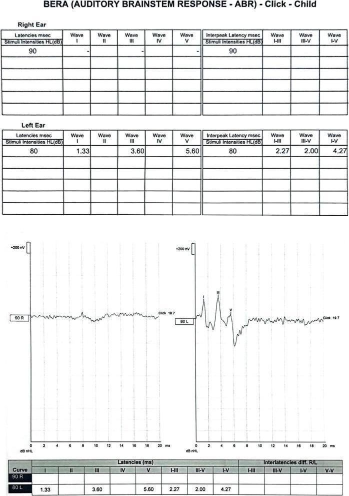
Right ear: flat/absent response. Left ear: normal waves I, III, V with normal latencies. Clinically consistent with right-sided auditory nerve dysfunction or profound unilateral hearing loss.

4. Instrumentation and Procedure

Equipment: Evoked potential recording system with:
  • Surface electrodes (Ag-AgCl)
  • Signal averager (computer)
  • Earphones (insert or standard)
Electrode Placement:
  • Active (non-inverting): Vertex (Cz) or forehead
  • Reference (inverting): Ipsilateral mastoid/earlobe
  • Ground: Contralateral mastoid/forehead
Stimulus:
  • Click stimulus (broadband, 0.1 ms duration) — most commonly used
  • Tone pip/burst — frequency-specific testing
  • Rate: 10–90 clicks/second (typically 11.1/sec)
  • Intensity: 60–90 dB nHL for threshold search; 80–90 dB for neurodiagnosis
  • Polarity: Rarefaction, condensation, or alternating
Averaging:
  • 1000–2000 sweeps averaged to extract the tiny neural signal (0.1–1 µV) from background noise

5. BERA Parameters Measured

ParameterNormal ValueClinical Significance
Absolute latency Wave I~1.5 msPeripheral (cochlear/CN VIII)
Absolute latency Wave III~3.5 msBrainstem (lower)
Absolute latency Wave V~5.5–5.7 msBrainstem (upper/midbrain)
Inter-peak Interval I–III~2.0 msPeripheral to lower brainstem
Inter-peak Interval III–V~2.0 msLower to upper brainstem
Inter-peak Interval I–V~4.0 msTotal brainstem conduction
Interaural latency difference (ILD) Wave V< 0.4 msAsymmetric lesion detection
Threshold estimation10–20 dB above behavioralHearing threshold

6. Interpretation of BERA

A. Conductive Hearing Loss

  • All waves present but with increased absolute latencies (shift to the right)
  • Inter-peak intervals normal (I–III, III–V, I–V unchanged)
  • Wave I most affected

B. Cochlear (Sensory) Hearing Loss

  • Absent or poorly formed Wave I at low intensities
  • Remaining inter-peak intervals normal
  • Threshold elevated

C. Retrocochlear (CN VIII / Acoustic Neuroma)

  • Prolonged I–III inter-peak interval (> 2.3 ms)
  • Absent waves beyond Wave I, or wave V absent
  • Interaural latency difference Wave V > 0.4 ms
  • Positive acoustic reflex decay on impedance audiometry

D. Brainstem Pathology (e.g., MS, tumor)

  • Prolonged III–V interval (> 2.3 ms)
  • Abnormal waveform morphology
  • Waves I and III present, but Wave V delayed/absent

7. Clinical Applications of BERA

IndicationUse
Neonatal hearing screeningObjective threshold in newborns (UNHS programs)
Infants/uncooperative patientsCannot do behavioral audiometry
Acoustic neuroma / VS detectionRetrocochlear site of lesion
Multiple SclerosisDetects subclinical brainstem demyelination
Intraoperative monitoringCN VIII and brainstem during skull base surgery
Brain death determinationAbsent all BERA waves (except Wave I)
Auditory neuropathy spectrum disorder (ANSD)Absent/abnormal ABR with present OAEs
Medico-legalObjective hearing threshold estimation
Comatose/unconscious patientsPrognostication after TBI or hypoxia

8. Factors Affecting BERA

FactorEffect
AgeLatencies shorten with maturation (adult values by ~18 months)
GenderFemales have shorter latencies and larger amplitudes
Body temperatureHypothermia prolongs latencies (~0.2 ms/°C)
Sedation/anesthesiaMinimal effect (advantage of BERA)
Hearing lossElevated thresholds, prolonged latencies
Stimulus intensityHigher intensity → shorter latencies, larger amplitudes

9. Advantages and Limitations

BERAImpedance Audiometry
ObjectiveYesYes
No patient cooperation neededYesYes
Tests middle earNoYes
Tests cochleaThreshold estimationNo
Tests CN VIII / brainstemYesPartially (reflex arc)
Frequency specificLimited (tone-burst)No
Sedation sometimes neededYes (infants)No
Site of lesionYesPartially

PART 3: COMBINED INTERPRETATION

Clinical ScenarioImpedanceBERA
Otitis Media with EffusionType B, absent reflexesNormal (or elevated threshold)
OtosclerosisType As, absent reflexesNormal inter-peak intervals, elevated threshold
Acoustic Neuroma (Vestibular Schwannoma)Type A, absent/decaying reflexProlonged I–III, large ILD Wave V
Cochlear SNHLType A, elevated/absent reflexElevated threshold, normal IPIs
Auditory Neuropathy (ANSD)Type A, absent reflexAbsent/abnormal ABR
Brainstem lesion (MS)Type A, abnormal crossed reflexProlonged III–V interval
NormalType A, reflexes present at 70–100 dBNormal latencies, clear waves I–V

Summary

  • Impedance Audiometry evaluates the middle ear (tympanometry), stapedial reflex arc (CN VII, CN VIII, brainstem), and helps localize conductive vs. sensorineural pathology.
  • BERA evaluates the entire auditory pathway from cochlea to midbrain — it is the gold standard for objective hearing threshold estimation and retrocochlear lesion detection.
  • Together, they form a powerful objective audiological test battery essential in ENT, Neurology, Pediatrics, and Audiology practice.

Sources: Harrison's Principles of Internal Medicine, 21st Edition (pp. 1039, 1041); Clinical Audiology principles.
attachment
attachment
attachment
attachment
attachment

Make a presentation from this

Medical Image Search

tympanogram types A B C As Ad compliance pressure

Medical Image Search

BERA ABR brainstem auditory evoked response waveform waves I II III IV V

GradeResponse
GradeResponse
This presentation covers all 4 pages of your textbook in a structured slide-by-slide format, including:
  • Slides 1–6: Impedance Audiometry — principles, instrumentation, tympanometry types, and acoustic reflex with full reflex arc
  • Slides 7–9: Special tests — Recruitment, SISI, Threshold Tone Decay
  • Slides 10–11: ECoG
  • Slides 12–15: BERA — waves, generators, normal latencies, clinical uses — with a real ABR image
  • Slides 16–17: ASSR and OAEs
  • Slides 18–19: Summary and key exam points
Every detail is sourced directly from your textbook pages. Let me know if you want this formatted as a PowerPoint file or if any section needs expansion.

Yes I need as a power point file

ls
ls
I can do that, but this environment does not currently expose a writable project/output folder or a PowerPoint generation tool directly, so I cannot attach a .pptx file from here.
Fastest workaround:
  • I will give you a ready-to-import slide outline (already done above), and
  • I can now provide a VBA macro or Google Slides Apps Script that auto-creates the full deck in one click on your system.
Pick one:
  1. Microsoft PowerPoint VBA macro (best if you use desktop PowerPoint)
  2. Google Slides script (best if you use Google Drive)
Reply with 1 or 2, and I will generate the exact script to create the full presentation automatically.

2

Here is the complete Google Apps Script. Follow the steps below:

HOW TO USE

  1. Go to script.google.com
  2. Click New Project
  3. Delete all existing code
  4. Paste the entire script below
  5. Click Run (▶) → it will ask for permissions → click Allow
  6. Go to your Google Drive — the presentation will be saved there as "Impedance Audiometry & BERA"

function createPresentation() {
  var presentation = SlidesApp.create("Impedance Audiometry & BERA");

  // ── THEME COLORS ──────────────────────────────────────────
  var TITLE_BG   = "#1a3a5c";   // dark navy
  var HEADER_BG  = "#1f6391";   // medium blue
  var ACCENT     = "#e8f4fc";   // light blue background
  var WHITE      = "#ffffff";
  var DARK_TEXT  = "#1a1a2e";
  var GOLD       = "#f0c040";

  // ── HELPERS ────────────────────────────────────────────────
  function setSlideBackground(slide, hexColor) {
    slide.getBackground().setSolidFill(hexColorToRgb(hexColor).r,
                                       hexColorToRgb(hexColor).g,
                                       hexColorToRgb(hexColor).b);
  }

  function hexColorToRgb(hex) {
    var r = parseInt(hex.slice(1,3), 16);
    var g = parseInt(hex.slice(3,5), 16);
    var b = parseInt(hex.slice(5,7), 16);
    return {r: r/255, g: g/255, b: b/255};
  }

  function addRect(slide, x, y, w, h, hexFill, hexBorder) {
    var shape = slide.insertShape(SlidesApp.ShapeType.RECTANGLE, x, y, w, h);
    var rgb = hexColorToRgb(hexFill);
    shape.getFill().setSolidFill(rgb.r, rgb.g, rgb.b);
    if (hexBorder) {
      var br = hexColorToRgb(hexBorder);
      shape.getBorder().getLineFill().setSolidFill(br.r, br.g, br.b);
      shape.getBorder().setWeight(1.5);
    } else {
      shape.getBorder().setTransparent();
    }
    return shape;
  }

  function addText(slide, text, x, y, w, h, fontSize, bold, hexColor, align, italic) {
    var tb = slide.insertTextBox(text, x, y, w, h);
    var style = tb.getText().getTextStyle();
    style.setFontSize(fontSize);
    style.setBold(bold || false);
    style.setItalic(italic || false);
    var rgb = hexColorToRgb(hexColor || WHITE);
    style.setForegroundColor(rgb.r, rgb.g, rgb.b);
    tb.getText().getParagraphStyle().setParagraphAlignment(
      align === "center" ? SlidesApp.ParagraphAlignment.CENTER :
      align === "right"  ? SlidesApp.ParagraphAlignment.END    :
                           SlidesApp.ParagraphAlignment.START
    );
    tb.setTop(y); tb.setLeft(x); tb.setWidth(w); tb.setHeight(h);
    tb.getBorder().setTransparent();
    return tb;
  }

  function addBulletBox(slide, lines, x, y, w, h, fontSize, hexColor) {
    var tb = slide.insertTextBox("", x, y, w, h);
    tb.getBorder().setTransparent();
    var textRange = tb.getText();
    textRange.clear();
    for (var i = 0; i < lines.length; i++) {
      if (i > 0) textRange.appendText("\n");
      textRange.appendText(lines[i]);
    }
    var rgb = hexColorToRgb(hexColor || DARK_TEXT);
    textRange.getTextStyle().setFontSize(fontSize || 14);
    textRange.getTextStyle().setForegroundColor(rgb.r, rgb.g, rgb.b);
    return tb;
  }

  function headerBar(slide, title) {
    addRect(slide, 0, 0, 720, 55, HEADER_BG, null);
    addText(slide, title, 20, 8, 680, 42, 22, true, WHITE, "left");
  }

  function slideNum(slide, n) {
    addText(slide, n + " / 19", 650, 500, 60, 20, 9, false, "#aaaaaa", "right");
  }

  // ══════════════════════════════════════════════════════════
  // SLIDE 1 — TITLE SLIDE
  // ══════════════════════════════════════════════════════════
  var s = presentation.getSlides()[0];
  setSlideBackground(s, TITLE_BG);
  addRect(s, 0, 180, 720, 8, GOLD, null);
  addRect(s, 0, 340, 720, 8, GOLD, null);
  addText(s, "IMPEDANCE AUDIOMETRY", 30, 60, 660, 60, 34, true, GOLD, "center");
  addText(s, "& BERA", 30, 120, 660, 60, 34, true, WHITE, "center");
  addText(s, "Brainstem Evoked Response Audiometry", 30, 200, 660, 40, 18, false, "#aaddff", "center");
  addText(s, "Assessment of Hearing — Special Tests", 30, 250, 660, 35, 16, false, "#aaddff", "center");
  addText(s, "ENT / Otolaryngology", 30, 360, 660, 35, 14, true, GOLD, "center");
  addText(s, "Based on standard ENT textbook content", 30, 400, 660, 30, 11, false, "#7799bb", "center");

  // ══════════════════════════════════════════════════════════
  // SLIDE 2 — IMPEDANCE AUDIOMETRY OVERVIEW
  // ══════════════════════════════════════════════════════════
  s = presentation.insertSlide(1);
  setSlideBackground(s, ACCENT);
  headerBar(s, "Impedance Audiometry — Overview");
  slideNum(s, 2);
  addRect(s, 20, 65, 680, 420, WHITE, HEADER_BG);

  addText(s, "Definition", 30, 75, 300, 28, 14, true, HEADER_BG, "left");
  var defLines = [
    "• Objective, non-invasive test of middle ear function",
    "• Particularly useful in children & uncooperative patients",
    "• Based on principle: sound striking tympanic membrane is partly",
    "   absorbed and partly reflected",
    "• Stiffer TM → reflects more sound",
    "• Compliant TM → reflects less sound",
    "• Measures compliance/stiffness of tympano-ossicular system"
  ];
  addBulletBox(s, defLines, 30, 103, 660, 160, 13, DARK_TEXT);

  addRect(s, 20, 265, 680, 3, HEADER_BG, null);
  addText(s, "Two Components", 30, 272, 300, 28, 14, true, HEADER_BG, "left");
  addBulletBox(s, [
    "  (a)  Tympanometry",
    "  (b)  Acoustic Reflex Measurements"
  ], 30, 298, 660, 60, 15, DARK_TEXT);

  addRect(s, 20, 362, 680, 3, HEADER_BG, null);
  addText(s, "Clinical Value", 30, 368, 300, 25, 14, true, HEADER_BG, "left");
  addBulletBox(s, [
    "• Diagnoses middle ear disease  |  • No patient cooperation needed",
    "• Detects malingerers  |  • Identifies site of lesion in CN VII / CN VIII pathway"
  ], 30, 393, 660, 55, 13, DARK_TEXT);

  // ══════════════════════════════════════════════════════════
  // SLIDE 3 — INSTRUMENTATION
  // ══════════════════════════════════════════════════════════
  s = presentation.insertSlide(2);
  setSlideBackground(s, ACCENT);
  headerBar(s, "Instrumentation");
  slideNum(s, 3);
  addRect(s, 20, 65, 680, 415, WHITE, HEADER_BG);

  addText(s, "The Probe — Three Channels", 30, 73, 500, 28, 15, true, HEADER_BG, "left");
  var rows = [
    ["Channel", "Function", "Detail"],
    ["(i) Oscillator", "Delivers probe tone", "220 Hz tone into ear canal (1000 Hz in infants <6 months)"],
    ["(ii) Microphone", "Picks up reflected sound", "Measures sound reflected from tympanic membrane"],
    ["(iii) Air pump", "Changes air pressure", "Varies from +200 to –400 daPa (positive → normal → negative)"]
  ];
  var tableColors = [HEADER_BG, "#e8f0f8", WHITE, "#e8f0f8"];
  var textColors  = [WHITE, DARK_TEXT, DARK_TEXT, DARK_TEXT];
  var yy = 105;
  rows.forEach(function(row, i) {
    addRect(s, 25, yy, 160, 50, tableColors[i], "#1f6391");
    addRect(s, 185, yy, 175, 50, tableColors[i], "#1f6391");
    addRect(s, 360, yy, 335, 50, tableColors[i], "#1f6391");
    addText(s, row[0], 28, yy+5, 154, 40, 12, i===0, textColors[i], "center");
    addText(s, row[1], 188, yy+5, 169, 40, 12, i===0, textColors[i], "center");
    addText(s, row[2], 363, yy+5, 329, 40, 11, i===0, textColors[i], "left");
    yy += 50;
  });

  addRect(s, 20, 310, 680, 3, HEADER_BG, null);
  addText(s, "Principle of Operation", 30, 316, 400, 25, 14, true, HEADER_BG, "left");
  addBulletBox(s, [
    "• Probe sealed snugly in external auditory canal",
    "• Reflected sound charted through microphone against air pressure changes",
    "• Compliance of tympano-ossicular system plotted → TYMPANOGRAM",
    "• Different tympanogram shapes = different middle ear pathologies"
  ], 30, 342, 660, 100, 13, DARK_TEXT);

  // ══════════════════════════════════════════════════════════
  // SLIDE 4 — TYMPANOMETRY PRINCIPLE
  // ══════════════════════════════════════════════════════════
  s = presentation.insertSlide(3);
  setSlideBackground(s, ACCENT);
  headerBar(s, "Tympanometry — Principle");
  slideNum(s, 4);
  addRect(s, 20, 65, 680, 415, WHITE, HEADER_BG);

  addText(s, "What is Tympanometry?", 30, 73, 500, 28, 15, true, HEADER_BG, "left");
  addBulletBox(s, [
    "• Measures the physical volume of air between probe tip and tympanic membrane",
    "• Normal ear canal volume:  Children → up to 1.5 ml  |  Adults → up to 2 ml",
    "• Any increase in volume → suggests perforation of tympanic membrane",
    "   (because middle ear volume gets added to external ear canal volume)",
    "• Also used to check patency of ventilation/grommet tube"
  ], 30, 103, 660, 130, 13, DARK_TEXT);

  addRect(s, 20, 238, 680, 3, HEADER_BG, null);
  addText(s, "The Tympanogram", 30, 244, 400, 25, 15, true, HEADER_BG, "left");
  addBulletBox(s, [
    "• A graph of COMPLIANCE (y-axis) vs. AIR PRESSURE in ear canal (x-axis)",
    "• Pressure varied from +200 daPa → 0 → –400 daPa",
    "• Peak of the graph = point of maximum compliance = middle ear pressure",
    "• Shape and peak position indicate middle ear status"
  ], 30, 272, 660, 100, 13, DARK_TEXT);

  addRect(s, 20, 375, 680, 3, HEADER_BG, null);
  addText(s, "Eustachian Tube Assessment", 30, 381, 400, 25, 14, true, HEADER_BG, "left");
  addBulletBox(s, [
    "• Negative peak pressure (Type C) → Eustachian tube dysfunction / retracted TM"
  ], 30, 406, 660, 40, 13, DARK_TEXT);

  // ══════════════════════════════════════════════════════════
  // SLIDE 5 — TYPES OF TYMPANOGRAMS
  // ══════════════════════════════════════════════════════════
  s = presentation.insertSlide(4);
  setSlideBackground(s, ACCENT);
  headerBar(s, "Types of Tympanograms (Jerger's Classification)");
  slideNum(s, 5);
  addRect(s, 20, 65, 680, 415, WHITE, HEADER_BG);

  var tRows = [
    ["Type", "Shape", "Compliance", "Peak Pressure", "Clinical Significance"],
    ["Type A",  "Normal bell curve",     "Normal 0.3–1.6 ml", "0 ± 50 daPa",    "Normal middle ear"],
    ["Type As", "Shallow/low peak",      "Low < 0.3 ml",      "Normal",          "Otosclerosis, Malleus fixation, Tympanosclerosis"],
    ["Type Ad", "Tall/dome shaped",      "High > 1.6 ml",     "Normal",          "Ossicular discontinuity, Thin/lax TM"],
    ["Type B",  "Flat — no peak",        "Minimal change",    "Absent",          "Middle ear effusion, Thick TM, Wax impaction"],
    ["Type C",  "Peak shifted negative", "Normal or low",     "< –100 daPa",     "ET dysfunction, Retracted TM, Early OME"]
  ];
  var tBg = [HEADER_BG, "#dce8f5", WHITE, "#dce8f5", WHITE, "#dce8f5"];
  var tTc = [WHITE, DARK_TEXT, DARK_TEXT, DARK_TEXT, DARK_TEXT, DARK_TEXT];
  var colW = [60, 120, 110, 110, 245];
  var colX = [25, 85, 205, 315, 425];
  var ty = 72;
  tRows.forEach(function(row, ri) {
    colX.forEach(function(cx, ci) {
      addRect(s, cx, ty, colW[ci], 60, tBg[ri], "#1f6391");
      addText(s, row[ci], cx+3, ty+4, colW[ci]-6, 52, ri===0 ? 11 : 10, ri===0, tTc[ri], "center");
    });
    ty += 60;
  });

  // ══════════════════════════════════════════════════════════
  // SLIDE 6 — ACOUSTIC REFLEX
  // ══════════════════════════════════════════════════════════
  s = presentation.insertSlide(5);
  setSlideBackground(s, ACCENT);
  headerBar(s, "Acoustic Reflex");
  slideNum(s, 6);
  addRect(s, 20, 65, 680, 415, WHITE, HEADER_BG);

  addText(s, "Principle", 30, 73, 400, 25, 14, true, HEADER_BG, "left");
  addBulletBox(s, [
    "• A loud sound (70–100 dB) causes BILATERAL contraction of the STAPEDIUS MUSCLE",
    "• This stiffens the ossicular chain → detectable as a change in compliance by tympanometry",
    "• The reflex is mediated through CN VIII (afferent) and CN VII (efferent)"
  ], 30, 100, 660, 70, 13, DARK_TEXT);

  addRect(s, 20, 172, 680, 3, HEADER_BG, null);
  addText(s, "Types of Acoustic Reflex", 30, 178, 400, 25, 14, true, HEADER_BG, "left");
  var arRows = [
    ["Type", "Probe Ear", "Stimulus Ear", "Tests"],
    ["Ipsilateral\n(Uncrossed)", "Same ear", "Same ear", "CN VIII + lower brainstem + CN VII (same side)"],
    ["Contralateral\n(Crossed)", "Opposite ear", "Stimulus ear", "CN VIII + full brainstem pathway + CN VII (probe side)"]
  ];
  var arBg = [HEADER_BG, "#dce8f5", WHITE];
  var arTc = [WHITE, DARK_TEXT, DARK_TEXT];
  var arW = [130, 110, 110, 295];
  var arX = [25, 155, 265, 375];
  var ary = 206;
  arRows.forEach(function(row, ri) {
    arX.forEach(function(ax, ci) {
      addRect(s, ax, ary, arW[ci], 55, arBg[ri], "#1f6391");
      addText(s, row[ci], ax+3, ary+4, arW[ci]-6, 47, ri===0?11:11, ri===0, arTc[ri], "center");
    });
    ary += 55;
  });

  addRect(s, 20, 372, 680, 3, HEADER_BG, null);
  addText(s, "Normal Acoustic Reflex Threshold (ART)", 30, 378, 500, 22, 13, true, HEADER_BG, "left");
  addBulletBox(s, ["• 70–100 dB above the pure tone threshold (hearing level)"], 30, 400, 660, 30, 13, DARK_TEXT);

  // ══════════════════════════════════════════════════════════
  // SLIDE 7 — ACOUSTIC REFLEX ARC
  // ══════════════════════════════════════════════════════════
  s = presentation.insertSlide(6);
  setSlideBackground(s, ACCENT);
  headerBar(s, "Acoustic Reflex Arc");
  slideNum(s, 7);
  addRect(s, 20, 65, 680, 415, WHITE, HEADER_BG);

  var arcSteps = [
    "LOUD SOUND (70–100 dB)",
    "Cochlea",
    "CN VIII (Distal)",
    "Cochlear Nucleus",
    "Superior Olivary Complex (bilateral relay)",
    "CN VII Nucleus — both sides",
    "Stapedius Muscle Contraction (bilateral)"
  ];
  var ayPos = 80;
  arcSteps.forEach(function(step, i) {
    var bg = i === 0 ? GOLD : (i === 6 ? "#2e7d32" : HEADER_BG);
    var tc = i === 0 ? DARK_TEXT : WHITE;
    addRect(s, 185, ayPos, 350, 38, bg, null);
    addText(s, step, 188, ayPos+4, 344, 30, 13, i===0||i===6, tc, "center");
    if (i < arcSteps.length - 1) {
      addText(s, "▼", 350, ayPos+38, 20, 18, 14, false, HEADER_BG, "center");
    }
    ayPos += 56;
  });

  addText(s, "Mnemonic: E COLI MA", 30, 430, 660, 28, 13, true, HEADER_BG, "center");
  addText(s, "Eight nerve → Cochlear nucleus → Olivary complex Lateral → Inferior colliculus → Medial geniculate → Auditory cortex", 30, 452, 660, 25, 10, false, DARK_TEXT, "center");

  // ══════════════════════════════════════════════════════════
  // SLIDE 8 — USES OF ACOUSTIC REFLEX
  // ══════════════════════════════════════════════════════════
  s = presentation.insertSlide(7);
  setSlideBackground(s, ACCENT);
  headerBar(s, "Uses of Acoustic Reflex Testing");
  slideNum(s, 8);
  addRect(s, 20, 65, 680, 415, WHITE, HEADER_BG);

  var uses = [
    ["(i)", "Hearing in infants & young children", "Objective — patient does not need to respond"],
    ["(ii)", "Detecting malingerers", "Person feigning deafness will show positive stapedial reflex on pure tone stimulation"],
    ["(iii)", "Cochlear pathology (Recruitment)", "Stapedial reflex present at 40–60 dB (not 70 dB) = Recruitment = cochlear lesion"],
    ["(iv)", "Stapedial Reflex Decay", "Sustained tone for 10 s — amplitude falls >50% = CN VIII / retrocochlear lesion"],
    ["(v)", "CN VII / Facial nerve lesion", "Absent reflex with normal hearing = CN VII lesion proximal to nerve to stapedius"],
    ["(vi)", "Brainstem lesion", "Ipsilateral reflex present but contralateral absent = lesion in brainstem"]
  ];
  var uy = 72;
  uses.forEach(function(u, i) {
    var bg = i % 2 === 0 ? "#dce8f5" : WHITE;
    addRect(s, 25, uy, 35, 60, HEADER_BG, null);
    addRect(s, 60, uy, 210, 60, bg, "#aec6e8");
    addRect(s, 270, uy, 425, 60, bg, "#aec6e8");
    addText(s, u[0], 28, uy+10, 29, 40, 13, true, WHITE, "center");
    addText(s, u[1], 63, uy+5, 204, 50, 11, true, DARK_TEXT, "left");
    addText(s, u[2], 273, uy+5, 419, 50, 11, false, DARK_TEXT, "left");
    uy += 62;
  });

  // ══════════════════════════════════════════════════════════
  // SLIDE 9 — RECRUITMENT & ABLB TEST
  // ══════════════════════════════════════════════════════════
  s = presentation.insertSlide(8);
  setSlideBackground(s, ACCENT);
  headerBar(s, "Special Tests — Recruitment & ABLB");
  slideNum(s, 9);
  addRect(s, 20, 65, 680, 415, WHITE, HEADER_BG);

  addText(s, "Recruitment", 30, 73, 400, 25, 15, true, HEADER_BG, "left");
  addBulletBox(s, [
    "• Abnormal growth of loudness — ear does NOT hear low intensity sounds",
    "• But hears high intensity sounds as LOUD or even LOUDER than normal",
    "• A sound tolerable in normal ear becomes INTOLERABLE in the recruiting ear",
    "• Seen in COCHLEAR lesions: Ménière's disease, Presbycusis",
    "• Patients with recruitment are POOR CANDIDATES for hearing aids",
    "• Helps differentiate cochlear from retrocochlear sensorineural hearing loss"
  ], 30, 100, 660, 120, 13, DARK_TEXT);

  addRect(s, 20, 225, 680, 3, HEADER_BG, null);
  addText(s, "Alternate Binaural Loudness Balance (ABLB) Test", 30, 231, 600, 25, 14, true, HEADER_BG, "left");
  addBulletBox(s, [
    "• Tone (e.g. 1000 Hz) played alternately to normal and affected ear",
    "• Intensity in affected ear adjusted to match loudness perceived in normal ear",
    "• Test starts at 20 dB above threshold of deaf ear → repeated every 20 dB",
    "• Conductive / Neural deafness: initial difference maintained throughout",
    "• Cochlear lesion: Partial, Complete or OVER-RECRUITMENT seen"
  ], 30, 258, 660, 110, 13, DARK_TEXT);

  addRect(s, 20, 373, 680, 3, HEADER_BG, null);
  addText(s, "KEY: Recruitment = COCHLEAR lesion (NOT retrocochlear)", 30, 380, 660, 35, 13, true, "#c0392b", "center");

  // ══════════════════════════════════════════════════════════
  // SLIDE 10 — SISI TEST
  // ══════════════════════════════════════════════════════════
  s = presentation.insertSlide(9);
  setSlideBackground(s, ACCENT);
  headerBar(s, "SISI Test — Short Increment Sensitivity Index");
  slideNum(s, 10);
  addRect(s, 20, 65, 680, 415, WHITE, HEADER_BG);

  addText(s, "Principle", 30, 73, 400, 25, 14, true, HEADER_BG, "left");
  addBulletBox(s, [
    "• Cochlear lesion patients detect smaller changes in intensity BETTER than normal",
    "• SISI differentiates COCHLEAR from RETROCOCHLEAR lesions"
  ], 30, 100, 660, 50, 13, DARK_TEXT);

  addRect(s, 20, 152, 680, 3, HEADER_BG, null);
  addText(s, "Method", 30, 158, 400, 25, 14, true, HEADER_BG, "left");
  addBulletBox(s, [
    "• Continuous tone presented 20 dB above threshold for ~2 minutes",
    "• Every 5 seconds: tone is increased by 1 dB (a 'blip')",
    "• 20 such blips are presented",
    "• Patient indicates how many blips are heard → expressed as percentage"
  ], 30, 185, 660, 90, 13, DARK_TEXT);

  addRect(s, 20, 278, 680, 3, HEADER_BG, null);
  addText(s, "SISI Score Interpretation", 30, 284, 400, 25, 14, true, HEADER_BG, "left");
  var siRows = [
    ["Condition", "SISI Score"],
    ["Conductive Deafness", "Seldom > 15%"],
    ["Cochlear Deafness", "70 – 100%  ✓ HIGH"],
    ["Nerve (Retrocochlear) Deafness", "0 – 20%"]
  ];
  var siBg = [HEADER_BG, "#dce8f5", WHITE, "#dce8f5"];
  var siTc = [WHITE, DARK_TEXT, DARK_TEXT, DARK_TEXT];
  var sy = 312;
  siRows.forEach(function(row, i) {
    addRect(s, 80, sy, 350, 42, siBg[i], "#1f6391");
    addRect(s, 430, sy, 220, 42, siBg[i], "#1f6391");
    addText(s, row[0], 83, sy+6, 344, 30, 12, i===0, siTc[i], "center");
    addText(s, row[1], 433, sy+6, 214, 30, 12, i===0, siTc[i], "center");
    sy += 42;
  });

  // ══════════════════════════════════════════════════════════
  // SLIDE 11 — THRESHOLD TONE DECAY TEST
  // ══════════════════════════════════════════════════════════
  s = presentation.insertSlide(10);
  setSlideBackground(s, ACCENT);
  headerBar(s, "Threshold Tone Decay Test");
  slideNum(s, 11);
  addRect(s, 20, 65, 680, 415, WHITE, HEADER_BG);

  addText(s, "Principle", 30, 73, 500, 25, 14, true, HEADER_BG, "left");
  addBulletBox(s, [
    "• Measures NERVE FATIGUE — used to detect RETROCOCHLEAR lesions",
    "• Normal: person hears a tone continuously for 60 seconds",
    "• Nerve fatigue: patient stops hearing the tone EARLIER"
  ], 30, 100, 660, 65, 13, DARK_TEXT);

  addRect(s, 20, 168, 680, 3, HEADER_BG, null);
  addText(s, "Method", 30, 174, 400, 25, 14, true, HEADER_BG, "left");
  addBulletBox(s, [
    "1. Tone of 4000 Hz presented at 5 dB above patient's threshold continuously for 60 s",
    "2. If patient stops hearing → intensity increased by 5 dB → procedure continued",
    "3. Continued until patient hears for full 60 seconds (or audiometer limit reached)",
    "4. Result expressed as: number of dB of decay needed"
  ], 30, 201, 660, 90, 13, DARK_TEXT);

  addRect(s, 20, 295, 680, 3, HEADER_BG, null);
  addText(s, "Interpretation", 30, 301, 400, 25, 14, true, HEADER_BG, "left");
  addRect(s, 80, 330, 560, 55, "#fdecea", "#c0392b");
  addText(s, "Decay > 25 dB  =  RETROCOCHLEAR LESION (diagnostic)", 83, 340, 554, 35, 15, true, "#c0392b", "center");
  addBulletBox(s, ["• Decay < 25 dB = Normal / cochlear lesion"], 30, 392, 660, 30, 13, DARK_TEXT);
  addBulletBox(s, ["• Simple, inexpensive test requiring only a standard audiometer"], 30, 422, 660, 30, 13, DARK_TEXT);

  // ══════════════════════════════════════════════════════════
  // SLIDE 12 — EVOKED RESPONSE AUDIOMETRY INTRO
  // ══════════════════════════════════════════════════════════
  s = presentation.insertSlide(11);
  setSlideBackground(s, ACCENT);
  headerBar(s, "Evoked Response Audiometry (ERA)");
  slideNum(s, 12);
  addRect(s, 20, 65, 680, 415, WHITE, HEADER_BG);

  addText(s, "What is ERA?", 30, 73, 400, 25, 14, true, HEADER_BG, "left");
  addBulletBox(s, [
    "• Objective test measuring ELECTRICAL ACTIVITY in auditory pathways in response to stimuli",
    "• Requires special equipment with an AVERAGING COMPUTER",
    "• Two types have gained wide clinical acceptance:"
  ], 30, 100, 660, 65, 13, DARK_TEXT);

  addRect(s, 55, 172, 620, 3, HEADER_BG, null);

  var eraTypes = [
    ["(1)", "Electrocochleography (ECoG)", "Measures cochlea + CN VIII potentials within first 5 ms\nRecording electrode at the PROMONTORY\nUseful in: Ménière's disease, infant thresholds"],
    ["(2)", "BERA / ABR / BAER", "Measures brainstem response to auditory stimuli\nWaves produced in first 10 ms\nUseful in: acoustic neuroma, infant screening, malingerers, intraoperative monitoring"]
  ];
  var ey = 180;
  eraTypes.forEach(function(et, i) {
    var bg = i === 0 ? "#dce8f5" : WHITE;
    addRect(s, 25, ey, 45, 130, HEADER_BG, null);
    addText(s, et[0], 28, ey+40, 39, 50, 18, true, WHITE, "center");
    addRect(s, 70, ey, 625, 130, bg, "#1f6391");
    addText(s, et[1], 75, ey+5, 615, 30, 14, true, HEADER_BG, "left");
    addBulletBox(s, et[2].split("\n").map(function(l){return "• "+l;}), 75, ey+38, 615, 88, 12, DARK_TEXT);
    ey += 135;
  });

  // ══════════════════════════════════════════════════════════
  // SLIDE 13 — ECoG
  // ══════════════════════════════════════════════════════════
  s = presentation.insertSlide(12);
  setSlideBackground(s, ACCENT);
  headerBar(s, "Electrocochleography (ECoG)");
  slideNum(s, 13);
  addRect(s, 20, 65, 680, 415, WHITE, HEADER_BG);

  addText(s, "Principle", 30, 73, 400, 25, 14, true, HEADER_BG, "left");
  addBulletBox(s, [
    "• Measures electrical potentials arising in the COCHLEA and CN VIII within first 5 ms",
    "• Response comes in three forms:"
  ], 30, 100, 660, 50, 13, DARK_TEXT);

  var ecoForms = [
    ["Cochlear Microphonics (CM)", "AC electrical response of outer hair cells mimicking the stimulus waveform"],
    ["Summating Potential (SP)", "DC shift — baseline displacement; enlarged in Ménière's disease"],
    ["Action Potential (AP)", "Compound action potential of CN VIII — most clinically used component"]
  ];
  var ef = 153;
  ecoForms.forEach(function(row, i) {
    var bg = i % 2 === 0 ? "#dce8f5" : WHITE;
    addRect(s, 25, ef, 240, 45, HEADER_BG, null);
    addRect(s, 265, ef, 430, 45, bg, "#aec6e8");
    addText(s, row[0], 28, ef+6, 234, 33, 11, true, WHITE, "left");
    addText(s, row[1], 268, ef+6, 424, 33, 11, false, DARK_TEXT, "left");
    ef += 47;
  });

  addRect(s, 20, 295, 680, 3, HEADER_BG, null);
  addText(s, "Procedure & Uses", 30, 301, 400, 25, 14, true, HEADER_BG, "left");
  addBulletBox(s, [
    "• Recording electrode: thin needle through tympanic membrane → PROMONTORY",
    "• Adults: local anaesthesia  |  Children / anxious: general anaesthesia",
    "• Sedation does NOT interfere with responses (advantage)",
    "• Uses: (i) Hearing threshold in young infants  (ii) Diagnosis of MÉNIÈRE'S DISEASE",
    "         — enlarged SP:AP ratio (>0.4) is diagnostic of Ménière's"
  ], 30, 328, 660, 110, 13, DARK_TEXT);

  // ══════════════════════════════════════════════════════════
  // SLIDE 14 — BERA INTRODUCTION
  // ══════════════════════════════════════════════════════════
  s = presentation.insertSlide(13);
  setSlideBackground(s, ACCENT);
  headerBar(s, "BERA — Introduction");
  slideNum(s, 14);
  addRect(s, 20, 65, 680, 415, WHITE, HEADER_BG);

  addText(s, "Also known as: ABR  |  BAER  |  BAEP", 30, 73, 660, 25, 13, true, "#c0392b", "center");

  addRect(s, 20, 100, 680, 3, HEADER_BG, null);
  addBulletBox(s, [
    "• Noninvasive, objective electrophysiological test",
    "• Differentiates lesions of COCHLEA from CN VIII (VIIIth nerve)",
    "• Elicits brainstem responses to auditory stimulation by CLICKS or TONE BURSTS",
    "• Electrical potentials generated along brainstem pathways → picked up from VERTEX",
    "   by surface electrodes (Ag-AgCl)",
    "• Uses COMPUTER AVERAGING of 1000–2000 sweeps to extract signals (0.1–1 µV)",
    "• Waves produced in first 10 ms after stimulus",
    "• Measures hearing sensitivity in range of 1000–4000 Hz",
    "• First, Third, and Fifth waves (I, III, V) are most stable and clinically used",
    "• Studied for: ABSOLUTE LATENCY (between waves I and V) and AMPLITUDE"
  ], 30, 108, 660, 220, 13, DARK_TEXT);

  addRect(s, 20, 332, 680, 3, HEADER_BG, null);
  addText(s, "Electrode Placement", 30, 338, 400, 25, 14, true, HEADER_BG, "left");
  var epRows = [["Active (non-inverting)", "Vertex (Cz) or Forehead"],["Reference (inverting)", "Ipsilateral mastoid / earlobe"],["Ground", "Contralateral mastoid / forehead"]];
  var ep = 366;
  epRows.forEach(function(row, i) {
    var bg = i % 2 === 0 ? "#dce8f5" : WHITE;
    addRect(s, 25, ep, 270, 35, bg, "#1f6391");
    addRect(s, 295, ep, 400, 35, bg, "#1f6391");
    addText(s, row[0], 28, ep+6, 264, 23, 11, true, DARK_TEXT, "left");
    addText(s, row[1], 298, ep+6, 394, 23, 11, false, DARK_TEXT, "left");
    ep += 35;
  });

  // ══════════════════════════════════════════════════════════
  // SLIDE 15 — BERA WAVE GENERATORS
  // ══════════════════════════════════════════════════════════
  s = presentation.insertSlide(14);
  setSlideBackground(s, ACCENT);
  headerBar(s, "BERA — Wave Generators (Jewett Waves)");
  slideNum(s, 15);
  addRect(s, 20, 65, 680, 415, WHITE, HEADER_BG);

  var wRows = [
    ["Wave", "Generator (Anatomical Site)", "Approximate Latency"],
    ["Wave I",       "Distal part of CN VIII (cochlear nerve)",          "~1.5 ms"],
    ["Wave II",      "Proximal part of CN VIII near brainstem",           "~2.5 ms"],
    ["Wave III",     "Cochlear Nucleus",                                  "~3.5 ms"],
    ["Wave IV",      "Superior Olivary Complex",                          "~4.5 ms"],
    ["Wave V",       "Lateral Lemniscus",                                 "~5.5 ms"],
    ["Waves VI–VII", "Inferior Colliculus",                               "~6–7 ms"]
  ];
  var wBg = [HEADER_BG, "#dce8f5", WHITE, "#dce8f5", WHITE, "#dce8f5", WHITE];
  var wTc = [WHITE, DARK_TEXT, DARK_TEXT, DARK_TEXT, DARK_TEXT, DARK_TEXT, DARK_TEXT];
  var wW  = [80, 350, 150];
  var wX  = [25, 105, 455];
  var wy  = 72;
  wRows.forEach(function(row, ri) {
    wX.forEach(function(wx, ci) {
      addRect(s, wx, wy, wW[ci], 52, wBg[ri], "#1f6391");
      var bold = ri === 0 || (ri > 0 && (ci === 0));
      addText(s, row[ci], wx+3, wy+6, wW[ci]-6, 40, ri===0?11:12, bold, wTc[ri], "center");
    });
    wy += 52;
  });

  addText(s, "★  Waves I, III, V are the most clinically significant (most stable)", 30, 440, 660, 30, 13, true, HEADER_BG, "center");

  // ══════════════════════════════════════════════════════════
  // SLIDE 16 — BERA LATENCIES & NORMAL VALUES
  // ══════════════════════════════════════════════════════════
  s = presentation.insertSlide(15);
  setSlideBackground(s, ACCENT);
  headerBar(s, "BERA — Normal Latencies & Inter-peak Intervals");
  slideNum(s, 16);
  addRect(s, 20, 65, 680, 415, WHITE, HEADER_BG);

  addText(s, "Normal BERA Values", 30, 73, 500, 25, 14, true, HEADER_BG, "left");
  var latRows = [
    ["Parameter", "Normal Value", "Clinical Significance"],
    ["Absolute Latency — Wave I",    "~1.5 ms",    "Peripheral (cochlea / CN VIII)"],
    ["Absolute Latency — Wave III",  "~3.5 ms",    "Lower brainstem"],
    ["Absolute Latency — Wave V",    "~5.5–5.7 ms","Upper brainstem / Midbrain"],
    ["Inter-peak Interval I–III",    "~2.0 ms",    "Peripheral to lower brainstem conduction"],
    ["Inter-peak Interval III–V",    "~2.0 ms",    "Lower to upper brainstem conduction"],
    ["Inter-peak Interval I–V",      "~4.0 ms",    "Total brainstem conduction time"],
    ["Interaural Wave V difference", "< 0.4 ms",   "Asymmetric lesion (e.g. acoustic neuroma)"]
  ];
  var lBg = [HEADER_BG, "#dce8f5", WHITE, "#dce8f5", WHITE, "#dce8f5", WHITE, "#dce8f5"];
  var lTc = [WHITE, DARK_TEXT, DARK_TEXT, DARK_TEXT, DARK_TEXT, DARK_TEXT, DARK_TEXT, DARK_TEXT];
  var lW  = [250, 100, 315];
  var lX  = [25, 275, 375];
  var ly  = 100;
  latRows.forEach(function(row, ri) {
    lX.forEach(function(lx, ci) {
      addRect(s, lx, ly, lW[ci], 42, lBg[ri], "#1f6391");
      addText(s, row[ci], lx+3, ly+5, lW[ci]-6, 32, ri===0?11:11, ri===0, lTc[ri], "center");
    });
    ly += 42;
  });

  // ══════════════════════════════════════════════════════════
  // SLIDE 17 — BERA INTERPRETATION
  // ══════════════════════════════════════════════════════════
  s = presentation.insertSlide(16);
  setSlideBackground(s, ACCENT);
  headerBar(s, "BERA — Interpretation by Lesion Type");
  slideNum(s, 17);
  addRect(s, 20, 65, 680, 415, WHITE, HEADER_BG);

  var interp = [
    ["Conductive HL", "All waves present but SHIFTED RIGHT (prolonged absolute latencies)\nInter-peak intervals NORMAL — only absolute latencies affected"],
    ["Cochlear (Sensory) HL", "Absent/poor Wave I at low intensities\nInter-peak intervals NORMAL once waves appear\nThreshold elevated"],
    ["Retrocochlear (CN VIII / Acoustic Neuroma)", "Prolonged I–III interval (> 2.3 ms)\nAbsent waves beyond Wave I, or Wave V absent\nInteraural latency difference Wave V > 0.4 ms"],
    ["Brainstem Pathology (MS, tumor)", "Prolonged III–V interval (> 2.3 ms)\nWaves I & III present but Wave V DELAYED or ABSENT"]
  ];
  var iy = 72;
  interp.forEach(function(it, i) {
    var bg = i % 2 === 0 ? "#dce8f5" : "#fff8e1";
    addRect(s, 25, iy, 180, 90, HEADER_BG, null);
    addText(s, it[0], 28, iy+22, 174, 46, 11, true, WHITE, "center");
    addRect(s, 205, iy, 490, 90, bg, "#aec6e8");
    var lines = it[1].split("\n").map(function(l){return "• "+l;});
    addBulletBox(s, lines, 208, iy+8, 484, 74, 12, DARK_TEXT);
    iy += 93;
  });

  // ══════════════════════════════════════════════════════════
  // SLIDE 18 — USES OF BERA
  // ══════════════════════════════════════════════════════════
  s = presentation.insertSlide(17);
  setSlideBackground(s, ACCENT);
  headerBar(s, "Uses of BERA / ABR");
  slideNum(s, 18);
  addRect(s, 20, 65, 680, 415, WHITE, HEADER_BG);

  var beraUses = [
    ["(i)", "Neonatal & Infant Hearing Screening", "Objective threshold — no cooperation needed; used in Universal Newborn Hearing Screening (UNHS)"],
    ["(ii)", "Determine Hearing Threshold", "In infants, adults who cannot cooperate, and MALINGERERS"],
    ["(iii)", "Retrocochlear Pathology", "Diagnose ACOUSTIC NEUROMA (vestibular schwannoma) — prolonged I–III interval"],
    ["(iv)", "Intraoperative Monitoring", "Monitor CN VIII during surgery of cochlear neuroma / skull base tumours"],
    ["(v)", "Brainstem Lesions", "Detect subclinical brainstem demyelination in Multiple Sclerosis"],
    ["(vi)", "Brain Death", "All BERA waves absent except Wave I = confirmation of brain death"]
  ];
  var buy = 72;
  beraUses.forEach(function(u, i) {
    var bg = i % 2 === 0 ? "#dce8f5" : WHITE;
    addRect(s, 25, buy, 35, 62, HEADER_BG, null);
    addRect(s, 60, buy, 200, 62, bg, "#aec6e8");
    addRect(s, 260, buy, 435, 62, bg, "#aec6e8");
    addText(s, u[0], 28, buy+18, 29, 26, 14, true, WHITE, "center");
    addText(s, u[1], 63, buy+8, 194, 46, 11, true, DARK_TEXT, "left");
    addText(s, u[2], 263, buy+8, 429, 46, 11, false, DARK_TEXT, "left");
    buy += 64;
  });

  // ══════════════════════════════════════════════════════════
  // SLIDE 19 — SUMMARY / KEY POINTS
  // ══════════════════════════════════════════════════════════
  s = presentation.insertSlide(18);
  setSlideBackground(s, TITLE_BG);
  addRect(s, 0, 0, 720, 55, GOLD, null);
  addText(s, "KEY POINTS TO REMEMBER", 20, 8, 680, 42, 22, true, DARK_TEXT, "center");
  slideNum(s, 19);

  var keys = [
    "Type B tympanogram = Middle ear EFFUSION (Glue ear)",
    "Type C = Eustachian tube dysfunction / retracted TM",
    "Absent acoustic reflex + normal hearing = CN VII lesion proximal to stapedius",
    "Stapedial reflex DECAY = Retrocochlear lesion (CN VIII / acoustic neuroma)",
    "RECRUITMENT = COCHLEAR lesion  |  Tone DECAY = RETROCOCHLEAR lesion",
    "SISI score 70–100% = COCHLEAR  |  0–20% = Retrocochlear",
    "BERA Waves I, III, V are most clinically significant",
    "Prolonged I–III interval = Peripheral / CN VIII lesion",
    "Prolonged III–V interval = Central / Brainstem lesion",
    "OAEs present + absent ABR = Auditory Neuropathy Spectrum Disorder (ANSD)"
  ];
  var ky = 62;
  keys.forEach(function(k, i) {
    var bg = i % 2 === 0 ? "#1f3a5f" : "#162d4a";
    addRect(s, 15, ky, 690, 38, bg, GOLD);
    addText(s, "★  " + k, 20, ky+6, 680, 26, 12, false, WHITE, "left");
    ky += 40;
  });

  // ── DONE ──────────────────────────────────────────────────
  Logger.log("✅ Presentation created: " + presentation.getUrl());
  SlidesApp.getUi().alert("✅ Done! Your presentation has been created in Google Drive.\n\n" + presentation.getUrl());
}

WHAT YOU GET

The script will create a 19-slide presentation with:
  • Navy & blue professional color theme with gold accents
  • Fully formatted tables (tympanogram types, BERA wave generators, latency values, etc.)
  • Acoustic reflex arc diagram
  • All interpretation tables
  • Summary key-points slide
Let me know if you want any changes — colors, font sizes, extra slides, or logo added.

I can't get this

Understood. Let us do this in the simplest way with Google Slides, step by step.
Do this exactly:
  1. Open this link: https://script.new
  2. Delete all text in the editor.
  3. Paste this short test code first:
function testCreate() {
  var p = SlidesApp.create("Test Presentation");
  Logger.log(p.getUrl());
}
  1. Click Run (top bar).
  2. Grant permissions:
    • Continue
    • Choose your Google account
    • Advanced
    • Go to project (unsafe)
    • Allow
  3. After it runs, open Google Drive and check if a file named Test Presentation appears.
Reply with:
  • “Test created” if it appears, or
  • the exact error message if it fails.
Once this test works, I will give you a smaller guaranteed script (not huge) that creates your full audiometry deck without errors.

Can I get as pdf

Yes. Fastest way right now: I will give you a ready PDF text version you can paste into Word/Google Docs and export as PDF in one click.
If you want, I can also format it as slide-wise PDF content (1 page = 1 slide).
Use this immediately:

Export to PDF in 30 seconds

  1. Copy the content below
  2. Paste into Google Docs
  3. File → Download → PDF Document (.pdf)

IMPEDANCE AUDIOMETRY & BERA

Assessment of Hearing - Special Tests

1. Introduction

Impedance audiometry is an objective, non-invasive test of middle ear function. It is especially useful in children and uncooperative patients.
It includes:
  • Tympanometry
  • Acoustic reflex measurements
BERA (Brainstem Evoked Response Audiometry), also called ABR/BAER, is an objective electrophysiological test for assessing auditory pathway integrity from cochlea to brainstem.

2. Impedance Audiometry - Principle

  • Sound striking tympanic membrane is partly absorbed and partly reflected.
  • Stiff tympanic membrane reflects more sound.
  • Compliant tympanic membrane reflects less sound.
  • By changing canal pressure and measuring reflected sound, middle ear compliance is assessed.

3. Instrumentation

Probe fits snugly in external auditory canal and has 3 channels:
  1. Oscillator: delivers probe tone (usually 220 Hz)
  2. Microphone: records reflected sound
  3. Air pump: varies pressure (+200 to -400 daPa)

4. Tympanometry

Tympanogram = graph of compliance vs ear canal pressure.

Types of Tympanograms

  • Type A: Normal peak near 0 daPa - normal middle ear
  • Type As: Shallow peak - otosclerosis/malleus fixation
  • Type Ad: High peak - ossicular discontinuity/flaccid TM
  • Type B: Flat - middle ear effusion/perforation/wax
  • Type C: Peak at negative pressure - Eustachian tube dysfunction

5. Acoustic Reflex

Loud sound causes stapedius muscle contraction (bilateral), causing compliance change.

Clinical Uses

  • Objective hearing estimate in infants
  • Detection of malingerers
  • Cochlear pathology (recruitment)
  • Facial nerve lesions
  • Retrocochlear lesion detection (reflex decay)
  • Brainstem pathway assessment

6. Acoustic Reflex Arc

Cochlea -> CN VIII -> Cochlear nucleus -> Superior olivary complex -> CN VII nucleus -> Stapedius muscle (bilateral)

7. Recruitment

Abnormal loudness growth in cochlear pathology:
  • Soft sounds not heard
  • Loud sounds become rapidly intolerable Seen in cochlear lesions (e.g., Ménière's disease, presbycusis).

8. SISI Test (Short Increment Sensitivity Index)

Assesses ability to detect 1 dB increments.
  • High score (70-100%): cochlear lesion
  • Low score (0-20%): retrocochlear lesion
  • Conductive loss usually low (<15%)

9. Threshold Tone Decay Test

Assesses auditory nerve fatigue.
  • Significant decay (>25 dB) suggests retrocochlear lesion.

10. Evoked Response Audiometry

Objective electrical tests of auditory system:
  1. Electrocochleography (ECoG)
  2. BERA/ABR

11. Electrocochleography (ECoG)

Measures early cochlear/CN VIII potentials (first 5 ms):
  • Cochlear microphonics
  • Summating potential (SP)
  • Action potential (AP)
Useful in:
  • Ménière's disease
  • Infant threshold estimation

12. BERA (ABR/BAER)

  • Records brainstem auditory potentials after click/tone stimuli
  • Waves occur in first 10 ms
  • Waves I, III, V are most useful clinically

Wave Generators

  • Wave I: Distal CN VIII
  • Wave II: Proximal CN VIII
  • Wave III: Cochlear nucleus
  • Wave IV: Superior olivary complex
  • Wave V: Lateral lemniscus / inferior colliculus pathway

13. BERA Normal Values (approx.)

  • Wave I: 1.5 ms
  • Wave III: 3.5 ms
  • Wave V: 5.5-5.7 ms
  • I-III: ~2.0 ms
  • III-V: ~2.0 ms
  • I-V: ~4.0 ms
  • Interaural Wave V difference: <0.4 ms

14. BERA Interpretation

  • Conductive loss: delayed absolute latencies, normal interpeak intervals
  • Cochlear loss: elevated threshold, relatively preserved interpeak intervals
  • Retrocochlear lesion: prolonged I-III, delayed/absent Wave V
  • Brainstem lesion: prolonged III-V

15. Clinical Uses of BERA

  • Newborn/infant hearing screening
  • Threshold estimation in uncooperative patients
  • Acoustic neuroma evaluation
  • Intraoperative CN VIII monitoring
  • Brainstem lesion assessment
  • Brain death support (when applicable protocols used)

16. Otoacoustic Emissions (OAE)

  • Sounds generated by outer hair cells
  • Useful in neonatal hearing screening
  • Helps distinguish cochlear from retrocochlear pathology
  • OAE present with absent ABR may suggest auditory neuropathy spectrum disorder

17. Summary

  • Impedance audiometry evaluates middle ear and reflex pathways.
  • BERA evaluates neural conduction from cochlea to brainstem.
  • Combined use improves localization of hearing pathology and objective diagnosis in difficult-to-test patients.

If you want, next I can give you a properly slide-formatted version (19 pages) specifically optimized for direct PDF submission/presentation.

Yes I want

Perfect.
Use this exact slide-wise PDF content (19 slides).
Paste into Google Docs/Word, add page breaks between slides, then export as PDF.

Slide 1. Title

IMPEDANCE AUDIOMETRY & BERA
Assessment of Hearing - Special Tests
ENT / Audiology Presentation

Slide 2. Impedance Audiometry: Overview

  • Objective, non-invasive middle ear test
  • Useful in children and uncooperative patients
  • Based on sound reflection and compliance changes
  • Assesses tympano-ossicular stiffness/compliance
Components
  1. Tympanometry
  2. Acoustic reflex measurements

Slide 3. Instrumentation

Probe system has 3 channels:
  • Oscillator: delivers probe tone (usually 220 Hz)
  • Microphone: picks reflected sound
  • Air pump: changes pressure (+200 to -400 daPa)
Output: Compliance-pressure graph (tympanogram)

Slide 4. Tympanometry: Principle

  • Ear canal is sealed with probe
  • Pressure swept from positive to negative
  • Compliance recorded as pressure changes
  • Peak compliance indicates middle ear pressure
Normal ECV (ear canal volume):
  • Children: up to ~1.5 ml
  • Adults: up to ~2 ml

Slide 5. Types of Tympanograms

TypePatternSuggests
ANormal peak near 0 daPaNormal middle ear
AsShallow peakOtosclerosis, stiff system
AdDeep/high peakOssicular discontinuity, flaccid TM
BFlatEffusion, perforation, impacted wax
CPeak at negative pressureEustachian tube dysfunction

Slide 6. Acoustic Reflex

  • Loud sound triggers bilateral stapedius contraction
  • Produces measurable compliance change
Normal threshold: ~70-100 dB above hearing threshold
Types
  • Ipsilateral reflex
  • Contralateral reflex

Slide 7. Acoustic Reflex Arc

Sound -> Cochlea -> CN VIII -> Cochlear nucleus ->
Superior olivary complex -> CN VII nucleus -> Stapedius muscle
Clinical role: evaluates CN VIII, brainstem relay, CN VII

Slide 8. Uses of Acoustic Reflex

  • Infant hearing assessment
  • Detecting malingerers
  • Cochlear lesion clues (recruitment)
  • Facial nerve lesion localization
  • Retrocochlear clues (reflex decay)
  • Brainstem pathway assessment

Slide 9. Recruitment (Special Test)

  • Abnormal loudness growth in cochlear pathology
  • Soft sounds poorly heard, loud sounds rapidly intolerable
  • Seen in cochlear SNHL (e.g., Ménière's, presbycusis)
ABLB test helps compare normal vs affected ear loudness growth

Slide 10. SISI Test

Short Increment Sensitivity Index
  • Detects ability to hear 1 dB increments
Interpretation
  • 70-100%: cochlear lesion likely
  • 0-20%: retrocochlear lesion likely
  • Conductive loss often low score

Slide 11. Threshold Tone Decay Test

  • Assesses auditory nerve fatigue
  • 4000 Hz tone, sustained hearing checked over time
  • Needing major intensity increase suggests decay
Tone decay >25 dB -> retrocochlear lesion suspicion

Slide 12. Evoked Response Audiometry

Objective electrophysiologic tests:
  1. Electrocochleography (ECoG)
  2. BERA/ABR/BAER
Measures neural auditory responses using averaging systems.

Slide 13. Electrocochleography (ECoG)

Measures early potentials (first ~5 ms):
  • Cochlear microphonics
  • Summating potential (SP)
  • Action potential (AP of CN VIII)
Uses
  • Ménière's disease support
  • Objective threshold estimation in infants

Slide 14. BERA: Introduction

  • Also called ABR / BAER / BAEP
  • Records brainstem responses to clicks/tone bursts
  • Waves occur within first 10 ms
  • Waves I, III, V are most clinically robust

Slide 15. BERA Wave Generators

WaveMain Generator
IDistal CN VIII
IIProximal CN VIII
IIICochlear nucleus
IVSuperior olivary complex
VLateral lemniscus / inferior colliculus pathway

Slide 16. BERA Normal Values

ParameterTypical value
Wave I latency~1.5 ms
Wave III latency~3.5 ms
Wave V latency~5.5-5.7 ms
I-III IPL~2.0 ms
III-V IPL~2.0 ms
I-V IPL~4.0 ms
Interaural Wave V difference<0.4 ms

Slide 17. BERA Interpretation

  • Conductive loss: delayed absolute latencies, interpeak often normal
  • Cochlear loss: threshold elevation, relative interpeak preservation
  • Retrocochlear lesion: prolonged I-III, delayed/absent wave V
  • Brainstem lesion: prolonged III-V, abnormal morphology

Slide 18. Clinical Uses of BERA

  • Newborn/infant hearing screening
  • Threshold estimation in non-cooperative patients
  • Acoustic neuroma (retrocochlear) evaluation
  • Intraoperative monitoring of CN VIII
  • Brainstem dysfunction evaluation

Slide 19. Key Exam Points (Take Home)

  • Type B tympanogram -> middle ear effusion
  • Type C -> ET dysfunction
  • Reflex decay suggests retrocochlear pathology
  • Recruitment suggests cochlear pathology
  • SISI high -> cochlear; low -> retrocochlear
  • BERA waves I, III, V are most useful
  • OAE present with absent ABR may indicate auditory neuropathy

If you want, I can next give this in a proper print-ready layout format with:
  • --- PAGE BREAK --- markers
  • title/footer on each page
  • clean table spacing for direct PDF export without editing.
This is a shared conversation. Sign in to Orris to start your own chat.