
Make a ppt of this topic minimum 20 slides First page topic Name : Talha Khan Group: 610A Cardiomegaly & Causes of cardiomegaly, types of cardiomegaly, specific clinical symptoms, ECG and radiological changes. Treatment, and specific features of using B-blockers and cardiac glycosides.
2025 cardiomegaly diagnosis ECG radiographic changes treatment beta blockers cardiac glycosides guideline
"cardiomegaly" AND "heart failure"
chest radiograph cardiomegaly increased cardiothoracic ratio pulmonary vascular congestion
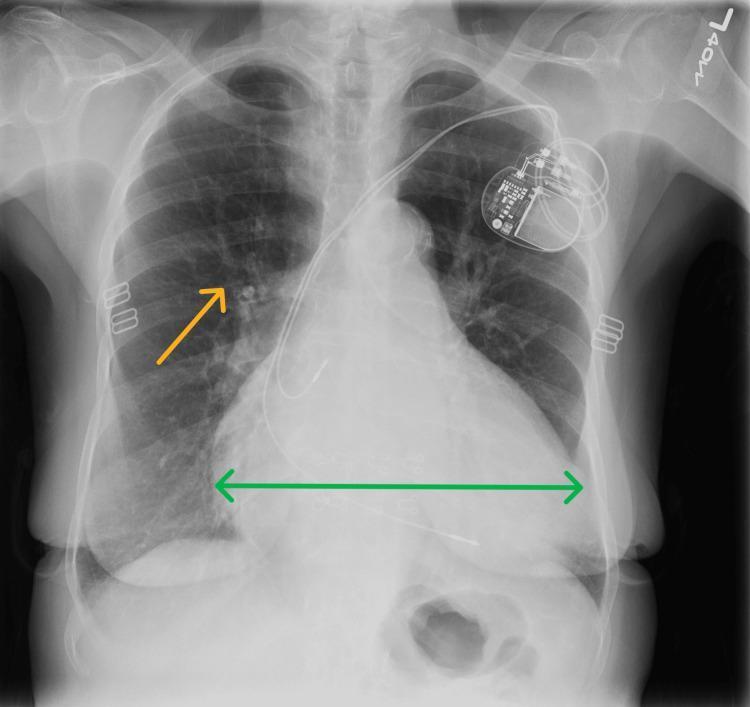
A frontal (posteroanterior) chest radiograph demonstrating significant cardiomegaly and signs of pulmonary congestion. The cardiac silhouette is markedly enlarged, with the cardiothoracic ratio exceeding 50%, as highlighted by a green double-headed horizontal arrow across the maximum transverse diameter of the heart. In the upper left chest wall, a permanent pacemaker is visible with leads extending toward the right atrium and right ventricle. The lung fields show increased interstitial markings and cephalization of the pulmonary vessels, particularly in the right mid-lung zone (indicated by an orange arrow), consistent with mild pulmonary vascular congestion or volume overload. Small metallic surgical clips are noted in the left axillary/chest wall region. This diagnostic image is intended to illustrate the radiographic presentation of heart failure with reduced ejection fraction (HFrEF) and chronic volume overload in a clinical setting.
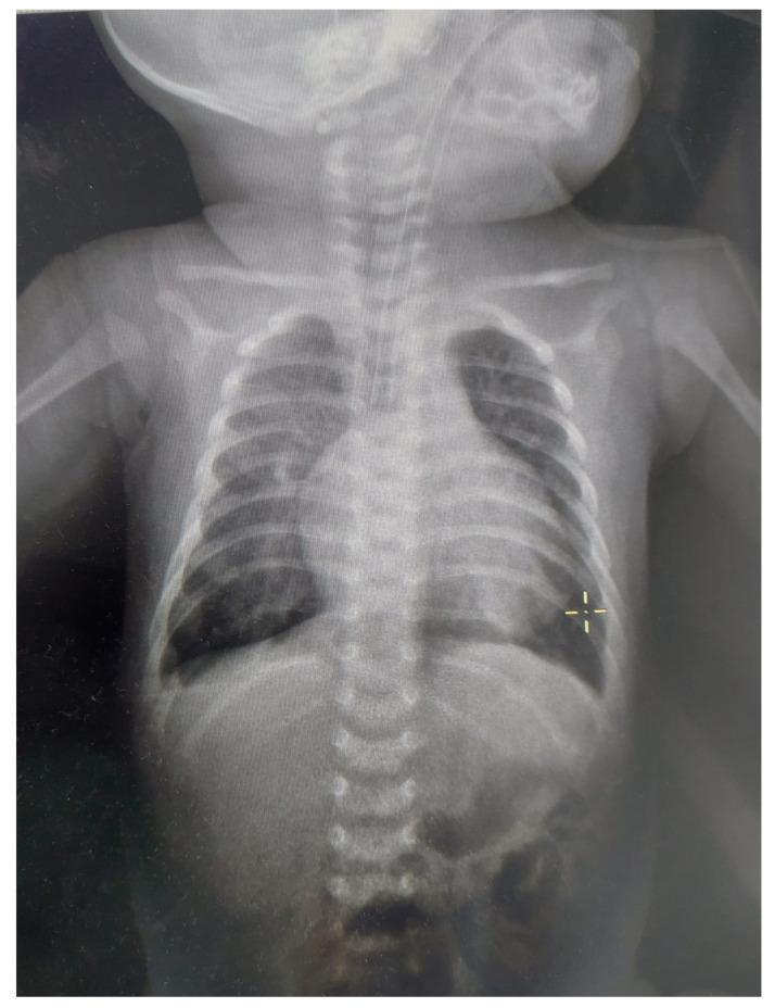
Anteroposterior (AP) chest radiograph of a one-month-old infant in a neonatal intensive care setting. The image demonstrates a significantly enlarged cardiac silhouette with a cardiothoracic ratio exceeding 0.6, suggestive of cardiomegaly. There is a visible endotracheal tube in situ, confirming mechanical ventilation. The lung fields show generalized increased opacity and decreased lung volumes consistent with pulmonary hypoventilation. Accentuation of the pulmonary hilar regions is noted, indicating increased pulmonary vascular markings and potential pulmonary congestion or overload. Bony structures, including the ribs, clavicles, and vertebral column, appear appropriate for age without gross fractures. The overall clinical picture is indicative of congenital heart disease, specifically a complete atrioventricular canal defect (CAVC), leading to cardiac enlargement and pulmonary circulatory overload.
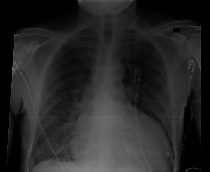
This diagnostic image is a posterior-anterior (PA) view chest radiograph. The primary anatomical findings include significant cardiomegaly, evidenced by an increased cardiothoracic ratio with a widened heart silhouette and lateral displacement of the left heart border. Both lung fields demonstrate diffuse, bilateral pulmonary infiltrates, characterized by hazy, increased opacification that obscures normal vascular markings. These findings are clinically suggestive of pulmonary edema and congestive heart failure. Additionally, visible medical artifacts include external monitoring leads and cables overlying the thoracic cage and axillary regions. The trachea is midline, and the visualized bony structures, including the clavicles and ribs, appear intact. This radiograph serves as a key educational example of cardiogenic pulmonary congestion in the context of cardiovascular pathology, such as syphilitic aortitis or aortic root aneurysm leading to heart failure.
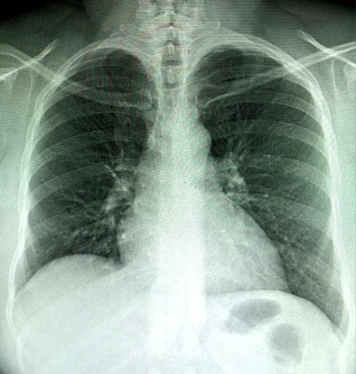
This diagnostic image is a frontal (posteroanterior) chest radiograph. The primary finding is a moderate enlargement of the cardiac silhouette, consistent with cardiomegaly, characterized by a cardiothoracic ratio of approximately 0.60. The lung fields show a diffuse accentuation of the bronchovascular pattern, appearing as branching linear densities radiating from the hila. This increased interstitial marking is most pronounced at the right lung base. Both costophrenic angles appear relatively sharp, and there is no evidence of discrete pulmonary consolidation, large pleural effusions, or pneumothorax. The mediastinal contours are intact, and the visualized bony structures including the ribs and clavicles appear unremarkable. This radiograph is an educational example used to demonstrate findings of early congestive heart failure or chronic pulmonary congestion, highlighting the visual assessment of the heart-to-thorax ratio and the distribution of pulmonary vascular markings.
ECG left ventricular hypertrophy strain pattern 12 lead
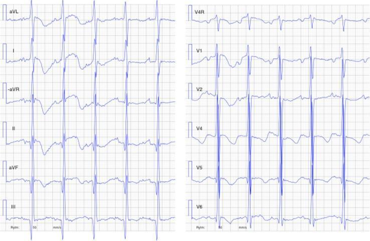
This diagnostic image is a 12-lead electrocardiogram (ECG) including right-sided lead V4R, obtained from a 4-month-old infant with dilated cardiomyopathy. The tracing demonstrates sinus tachycardia with a high ventricular rate. Key pathological features include markedly enlarged QRS amplitudes, particularly evident in the precordial leads (V1-V6) and limb leads (I, aVL), consistent with severe left ventricular hypertrophy. There are widespread ST-segment abnormalities characterized by down-sloping ST-segment depression and T-wave inversion/flattening in the lateral and inferior leads (I, aVL, II, aVF, V4-V6), representing a ventricular strain pattern. The P-waves are enlarged, suggesting atrial enlargement. Additionally, the QT interval is corrected (QTc) and appears prolonged. This ECG illustrates significant myocardial stress and structural remodeling associated with severe heart failure and pediatric cardiomyopathy.
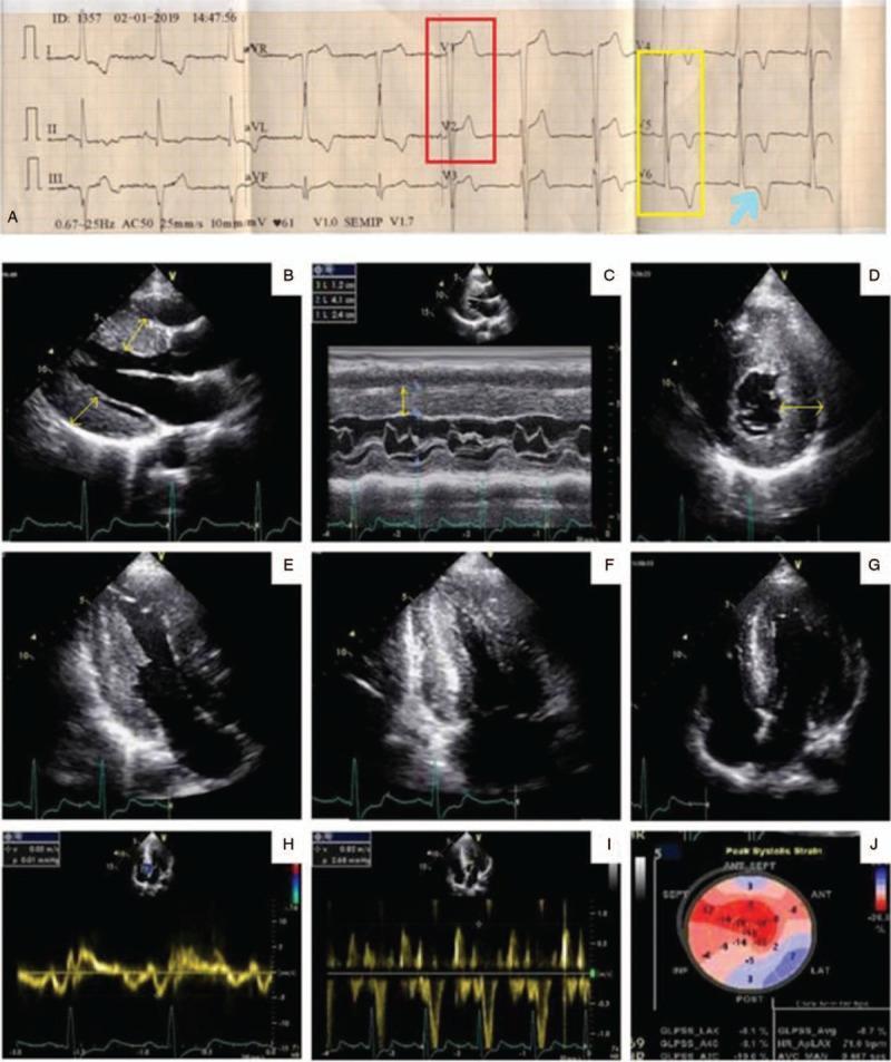
A multi-panel clinical diagnostic figure comprising a surface electrocardiogram (ECG) and transthoracic echocardiography (TTE) images related to cardiac amyloidosis evaluation. Panel A displays a 12-lead ECG showing signs of left ventricular hypertrophy (LVH), with high-amplitude QRS complexes in V2 (red box) and V5-V6 (yellow box) accompanied by a classic ST-segment depression and T-wave inversion indicative of a strain pattern (blue arrow). Panels B-G show 2D and M-mode echocardiography images illustrating significant asymmetric septal hypertrophy, with the interventricular septum measuring 2.4 cm. A distinctive 'speckled' or granular myocardial texture is visible in the short-axis views (D-E). Panels H-I utilize Tissue Doppler imaging to evaluate diastolic dysfunction, showing an E/e' ratio suggestive of grade 2 dysfunction. Panel J presents a bull's-eye map from speckle-tracking strain echocardiography, demonstrating a characteristic 'apical sparing' pattern (marked diminution of longitudinal strain in the basal and mid-segments with preserved apical function), which is highly suggestive of cardiac amyloidosis.
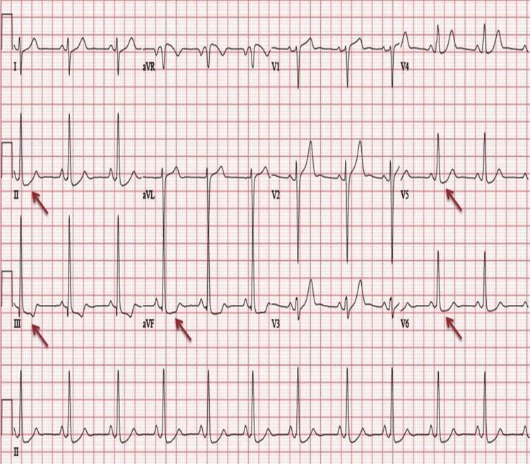
This diagnostic image is a 12-lead electrocardiogram (ECG) demonstrating classic features of Left Ventricular Hypertrophy (LVH) with associated strain patterns. The tracing shows a sinus rhythm with high QRS voltage, characterized by tall R-waves in the lateral leads (V5, V6) and deep S-waves in the right precordial leads (V1, V2), satisfying Sokolow-Lyon criteria for LVH. Red arrows highlight significant ST-segment depression and T-wave inversions in the inferolateral leads (II, III, aVF, V5, and V6). This repolarization abnormality is characteristic of a 'strain pattern' often seen in severe chronic pressure or volume overload of the left ventricle. Clinical relevance includes the assessment of hypertensive heart disease and cardiomyopathies, such as left ventricular non-compaction (LVNC). The ECG serves as a primary screening tool for identifying structural heart changes and associated ischemic or metabolic disturbances.
2022 AHA ACC HFSA guideline beta blockers digoxin heart failure recommendations
~/
mkdir -p "/tmp/workspace/cardiomegaly-ppt/src" "/tmp/workspace/cardiomegaly-ppt/output" "/tmp/workspace/cardiomegaly-ppt/assets" && python /tmp/skills/shared/scripts/fetch_images.py "https://cdn.orris.care/cdss_images/pmc_clinical_VQA_4ee8d8c1a9e409dcc3d45aef14b4e850b78c1d7c05459012be5ab562929b6e71.jpg" "https://cdn.orris.care/cdss_images/pmc_clinical_VQA_03152b722b1cb86ef1d57f4d755aa687989488df7da9defa36d7d35f9ab80e28.jpg" > "/tmp/workspace/cardiomegaly-ppt/assets/image_data.json"
python - <<'PY' import json,base64,os p='/tmp/workspace/cardiomegaly-ppt/assets/image_data.json' with open(p) as f: d=json.load(f) for i,x in enumerate(d): raw=base64.b64decode(x['base64'].split(',',1)[1]) open(f'/tmp/workspace/cardiomegaly-ppt/assets/img{i+1}.jpg','wb').write(raw) print([(i+1,len(base64.b64decode(x['base64'].split(',',1)[1]))) for i,x in enumerate(d)]) PY
cat > "/tmp/workspace/cardiomegaly-ppt/src/create_ppt.py" <<'PY' from pptx import Presentation from pptx.util import Inches, Pt from pptx.dml.color import RGBColor from pptx.enum.text import PP_ALIGN, MSO_ANCHOR from pptx.enum.shapes import MSO_SHAPE from pptx.enum.dml import MSO_THEME_COLOR from pptx.enum.text import MSO_AUTO_SIZE from pptx.enum.shapes import MSO_CONNECTOR from pptx import Presentation from pathlib import Path OUT = Path('/tmp/workspace/cardiomegaly-ppt/output/Cardiomegaly_Talha_Khan_610A.pptx') ASSET = Path('/tmp/workspace/cardiomegaly-ppt/assets') prs = Presentation(); prs.slide_width=Inches(13.333); prs.slide_height=Inches(7.5) blank=prs.slide_layouts[6] NAVY=RGBColor(11,31,52); BLUE=RGBColor(25,116,171); TEAL=RGBColor(25,150,152); RED=RGBColor(202,66,67); GOLD=RGBColor(237,171,42); PALE=RGBColor(238,246,249); WHITE=RGBColor(255,255,255); TEXT=RGBColor(34,49,63); MUTED=RGBColor(91,108,122); GREEN=RGBColor(46,139,87); LIGHTRED=RGBColor(252,235,235) def rect(s,x,y,w,h,fill,line=None,r=False): sh=s.shapes.add_shape(MSO_SHAPE.ROUNDED_RECTANGLE if r else MSO_SHAPE.RECTANGLE, Inches(x), Inches(y), Inches(w), Inches(h)) sh.fill.solid(); sh.fill.fore_color.rgb=fill sh.line.color.rgb = line if line else fill return sh def textbox(s,x,y,w,h,text,size=18,color=TEXT,bold=False,align=PP_ALIGN.LEFT,font='Aptos',val=MSO_ANCHOR.TOP): sh=s.shapes.add_textbox(Inches(x),Inches(y),Inches(w),Inches(h)); tf=sh.text_frame tf.clear(); tf.word_wrap=True; tf.margin_left=Inches(.06); tf.margin_right=Inches(.06); tf.margin_top=Inches(.03); tf.margin_bottom=Inches(.02); tf.vertical_anchor=val lines=text.split('\n') for i,line in enumerate(lines): p=tf.paragraphs[0] if i==0 else tf.add_paragraph(); p.text=line; p.alignment=align; p.space_after=Pt(4) for r in p.runs: r.font.name=font; r.font.size=Pt(size); r.font.bold=bold; r.font.color.rgb=color return sh def title(s,n,t,sub=None): rect(s,0,0,13.333,.48,NAVY); textbox(s,.55,.61,11.8,.48,t,27,NAVY,True) if sub: textbox(s,.58,1.10,11.7,.28,sub,11,MUTED) textbox(s,12.18,.12,.65,.2,f'{n:02}',10,WHITE,True,PP_ALIGN.RIGHT) rect(s,.56,1.46,1.05,.05,TEAL) def footer(s): rect(s,0,7.22,13.333,.28,NAVY); textbox(s,.55,7.255,8,.16,'Cardiomegaly | Talha Khan | Group 610A',8,WHITE) def bullets(s, items, x=.75,y=1.75,w=6.0,h=4.9,size=19,accent=TEAL): for i,item in enumerate(items): yy=y+i*(h/len(items)) rect(s,x,yy+.10,.12,.12,accent) textbox(s,x+.27,yy,w-.27,h/len(items)-.03,item,size,TEXT,False) def card(s,x,y,w,h,head,body,color=BLUE): rect(s,x,y,w,h,WHITE,RGBColor(210,222,230),True); rect(s,x,y,w,.12,color) textbox(s,x+.22,y+.25,w-.44,.34,head,16,color,True) textbox(s,x+.22,y+.70,w-.44,h-.82,body,13,TEXT) def add_notes(slide, note): notes=slide.notes_slide.notes_text_frame notes.text=note def image(s,path,x,y,w,h,cap=None): s.shapes.add_picture(str(path), Inches(x), Inches(y), width=Inches(w), height=Inches(h)) if cap: textbox(s,x,y+h+.03,w,.30,cap,9,MUTED,False,PP_ALIGN.CENTER) def cover(): s=prs.slides.add_slide(blank); rect(s,0,0,13.333,7.5,NAVY); rect(s,0,0,.22,7.5,TEAL); rect(s,8.9,0,4.43,7.5,RGBColor(16,55,79)) # stylized cardiac silhouette for x,y,sz,c in [(9.65,1.05,2.2,TEAL),(10.7,2.3,1.55,BLUE),(9.2,3.3,1.9,RED)]: sh=s.shapes.add_shape(MSO_SHAPE.HEART,Inches(x),Inches(y),Inches(sz),Inches(sz)); sh.fill.solid();sh.fill.fore_color.rgb=c;sh.line.color.rgb=c textbox(s,.8,1.2,7.5,.55,'CARDIOMEGALY',36,WHITE,True) textbox(s,.82,1.93,7.4,.8,'Causes, types, clinical features, ECG and radiological changes, treatment, beta-blockers and cardiac glycosides',20,RGBColor(211,229,238)) rect(s,.82,3.35,2.0,.06,GOLD); textbox(s,.82,4.0,4.8,.35,'Name: Talha Khan',20,WHITE,True); textbox(s,.82,4.48,4.8,.35,'Group: 610A',20,WHITE,True) textbox(s,.82,6.65,6,.24,'Educational presentation | September 2026',10,RGBColor(185,208,220)) add_notes(s,'Opening slide. Introduce cardiomegaly as an enlarged heart silhouette or true chamber/wall enlargement, not a diagnosis by itself.') def standard(n,t,items,sub=None, note=''): s=prs.slides.add_slide(blank); title(s,n,t,sub); bullets(s,items); footer(s); add_notes(s,note); return s cover() standard(2,'Learning objectives',['Define cardiomegaly and distinguish it from pseudo-cardiomegaly.','Classify cardiomegaly by chamber, mechanism, and time course.','Recognize clinical, ECG, and chest radiographic features.','Outline cause-directed treatment and safe use of beta-blockers and cardiac glycosides.'],note='State that the presentation focuses on adult principles; individual therapy needs clinical assessment and echocardiography.') standard(3,'What is cardiomegaly?',['Cardiomegaly means enlargement of the heart caused by chamber dilatation, myocardial hypertrophy, or both.','It may be physiological, for example in trained athletes, or pathological.','On an upright posteroanterior chest radiograph, a cardiothoracic ratio (CTR) greater than 0.50 suggests an enlarged cardiac silhouette.','Chest radiography is a screening tool. Echocardiography determines chamber size, wall thickness, valves, and ventricular function.'],note='Mention that CTR may mislead in AP portable films, poor inspiration, obesity, pericardial effusion, or chest-wall deformity.') standard(4,'Why the heart enlarges',['Pressure overload causes hypertrophy: myocytes thicken to generate higher pressure.','Volume overload causes dilatation: increased end-diastolic volume enlarges a chamber.','Myocardial injury or genetic disease can reduce contractility and promote remodeling.','Pericardial fluid can mimic cardiomegaly on radiography without true cardiac enlargement.'],note='Use the terms concentric hypertrophy, eccentric hypertrophy and dilatation.') standard(5,'Types: by anatomical pattern',['Left ventricular enlargement: hypertension, aortic valve disease, ischemic/dilated cardiomyopathy.','Right ventricular enlargement: pulmonary hypertension, chronic lung disease, pulmonary valve disease, congenital shunts.','Biatrial enlargement: chronic mitral disease, restrictive cardiomyopathy, long-standing atrial arrhythmias.','Global enlargement: dilated cardiomyopathy, advanced valvular disease, large shunts, or combined heart failure.'],note='Emphasize that chamber pattern helps suggest etiology but is confirmed by echo or cardiac MRI.') standard(6,'Types: by mechanism and time course',['Concentric hypertrophy: increased wall thickness with relatively small/normal cavity, classically pressure overload.','Eccentric hypertrophy/dilatation: increased cavity size with increased mass, classically volume overload or systolic failure.','Acute enlargement can occur with acute valvular regurgitation or myocarditis, but marked radiographic enlargement usually takes time.','Physiological remodeling in athletes should be differentiated from disease using history, examination, ECG, and imaging.'],note='Avoid calling all large hearts “hypertrophy”. Dilatation and hypertrophy have different causes.') standard(7,'Major causes: pressure overload',['Systemic hypertension: common cause of left ventricular hypertrophy.','Aortic stenosis or coarctation: chronic left ventricular pressure load.','Pulmonary hypertension: right ventricular hypertrophy and later dilatation.','Pulmonic stenosis or chronic thromboembolic disease: right-sided pressure overload.'],note='Relate pressure load to concentric hypertrophy, then eventual decompensation/dilatation.') standard(8,'Major causes: volume overload',['Valvular regurgitation: aortic, mitral, tricuspid, or pulmonary regurgitation.','Congenital shunts: atrial septal defect, ventricular septal defect, patent ductus arteriosus.','High-output states: severe anemia, thyrotoxicosis, arteriovenous fistula, and some nutritional disorders.','Chronic kidney disease and salt-water retention can worsen volume load and heart failure.'],note='Explain that shunts may create right-sided or biventricular enlargement depending on the lesion.') standard(9,'Major causes: myocardial disease',['Ischemic heart disease and prior myocardial infarction with ventricular remodeling.','Dilated cardiomyopathy: idiopathic, genetic, viral/inflammatory, toxin-related, or peripartum.','Hypertrophic cardiomyopathy and infiltrative/storage disease such as amyloidosis or Fabry disease.','Myocarditis, tachycardia-induced cardiomyopathy, and stress-related cardiomyopathy.'],note='A cause-based diagnosis determines management. Cardiomegaly itself is not treated as a stand-alone disorder.') standard(10,'Specific clinical symptoms',['Breathlessness on exertion, orthopnea, paroxysmal nocturnal dyspnea, and reduced exercise tolerance.','Fatigue, weakness, early satiety, and reduced concentration due to low cardiac output.','Palpitations, dizziness, presyncope/syncope, or chest pain depending on rhythm and cause.','Leg swelling, abdominal distension, weight gain, and nocturia from congestion.'],note='Symptoms can be absent early. Ask about rapid symptom change, which may signify acute heart failure or arrhythmia.') standard(11,'Key physical signs',['Displaced and diffuse apical impulse; heaves may suggest ventricular hypertrophy.','S3 gallop may indicate volume overload/systolic dysfunction; S4 may occur with a stiff hypertrophied ventricle.','Murmurs point to valvular disease; irregular pulse may indicate atrial fibrillation.','Raised JVP, basal crackles, hepatomegaly, ascites, and peripheral edema indicate congestion.'],note='Signs must be interpreted together with history, ECG, imaging and biomarkers.') # image slide CXR s=prs.slides.add_slide(blank); title(s,12,'Radiological changes: chest radiograph','A chest X-ray supports, but does not establish, the diagnosis.'); image(s,ASSET/'img1.jpg',.65,1.75,6.1,4.55,'PA radiograph: enlarged cardiac silhouette with pulmonary vascular congestion (educational image).'); bullets(s,['CTR >0.50 on an upright PA film suggests cardiomegaly.','Assess shape: left ventricular prominence, right-sided contour changes, or globular silhouette.','Look for pulmonary venous redistribution, interstitial/alveolar edema, pleural effusions, and vascular congestion.','Confirm with echocardiography; AP portable films can magnify the heart.'],7.05,1.75,5.55,4.9,15,RED); footer(s); add_notes(s,'Read the film systematically: projection, inspiration, CTR, cardiac contours, pulmonary vessels, edema, and pleura.') standard(13,'Radiological changes: beyond X-ray',['Echocardiography is first-line to measure chamber dimensions, ejection fraction, wall thickness, valves, filling pressures, and pericardial effusion.','Cardiac MRI characterizes myocardial morphology, function, fibrosis, inflammation, and infiltrative disease.','CT may define coronary, aortic, pericardial, or congenital structural disease when indicated.','Imaging must be integrated with clinical findings and ECG.'],note='No test should be ordered in isolation. Echo answers the most common questions after an enlarged silhouette is seen.') # ECG image slide s=prs.slides.add_slide(blank); title(s,14,'ECG changes associated with cardiomegaly','ECG findings depend on the enlarged chamber and underlying disease.'); image(s,ASSET/'img2.jpg',.55,1.65,5.8,4.95,'Example: LVH voltage with lateral ST-T “strain” pattern (educational image).'); bullets(s,['LVH: increased QRS voltage, left-axis deviation, and lateral ST depression/T-wave inversion (“strain”).','RVH: right-axis deviation, dominant R in V1, and right-precordial strain.','Atrial enlargement: broad/notched P waves in LAE or peaked P waves in RAE.','Other possible findings: atrial fibrillation, bundle branch block, Q waves from infarction, or low voltage with effusion/infiltration.'],6.65,1.75,5.95,4.75,15,TEAL); footer(s); add_notes(s,'ECG cannot diagnose cardiomegaly reliably. It detects electrical effects and clues to etiology.') standard(15,'Diagnostic approach',['History: hypertension, coronary disease, valvular/rheumatic disease, alcohol/toxins, family history, pregnancy, systemic symptoms.','Examination: signs of heart failure, valvular lesions, arrhythmia, and systemic disease.','Initial tests: ECG, chest X-ray, blood tests such as CBC, renal function, electrolytes, thyroid tests, and natriuretic peptides when appropriate.','Confirm and phenotype with echocardiography. Consider MRI, ischemia evaluation, genetic or disease-specific tests when indicated.'],note='Include red flags: syncope, severe dyspnea, hypotension, acute pulmonary edema, ongoing chest pain, or sustained ventricular arrhythmia need urgent assessment.') standard(16,'Treatment principles',['Treat the cause: control blood pressure, repair/replace significant valves, treat ischemia, correct endocrine or hematologic causes, and manage congenital lesions.','Treat heart failure phenotype: diuretics for congestion plus evidence-based long-term therapy when HFrEF is present.','Manage arrhythmias, especially atrial fibrillation, with rate/rhythm strategy and anticoagulation when indicated.','Supportive care: salt moderation when advised, fluid plan for selected patients, smoking cessation, activity/cardiac rehabilitation, and vaccination.'],note='Do not imply every cardiomegaly needs the same drugs. Therapy is phenotype and cause specific.') standard(17,'HFrEF therapy: a practical framework',['For symptomatic HFrEF, guideline-directed therapy typically includes an ARNI/ACE inhibitor/ARB, evidence-based beta-blocker, mineralocorticoid receptor antagonist, and SGLT2 inhibitor.','Loop diuretics relieve fluid congestion but do not replace disease-modifying therapy.','Iron deficiency treatment, device therapy, revascularization, valve intervention, or advanced HF referral may be appropriate in selected patients.','Titrate medications gradually with blood pressure, pulse, renal function, potassium, symptoms, and volume status monitoring.'],note='This is a high-level educational framework, not a prescription. The four foundational drug classes are for HFrEF, not automatically for all cardiomegaly.') standard(18,'Beta-blockers: specific features',['In stable symptomatic HFrEF, the mortality-reducing agents are bisoprolol, carvedilol, and sustained-release metoprolol succinate. Start low and titrate slowly when euvolemic and clinically stable.','Benefits: lower mortality and hospitalization, improve remodeling, slow heart rate, and help control rate in atrial fibrillation.','Monitor heart rate, blood pressure, fatigue, fluid retention, AV block, bronchospasm risk, and glucose masking in diabetes.','Avoid initiation or dose escalation during acute decompensated heart failure, shock, marked bradycardia, or high-grade AV block unless paced/specialist-directed.'],note='Beta-blockers can transiently worsen symptoms at initiation. Reassess volume status before increasing dose.') standard(19,'Cardiac glycosides: digoxin',['Digoxin inhibits Na+/K+-ATPase, increasing intracellular calcium and contractility; it also increases vagal tone and slows AV nodal conduction.','Main roles: selected symptomatic HFrEF despite guideline-directed therapy to reduce HF hospitalization, and ventricular rate control in atrial fibrillation when appropriate.','It does not provide the mortality benefit of evidence-based beta-blockers in HFrEF.','Use caution with renal impairment, older age, low body mass, hypokalemia/hypomagnesemia, interacting drugs, sinus-node disease, and AV block.'],note='Discuss digoxin as an adjunct in selected patients, never a replacement for foundational HFrEF treatment.') standard(20,'Digoxin toxicity and safe monitoring',['Typical toxicity: nausea, vomiting, anorexia, confusion, visual disturbance (yellow/blurred vision), bradycardia, AV block, atrial tachycardia with block, or ventricular arrhythmias.','Check renal function, potassium, magnesium, calcium, heart rate, ECG, and drug interactions. Serum level timing and targets follow local protocol.','Hypokalemia increases toxicity risk; amiodarone, verapamil, macrolides, and other drugs may raise levels.','Suspected severe toxicity or life-threatening arrhythmia requires urgent care; digoxin immune Fab may be indicated under specialist management.'],note='End with safety message: digoxin has a narrow therapeutic index. Never recommend a dose in a generic slide deck.') # sources slide s=prs.slides.add_slide(blank); title(s,21,'Key take-home points and sources'); card(s,.65,1.7,5.9,4.7,'TAKE-HOME POINTS','• Cardiomegaly is a sign, not a diagnosis.\n• Define the chamber pattern and cause with echocardiography.\n• ECG and radiography provide clues, but have limitations.\n• Treat the cause and the heart-failure/rhythm phenotype.\n• Beta-blockers are foundational in stable HFrEF; digoxin is selective and requires close monitoring.',TEAL); card(s,6.85,1.7,5.8,4.7,'SELECTED SOURCES','• Fuster & Hurst’s The Heart, 15th ed.: cardiac size; chest radiography; ECG.\n• Goldman-Cecil Medicine: radiographic assessment of heart size; digoxin.\n• 2022 AHA/ACC/HFSA Guideline for the Management of Heart Failure.\n• Images: indexed educational clinical images from PMC clinical VQA library.\n\nFor education only. Clinical decisions require clinician assessment and current local guidelines.',BLUE); footer(s); add_notes(s,'Summarize and invite questions. Cite 2022 AHA/ACC/HFSA: beta blockers are Class I in symptomatic HFrEF; digoxin may be considered to reduce HF admissions in selected patients.') # meta settings prs.core_properties.title='Cardiomegaly: causes, diagnosis, and treatment' prs.core_properties.author='Talha Khan' prs.core_properties.subject='Group 610A' prs.save(OUT) print(OUT) PY python "/tmp/workspace/cardiomegaly-ppt/src/create_ppt.py"
python - <<'PY' from pptx import Presentation from pathlib import Path p=Path('/tmp/workspace/cardiomegaly-ppt/output/Cardiomegaly_Talha_Khan_610A.pptx') print('exists',p.exists(),'bytes',p.stat().st_size) prs=Presentation(p) print('slides',len(prs.slides)) for i,s in enumerate(prs.slides,1): text=' | '.join(sh.text.replace('\n',' / ') for sh in s.shapes if hasattr(sh,'text_frame') and sh.text.strip()) print(i, text[:150]) PY
Cardiomegaly Talha Khan 610A
Presentation · PPTX